section name header

Disease Prologue

Summary

Cancers Staged Using This Staging System

Squamous cell carcinoma and salivary gland carcinoma of all head and neck sites except HPV-related oropharynx cancer, nasopharynx cancer, melanoma, thyroid carcinoma, and sarcoma. Staging of the patient who presents with an occult primary tumor and EBV-unrelated and HPV-unrelated metastatic cervical lymphadenopathy is also included.

Cancers Not Staged Using This Staging System

These histopathologic types of cancer…Are staged according to the classification for…and can be found in chapter…
Nasopharyngeal cancerNasopharynx9
HPV-related oropharynx cancerHPV-mediated (p16+) oropharyngeal cancer10
MelanomaMelanoma of the skin47
Mucosal melanomaMucosal melanoma of the head and neck14
Thyroid carcinomaThyroid carcinoma73-74
Soft tissue sarcomaSoft tissue sarcoma of the head and neck40
EyelidEyelid carcinoma64

Summary of Changes

ChangeDetails of ChangeLevel of Evidence
Definition of Regional Lymph Node (N)Separate N staging approaches have been described for HPV-related and HPV-unrelated cancers.II1,2
Definition of Regional Lymph Node (N)Separate N category approaches have been described for patients treated without cervical lymph node dissection (clinical N) and patients treated with cervical lymph node dissection (pathological N).II1,2
Definition of Regional Lymph Node (N)Extranodal extension (ENE) is introduced as a descriptor in all HPV-unrelated cancers.II2
ENE in HPV-negative cancersOnly clinically and radiographically overt ENE should be used for cN.II2
ENE in HPV-negative cancersAny pathologically detected ENE is considered ENE(+) and is used for pN.II2
ENE in HPV-negative cancersPresence of ENE is designated pN2a for a single ipsilateral node less than 3 cm and pN3b for all other node(s).II2
Classification of ENEClinically overt ENE is classified as ENEc and is considered ENE(+) for cN.III3
Classification of ENEPathologically detected ENE is classified as either ENEmi (less than or equal to 2 mm) or ENEma (greater than 2mm) for data collection purposes only but both are considered ENE(+) for definition of pN.III3
Occult Primary TumorStaging of the patient who presents with EBV-unrelated and HPV-unrelated metastatic cervical lymphadenopathy is now included in this chapter.IV

ICD-O-3 Topography Codes

CodeDescription
C00.0External upper lip
C00.1External lower lip
C00.2External lip, NOS
C00.3Mucosa of upper lip
C00.4Mucosa of lower lip
C00.5Mucosa of lip, NOS
C00.6Commissure of lip
C00.8Overlapping lesion of lip
C00.9Lip, NOS
C01.9Base of tongue, NOS
C02.0Dorsal surface of tongue, NOS
C02.1Border of tongue
C02.2Ventral surface of tongue, NOS
C02.3Anterior two-thirds of tongue, NOS
C02.4Lingual tonsil
C02.8Overlapping lesion of tongue
C02.9Tongue, NOS
C03.0Upper gum
C03.1Lower gum
C03.9Gum, NOS
C04.0Anterior floor of mouth
C04.1Lateral floor of mouth
C04.8Overlapping lesion of floor of mouth
C04.9Floor of mouth, NOS
C05.0Hard palate
C05.1Soft palate, NOS
C05.2Uvula
C05.8Overlapping lesion of palate
C05.9Palate, NOS
C06.0Cheek mucosa
C06.1Vestibule of mouth
C06.2Retromolar area
C06.8Overlapping lesion of other and unspecified parts of mouth
C06.9Mouth, NOS
C07.9Parotid gland
C08.0Submand ibular gland
C08.1Sublingual gland
C08.8Overlapping lesion of major salivary gland s
C08.9Major salivary gland , NOS
C09.0Tonsillar fossa
C09.1Tonsillar pillar
C09.8Overlapping lesion of tonsil
C09.9Tonsil, NOS
C10.0Vallecula
C10.1Anterior surface of epiglottis
C10.2Lateral wall of oropharynx
C10.3Posterior wall of oropharynx
C10.4Branchial cleft
C10.8Overlapping lesions of oropharynx
C10.9Oropharynx, NOS
C12.9Pyriform sinus
C13.0Postcricoid region
C13.1Hypopharyngeal aspect of aryepiglottic fold
C13.2Posterior wall of hypopharynx
C13.8Overlapping lesion of hypopharynx
C13.9Hypopharynx, NOS
C14.0Pharynx, NOS
C14.2Waldeyer ring
C14.8Overlapping lesion of lip, oral cavity, and pharynx
C30.0Nasal cavity
C30.1Middle ear
C31.0Maxillary sinus
C31.1Ethmoid sinus
C31.2Frontal sinus
C31.3Sphenoid sinus
C31.8Overlapping lesion of accessory sinuses
C31.9Accessory sinus, NOS
C32.0Glottis
C32.1Supraglottis
C32.2Subglottis
C32.3Laryngeal cartilage
C32.8Overlapping lesion of larynx
C32.9Larynx, NOS
C44.0Skin of lip, NOS
C44.2External ear
C44.3Skin of other and unspecified parts of face
C44.4Skin of scalp and neck
C44.8Overlapping lesion of skin
C76.0Head, face or neck, NOS
C80.9Unknown primary site

WHO Classification of Tumors

This list includes histology codes and preferred terms from the WHO Classification of Tumors and the International Classification of Diseases for Oncology (ICD-O). Most of the terms in this list represent malignant behavior. For cancer reporting purposes, behavior codes /3 (denoting malignant neoplasms), /2 (denoting in situ neoplasms), and in some cases /1 (denoting neoplasms with uncertain and unknown behavior) may be appended to the 4-digit histology codes to create a complete morphology code.

CodeDescription
8010Carcinoma, NOS
8046Non-small cell carcinoma
8051Verrucous squamous cell carcinoma
8052Papillary squamous cell carcinoma
8070Squamous cell carcinoma
8071Keratinizing squamous cell carcinoma
8072Non-keratinizing squamous cell carcinoma
8073Squamous cell carcinoma, small cell, nonkeratinizing
8074Spindle cell squamous cell carcinoma
8082Lymphoepithelial carcinoma
8083Basaloid squamous cell carcinoma
8084Squamous cell carcinoma, clear cell type
8121Schneiderian carcinoma
8140Adenocarcinoma, NOS
8147Basal cell adenocarcinoma
8200Adenoid cystic carcinoma
8310Clear cell carcinoma
8430Mucoepidermoid carcinoma
8450Papillary cystadenocarcinoma, NOS
8480Mucinous carcinoma
8525Polymorphous adenocarcinoma
8550Acinic cell carcinoma
8562Epithelial-myoepithelial carcinoma
8941Carcinoma ex pleomorphic adenoma

Histology is not ideal for clinical care, as the staging system was not developed using these cases. Data collectors may use this code only if there is not enough information in the medical record to document a more specific diagnosis.

El-Naggar AK, Chan JKC, Grand is JR, Takata T, Slootweg PJ, eds. World Health Organization Classification of Head and Neck Tumours. Lyon: IARC; 2017. Used with permission.

International Agency for Research on Cancer, World Health Organization. International Classification of Diseases for Oncology. ICD-O-3-Online.http://codes.iarc.fr/home. Accessed September 29, 2017. Used with permission.

Introduction

The presence of cervical lymph node metastases is the most important adverse prognostic feature for most cancers of the head and neck. However, the degree of impact on prognosis varies depending on host and tumor characteristics such as interplay of Human Papilloma Virus (HPV) initiation and smoking status in cancers of the oropharynx, and the presence of extranodal extension (ENE) for most other sites. The natural history and response to treatment of cervical nodal metastases from Epstein Barr Virus (EBV)-associated nasopharynx and HPV-related oropharynx primary sites are different in terms of their impact on prognosis, so they warrant separate N classification schemes. This difference also has led to reclassification of the T0 category (occult primary tumor) based on the EBV and HPV status of the metastatic cervical nodes. Current understand ing of other nodal features—such as the size of the largest metastatic node, number of metastatic nodes, and laterality—has improved based on an analysis of large multi-institutional datasets for oral cavity cancer. The American Joint Committee on Cancer (AJCC) Cancer Staging Manual, 8th Edition (8th Edition) incorporates these data while striving to maintain the balance between increased complexity and ease of use.

Two major changes are instituted in the 8th Edition for staging cervical nodal metastasis.

  1. For the first time, different clinical and pathological N classifications are proposed for definition of regional lymph node metastasis. This breaks with tradition in head and neck cancer, although distinct clinical and pathological classifications have been promulgated for other tumor sites (e.g., breast cancer). It is helpful to consider that clinical TNM (cTNM) and pathological TNM (pTNM) have different purposes. cTNM classification is required for all patients, including those undergoing surgery. pTNM classification provides additional information, but only for patients undergoing surgery; this guides the use of adjuvant treatment based on factors that become available after histopathological examination of the specimen (e.g., high-risk features of the primary site, ENE, laterality, and volume of metastatic nodal disease).
  2. A second significant change is in the use of ENE in categorizing metastatic cancer to neck nodes. The effect of ENE in prognosis in head and neck cancers is profound, except for those tumors associated with HPV. Most of the data supporting ENE as an adverse prognostic factor is based on histopathological characterization of ENE, especially the distinction between microscopic and macroscopic ENE. Only unquestionable clinically evident ENE that is supported by radiological evidence is to be used for clinical staging. The guiding principle is to assign the lesser attribute (ENE negative) if there are doubts in a particular case to avoid stage migration, in accordance with the uncertain rule, which articulates the concept that whenever there is a question, the lesser stage should be chosen. For clinical ENE, the known limitations of current imaging modalities to define ENE accurately demand that stringent criteria be met prior to assigning a clinical diagnosis of ENE if the patient is treated with nonsurgical therapy for neck metastasis. However, unambiguous evidence of gross ENE on clinical examination (e.g., invasion of skin, infiltration of musculature, dense tethering or fixation to adjacent structures, or cranial nerve, brachial plexus, sympathetic trunk, or phrenic nerve invasion with dysfunction) supported by strong radiographic evidence permits classification of disease as ENEc. Pathological ENE also will be clearly defined as extension of metastatic tumor (beyond the confines of the lymph node, through the lymph node capsule into the surrounding connective tissue, with or without associated stromal reaction). Again when there is doubt, recall the uncertain rule and assign a lower stage.

Anatomy

Regional Lymph Nodes

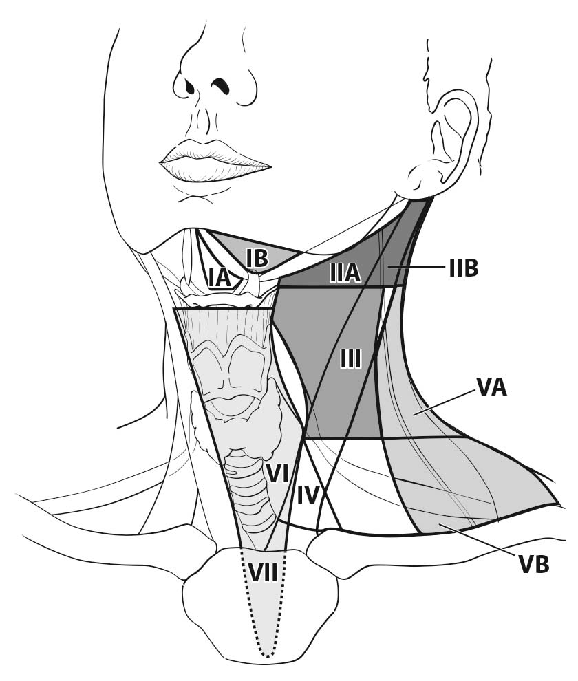
The lymph nodes in the neck may be subdivided into specific anatomic subsites and grouped into seven levels for ease of description (Figure 6.1, Tables 6.1 and 6.2).

Other groups

  • Suboccipital
  • Retropharyngeal
  • Parapharyngeal
  • Buccinator (facial)
  • Preauricular
  • Periparotid and intraparotid

6.1 Schematic indicating the location of the lymph node levels in the neck.

6.1 Anatomical structures defining the boundaries of the neck levels and sublevels.

Boundary LevelSuperiorInferiorAnterior (medial)Posterior (lateral)
IASymphysis of the mand ibleBody of the hyoidAnterior belly of the contralateral digastric muscleAnterior belly of the ipsilateral digastric muscle
IBBody of the mand iblePosterior belly of the diagastric muscleAnterior belly of the digastric muscleStylohyoid muscle
IIASkull baseHorizontal plane defined by the inferior border of the hyoid boneStylohyoid muscleVertical plane defined by the spinal accessory nerve
IIBSkull baseHorizontal plane defined by the inferior body of the hyoid boneVertical plane defined by the spinal accessory nerveLateral border of the sternocleidomastoid muscle
IIIHorizontal plane defined by the inferior body of the hyoidHorizontal plane defined by the inferior border of the cricoid cartilageLateral border of the sternohyoid muscleLateral border of the sternocleidomastoid or sensory branches of the cervical plexus
IVHorizontal plane defined by the inferior border of the cricoid cartilageClavicleLateral border of the sternohyoid muscleLateral border of the sternocleidomastoid or sensory branches of the cervical plexus
VAApex of the convergence of the sternocleidomastoid and trapezius musclesHorizontal plane defined by the lower border of the cricoid cartilagePosterior border of the sternocleido¬mastoid muscle or sensory branches of the cervical plexusAnterior border of the trapezius muscle
VBHorizontal plane defined by the lower border of the cricoid cartilageClaviclePosterior border of the sternocleido¬mastoid muscleAnterior border of the trapezius muscle
VIHyoid boneSuprasternal notchCommon carotid arteryCommon carotid artery
VIISuprasternal notchInnominate arterySternumTrachea, esophagus, and prevertebral fascia
Modified from Robbins KT, Clayman G, Levine PA, et al.,4 with permission of the American Medical Association.

6.2 Lymph node groups found within the seven levels and sublevels of the neck

Lymph Node GroupDescription
Submental (sublevel IA)Lymph nodes within the triangular boundary of the anterior belly of the digastric muscles and the hyoid bone. These nodes are at greatest risk for harboring metastases from cancers arising from the floor of mouth, anterior oral tongue, anterior mand ibular alveolar ridge, and lower lip.
Submand ibular (sublevel IB)Lymph nodes within the boundaries of the anterior and posterior bellies of the digastric muscle, the stylohyoid muscle, and the body of the mand ible. These include the pregland ular and the postgland ular nodes and the prevascular and postvascular nodes. The sub¬mand ib¬ular gland is included in the specimen when the lymph nodes within the triangle are removed. These nodes are at greatest risk for harboring metastases from cancers arising from the oral cavity, anterior nasal cavity, skin, and soft tissue structures of the midface, as well as from the submand ibular gland .
Upper Jugular (sublevels IIA and IIB)Lymph nodes located around the upper third of the internal jugular vein and adjacent spinal accessory nerve, extending from the level of the skull base (above) to the level of the inferior border of the hyoid bone (below). The anterior (medial) boundary is stylohyoid muscle (the radiologic correlate is the vertical plane defined by the posterior surface of the submand ibular gland ) and the posterior (lateral) boundary is the posterior border of the sternocleido¬mastoid muscle. Sublevel IIA nodes are located anterior (medial) to the vertical plane defined by the spinal accessory nerve. Sublevel IIB nodes are located posterior lateral to the vertical plane defined by the spinal accessory nerve. (The radiologic correlate is the lateral border of the internal jugular on a contrast-enhanced CT scan.) The upper jugular nodes are at greatest risk for harboring metastases from cancers arising from the oral cavity, nasal cavity, nasopharynx, oropharynx, hypopharynx, larynx, and parotid gland .
Middle Jugular (level III)Lymph nodes located around the middle third of the internal jugular vein, extending from the inferior border of the hyoid bone (above) to the inferior border of the cricoid cartilage (below). The anterior (medial) boundary is the lateral border of the sternohyoid muscle, and the posterior (lateral) boundary is the posterior border of the sterno¬cleidomastoid muscle. These nodes are at greatest risk for harboring metastases from cancers arising from the oral cavity, nasopharynx, oropharynx, hypopharynx, and larynx.
Lower Jugular (level IV)Lymph nodes located around the lower third of the internal jugular vein, extending from the inferior border of the cricoid cartilage (above) to the clavicle below. The anterior (medial) boundary is the lateral border of the sternohyoid muscle and the posterior (lateral) boundary is the posterior border of the sternocleido¬mastoid muscle. These nodes are at greatest risk for harboring metastases from cancers arising from the hypopharynx, thyroid, cervical esophagus, and larynx.
Posterior Triangle (sublevels VA and VB)This group is composed predominantly of the lymph nodes located along the lower half of the spinal accessory nerve and the transverse cervical artery. The supraclavicular nodes also are included in the posterior triangle group. The superior boundary is the apex formed by the convergence of the sternocleidomastoid and trapezius muscles; the inferior boundary is the clavicle; the anterior (medial) boundary is the posterior border of the sternocleidomastoid muscle; and the posterior (lateral) boundary is the anterior border of the trapezius muscle. Thus, sublevel VA includes the spinal accessory nodes, whereas sublevel VB includes the nodes following the transverse cervical vessels and the supraclavicular nodes, with the exception of the Virchow node, which is located in level IV. The posterior triangle nodes are at greatest risk for harboring metastases from cancers arising from the nasopharynx, oropharynx, and cutaneous structures of the posterior scalp and neck.
Anterior Compartment (level VI)Lymph nodes in this compartment include the pretracheal and paratracheal nodes, precricoid (Delphian) node, and the perithyroidal nodes, including the lymph nodes along the recurrent laryngeal nerves. The superior boundary is the hyoid bone; the inferior boundary is the suprasternal notch; and the lateral boundaries are the common carotid arteries. These nodes are at greatest risk for harboring metastases from cancers arising from the thyroid gland , glottic and subglottic larynx, apex of the piriform sinus, and cervical esophagus.
Superior Mediastinal (level VII)Lymph nodes in this group include pretracheal, paratracheal, and esophageal groove lymph nodes, extending from the level of the suprasternal notch cephalad and up to the innominate artery caudad. These nodes are at greatest risk of involvement by thyroid cancer and cancer of the esophagus.
Modified from Robbins KT, Clayman G, Levine PA, et al.,4 with permission of the American Medical Association.

Metastatic Sites

Classification Rules

Clinical Classification

When enlarged lymph nodes are detected, the size of the nodal mass(es) should be measured and their location should be recorded in terms of the levels of the neck (Tables 6.1 and 6.2). The presence of ENE can be diagnosed clinically by the presence of involvement of overlying skin, fixity to adjacent soft tissue, or clinical signs of cranial nerve or brachial plexus, sympathetic chain or phrenic nerve invasion. The presence of clinically evident gross ENE is designated ENEc(+).

Pathological confirmation of metastasis by a fine-needle aspirate, needle biopsy, excisional biopsy of a lymph node, or a sentinel node procedure should be assigned cN.

The Occult Primary Tumor (T0)

The head and neck region is unique among solid tumor sites because several different staging classifications are predicated upon anatomic site of the primary tumor. The AJCC Cancer Staging Manual, 7th Edition, T classifications for head and neck sites included T0 (primary site cannot be identified). This concept is not consistent with anatomic site staging and represents a problem if a primary tumor cannot be identified on clinical examination and with currently available radiographic imaging techniques. This dilemma of the occult primary has been partially resolved by improved understand ing of tumorigenesis and availability of cytologic and histologic methods to identify EBV- and HPV-related tumors, which are known to predominantly arise in the nasopharynx and oropharynx, respectively. In spite of modern technology, the origin of the primary tumor does remain unknown in all other patients whose primary tumor is clinically and radiographically occult and who present with EBV-negative and HPV-negative metastatic cervical node(s). Furthermore, T0 will still exist in salivary gland primary sites based on histology of the lymph node.

Three separate approaches are employed to stage patients who present with an occult primary tumor. The primary T category is described as T0 and the N category is designated according to the respective anatomic site based on EBV and HPV status: (1) patients with EBV-related cervical adenopathy are staged according to Chapter 9 (Nasopharynx); (2) patients with HPV-related cervical adenopathy are staged according to Chapter 10 (HPV-mediated oropharyngeal cancer (p16+)); and (3) all other patients with EBV-unrelated and HPV-unrelated cervical adenopathy are staged according to the N category described in this chapter. The stage groupings for these specific patients with occult primary tumors (T0) take into account the varying prognostic impact of metastatic cervical adenopathy for their different diseases. The stage groupings for EBV-related nasopharynx and HPV-related oropharynx cancer are described separately in the relevant chapters. Stage grouping for the patient with EBV-unrelated HPV-unrelated occult primary tumor is described in AJCC Prognostic Stage Groups in this chapter.

Imaging

Abnormal lymph nodes should be described according to the level of the neck that is involved, using the stand ard imaging-based classification (Table 6.1).

Ultrasonography (US) is a convenient and commonly used modality for assessment of the neck. However, interpretation is observer dependent and certain areas, such as the retropharyngeal nodes and mediastinal nodes, cannot be assessed with US. Currently, its utility is being defined in the assessment of ENE. Computed tomography (CT) or magnetic resonance (MR) imaging can be used for evaluating all nodal levels for metastatic involvement, with particular attention to the expected drainage pattern of the primary tumor site. Fluorodeoxyglucose (FDG) positron emission tomography (PET) may increase sensitivity and specificity over cross-sectional imaging alone for nodal detection, although small and cystic nodes may be falsely negative while reactive nodes may be falsely positive.

A maximum short-axis diameter of 10 mm for lymph nodes is commonly used to describe abnormal nodes, although this results in a high false-negative rate. Because size criteria alone are not reliable, small nodes, particularly in the expected drainage levels of the primary site, should be carefully evaluated. Enlarged or round nodes with loss of normal oval contour and /or loss of the fatty hilus, and nodes with focal nodal inhomogeneity suggestive of necrosis or cystic change should be sought.

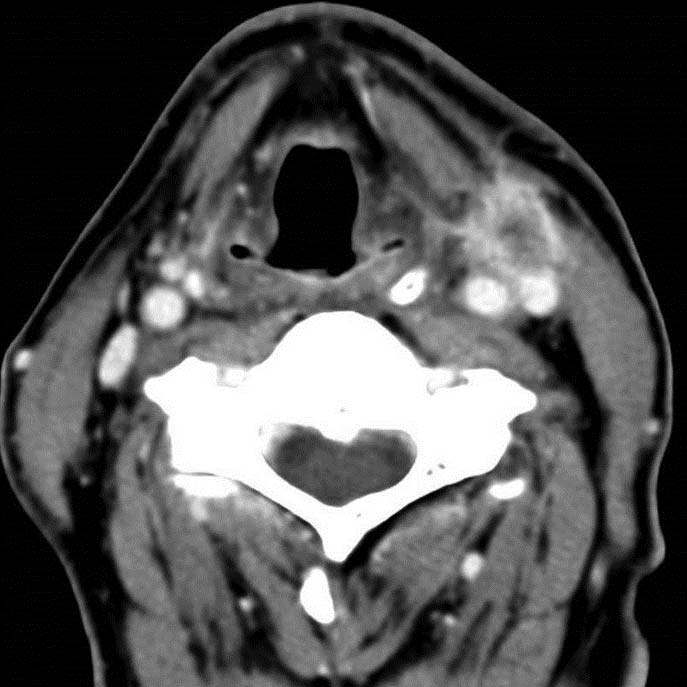
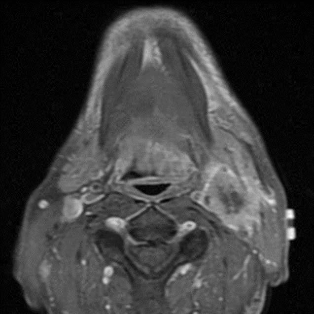
Cross-sectional imaging (CT or MR imaging) generally has low sensitivity (65-80%) but high specificity (86-93%) for the detection of ENE. US appears to be less accurate in assessment of ENE than CT and MR imaging, and its utility is currently being defined. ENE is suggested by interrupted or undefined nodal contours with high-resolution US imaging. Several CT or MR imaging features suggest ENE, such as indistinct nodal margins and irregular nodal capsular enhancement; however, the strongest imaging feature supporting the clinical diagnosis for ENE is clear infiltration into the adjacent fat or muscle. (Figures 6.2 and 6.3) This limitation of current technology limits the definitive nonsurgical diagnosis of ENE to only those patients who have clinically obvious signs described above. As noted, the presence of clinically obvious ENE is designated ENEc.

6.2 Axial contrast enhanced CT scan in a patient with a left level III enlarged heterogeneous node with ill-defined margins, infiltrating the adjacent fat and sternocleidomastoid muscle.

6.3 Axial T1 post-contrast fat-saturated MR imaging demonstrates a large heterogeneous and enhancing left level II mass with ill-defined margins and infiltration into the adjacent fat and sternocleidomastoid muscle. The primary left base of tongue squamous cell carcinoma is also evident on this image.

Pathological Classification

Histopathological examination is necessary to exclude the presence of tumor in lymph nodes because no imaging study as yet is accurate enough to identify microscopic tumor foci in regional nodes or distinguish between small reactive nodes and small malignant nodes. When a histopathologically involved node is identified, pN is designated based on measurement of the largest dimension of the metastatic deposit and not of the entire lymph node.

An excisional biopsy of a lymph node does not qualify for full evaluation of the pN category and should be assigned cN.

Pathological examination is necessary for documentation of tumor extent in terms of the location or level of the lymph node(s) involved, the number of nodes that contain metastases, and the presence, absence, and extent of ENE.

Minimum number of lymph nodes harvested in an adequate neck dissection

For assessment of pN, an adequate neck dissection ordinarily will include 15 or more lymph nodes in a previously untreated patient. Examination of fewer tumor-free nodes in a neck dissection specimen still mand ates a pN0 designation.

Sentinel node(s)

Sentinel lymph nodes (SLN) are described as the first nodes directly draining lymph from the primary tumor. SLN biopsy has been used as a staging procedure for the clinically negative neck in certain sites, such as oral cancer. Negative histopathological examination of SLNs justifies cN0 when sentinel node biopsy is part of the diagnostic workup and also is used for pathological categorization (pN0) in those cases that meet other criteria for pathological staging (e.g., resection of the primary tumor). The positive sentinel node also may be used to classify as pN1, though patients with positive SLNs usually undergo lymphadenectomy, and pN status is assigned based on assessment of the neck dissection specimen if performed.

Micrometastases

Micrometastases are labeled as pN1(mi), pN2b(mi), or pN2c(mi) for 2 mm deposits detected in single or multiple nodes detected exclusively on histopathological examination. These nodes are considered positive for the definition of pN. Although this designation will not influence staging, it is recommended for data collection and future analysis of the impact of incidentally detected micrometastases on outcomes.

Terminology for disease extension outside lymph nodes

Although terms such as extracapsular spread (ECS), extracapsular extension (ECE), or extranodal involvement (ENI) have been used to denote tumor extension outside the capsule of a metastatic node, extranodal extension (ENE) is the preferred wording.

Definition of ENE and description of its extent

All surgically resected metastatic nodes should be examined for the presence and extent of ENE. The precise definition of ENE has varied in the literature over the course of time. The College of American Pathologists defines ENE as extension of metastatic tumor, present within the confines of the lymph node, through the lymph node capsule into the surrounding connective tissue, with or without associated stromal reaction.

Gross ENE that is evident on clinical examination is designated ENEc and qualifies as ENE(+) for definition of cN. ENE detected on histopathologic examination is designated as ENEmi (microscopic ENE less than or equal to 2 mm) or ENEma (major ENE greater than 2 mm). Both ENEmi and ENEma qualify as ENE(+) for definition of pN. These descriptors of ENE will not be required for current pN definition, but data collection is recommended to allow stand ardization of data collection and future analysis.

Tumor deposits in the lymph drainage area of a primary carcinoma without histological evidence of residual lymph node tissue may represent a lymph node totally replaced by metastatic tumor. Such a nodule should be recorded as a positive lymph node with ENE(+).

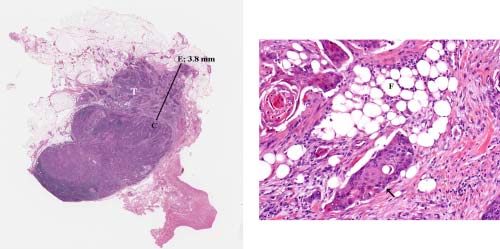
Extent of ENE is defined as the maximal distance in millimeters between the outer aspect of the intact or reconstructed nodal capsule and the farthest point of invasion into the extranodal tissue. Figure 6.4 illustrates the method for measurement of extent of ENE.

6.4 Histologic appearance of major extranodal extension (ENE). (A) Lymph node with metastatic tumor (T) invading perinodal fat. The extent of ENE (3.8 mm) is measured (solid bar) from the outer aspect of the lymph node capsule (C) to the most distant point of perinodal invasion (E). A 1-mm bar is shown for size comparison. (B) Higher power showing the squamous cell carcinoma (arrow) infiltrating in between adipose cells (F). From Wreesmann VB, et. al.3 with permission.

TNM Definitions

Definition of Primary Tumor (T)

T CategoryT Criteria
T0No evidence of primary tumor

Definition of Regional Lymph Node (N)

Clinical N (cN)
N CategoryN Criteria
NXRegional lymph nodes cannot be assessed
N0No regional lymph node metastasis
N1Metastasis in a single ipsilateral lymph node, 3 cm or smaller in greatest dimension and ENE(-)
N2Metastasis in a single ipsilateral node larger than 3 cm but not larger than 6 cm in greatest dimension and ENE(-); or metastases in multiple ipsilateral lymph nodes, none larger than 6 cm in greatest dimension and ENE(-); or in bilateral or contralateral lymph nodes, none larger than 6 cm in greatest dimension, ENE(-)
N2aMetastasis in a single ipsilateral node larger than 3 cm but not larger than 6 cm in greatest dimension and ENE(-)
N2bMetastases in multiple ipsilateral nodes, none larger than 6 cm in greatest dimension and ENE(-)
N2cMetastases in bilateral or contralateral lymph nodes, none larger than 6 cm in greatest dimension and ENE(-)
N3Metastasis in a lymph node larger than 6 cm in greatest dimension and ENE(-); or metastasis in any node(s) with clinically overt ENE(+) (ENEc)
N3aMetastasis in a lymph node larger than 6 cm in greatest dimension and ENE(-)
N3bMetastasis in any node(s) with clinically overt ENE(+) (ENEc)

Clinical N (cN) classification applies to patients who are treated with primary nonsurgical treatment without a cervical lymph node dissection.

Midline nodes are considered ipsilateral nodes.

ENEc is defined as invasion of skin, infiltration of musculature, dense tethering or fixation to adjacent structures, or cranial nerve, brachial plexus, sympathetic trunk, or phrenic nerve invasion with dysfunction.

A designation of “U” or “L” may be used for any N category to indicate metastasis above the lower border of the cricoid (U) or below the lower border of the cricoid (L). Similarly, clinical and pathological ENE should be recorded as ENE(-) or ENE(+).

Pathological N (pN)
N CategoryN Criteria
NXRegional lymph nodes cannot be assessed
N0No regional lymph node metastasis
N1Metastasis in a single ipsilateral lymph node, 3 cm or smaller in greatest dimension and ENE(-)
N2Metastasis in a single ipsilateral lymph node, 3 cm or smaller in greatest dimension and ENE(+); or single ipsilateral node larger than 3 cm but not larger than 6 cm in greatest dimension and ENE(-); or metastases in multiple ipsilaterallymph nodes, none larger than 6 cm in greatest dimension and ENE(-);or in bilateral or contralateral lymph node(s), none larger than 6 cm in greatest dimension and ENE(-)
N2aMetastasis in a single ipsilateral node 3 cm or less in greatest dimension and ENE(+); or a single ipsilateral node larger than 3 cm but not larger than 6 cm in greatest dimension and ENE(-)
N2bMetastases in multiple ipsilateral nodes, none larger than 6 cm in greatest dimension and ENE(-)
N2cMetastases in bilateral or contralateral lymph node(s), none larger than 6 cm in greatest dimension and ENE(-)
N3Metastasis in a lymph node larger than 6 cm in greatest dimension and ENE(-); or metastasis in a single ipsilateral node larger than 3 cm in greatest dimension and ENE(+); or multiple ipsilateral, contralateral, or bilateral nodes any size and ENE(+) in any node; or a single contralateral node of any size and ENE(+)
N3aMetastasis in a lymph node larger than 6 cm in greatest dimension and ENE(-)
N3bMetastasis in a single ipsilateralnode larger than 3 cm in greatest dimension and ENE(+);or multiple ipsilateral, contralateral, or bilateral nodes any size and ENE(+)in any node; or a single contralateral node of any size and ENE(+)

Pathological N (pN) classification applies to patients who are treated surgically with a cervical lymph node dissection.

Midline nodes are considered ipsilateral nodes.

ENE detected on histopathologic examination is designated as ENEmi (microscopic ENE less than or equal to 2 mm) or ENEma (major ENE greater than 2 mm). Both ENEmi and ENEma qualify as ENE(+) for definition of pN.

A designation of “U” or “L” may be used for any N category to indicate metastasis above the lower border of the cricoid (U) or below the lower border of the cricoid (L). Similarly, clinical and pathological ENE should be recorded as ENE(-) or ENE(+).

Definition of Distant Metastasis (M)

M CategoryM Criteria
M0No distant metastasis
M1Distant metastasis

Stage Prognostic

!!Calculator!!

AJCC PROGNOSTIC STAGE GROUPS

When T is…and N is…and M is…Then the stage group is…
T0N1M0III
T0N2M0IVA
T0N3M0IVB
T0Any NM1IVC

Prognostic Stage Groups for metastatic cervical adenopathy and unknown primary tumor except for EBV-related and HPV-related tumors.

Registry Data

Registry Data Collection Variables

  1. Extranodal extension for all anatomic sites with the exception of HPV-related oropharynx cancer, nasopharynx cancer, melanoma, sarcoma, and thyroid carcinoma
  2. Size of largest metastatic node
  3. Number of metastatic lymph nodes
  4. Laterality of metastatic nodes; not that midline nodes are considered ipsilateral nodes.
  5. Level of nodal involvement
  6. ENE clinical (+ or -)
  7. ENE pathological (+ or -)

Survival

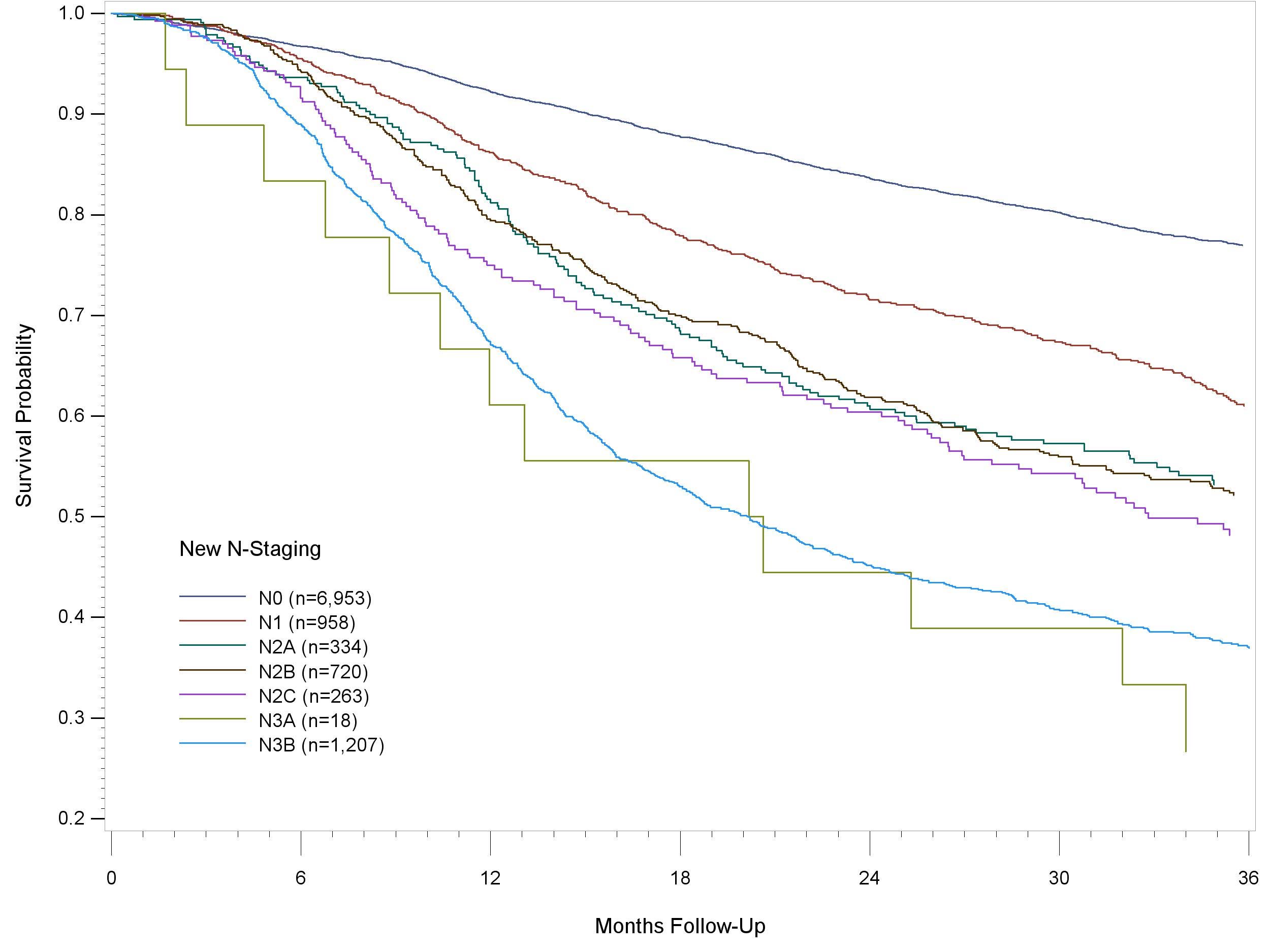
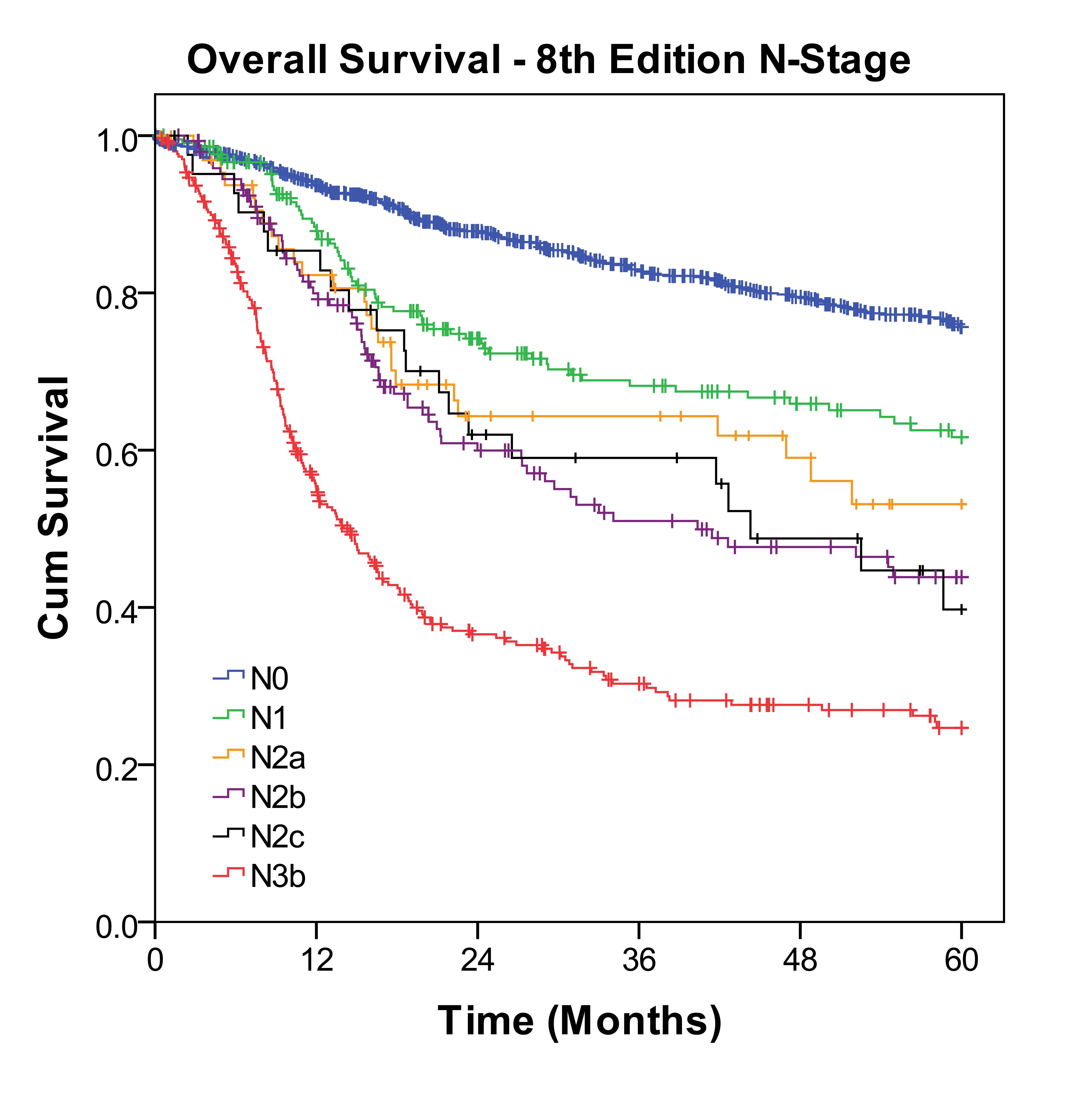
The data underpinning inclusion of ENE in the staging system are derived from histopathological examination of neck dissection specimens in patients treated surgically for their head and neck cancer. The modification is based on analysis of a large National Cancer Data Base (NCDB) data set, including cases with squamous cell carcinoma of the head and neck, with the exception of HPV-related oropharynx cancer and nasopharynx cancer (Figure 6.5). The new N category was then tested for validation on another large collaborative data set from Memorial Sloan Kettering Cancer Center, New York, and Princess Margaret Hospital, Toronto, for surgically treated oral cancer patients (Figure 6.6 and Table 6.3). The lack of ENE data in patients treated by nonsurgical modalities is problematic because currently available radiographic techniques are not sensitive enough to detect microscopic or less than gross ENE. Therefore, only clinically obvious ENE should be used for definition of cN when the patient is treated with nonsurgical therapy. This inability of current technology to reliably identify minimal or microscopic ENE without pathological examination of lymph node dissection specimens was the basis for separate cN and pN approaches for staging the neck. Clinically overt ENE (ENEc) will be designated cN3b irrespective of any other nodal characteristic in patients treated without neck dissection. Histologically identified ENE (ENEmi or ENEma) in a neck dissection specimen will be used in conjunction with node size and laterality for pN: Histopathologically confirmed ENE in a single ipsilateral or contralateral metastatic node 3 cm or smaller in largest dimension upstages the patient to pN2a, while all other nodes with histopathologically detected ENE are categorized pN3b.

Up-to-date cancer registry data on the influence of the new N criteria on outcomes are not available because ENE is only now being introduced into the nodal staging system. Limited data are available, however, from the NCDB for patients treated in 2010-11 for squamous cell carcinoma at sites other than nasopharynx and HPV-related oropharynx cancer (Figure 6.5). The proposed new N classification was then validated in a large dataset of patients with oral cancer treated at two tertiary cancer care centers in North America (Figure 6.6 and Table 6.3).

6.5 Overall Survival in squamous cell carcinoma of the head and neck based on the 8thEdition N criteria that incorporate ENE as a prognostic factor. Kaplan Meier methods were used to perform cancer-specific analyses predicting overall survival as the endpoint on a population of oral cavity cancer patients from NCDB.

6.6 Overall Survival based on 8thEdition N criteria that incorporate ENE as a prognostic factor. Kaplan Meier methods were used to perform cancer-specific analyses predicting overall survival as the endpoint on a population of oral cavity cancer patients from MSKCC and PMH.

6.3 Overall survival based on 8thEdition N criteria (MSKCC-PMH Data).

# of pts at Risk0 Months12 Months24 Months36 Months48 Months60 Months
N010188700710596513421
N1211168119978270
N2a665030282113
N2b14810765493929
N2c42342219128
N3b30314681594331

Bibliography

  1. O'Sullivan B, Huang SH, Su J, et al. Development and validation of a staging system for HPV-related oropharyngeal cancer by the International Collaboration on Oropharyngeal cancer Network for Staging (ICON-S): a multicentre cohort study. The lancet oncology. Feb 26 2016.
  2. Patel S. Personal Communication. In: Lydiatt W, Shah JP, eds2015.
  3. Wreesmann VB, Katabi N, Palmer FL, et al. Influence of extracapsular nodal spread extent on prognosis of oral squamous cell carcinoma. Head & neck. Oct 30 2015.
  4. Robbins KT, Clayman G, Levine PA, et al. Neck dissection classification update: revisions proposed by the American Head and Neck Society and the American Academy of Otolaryngology-Head and Neck Surgery. Archives of otolaryngology--head & neck surgery. Jul 2002;128(7):751-758.
  5. Agrawal A, Civantos FJ, Brumund KT, et al. [99mTc] Tilmanocept Accurately Detects Sentinel Lymph Nodes and Predicts Node Pathology Status in Patients with Oral Squamous Cell Carcinoma of the Head and Neck: Results of a Phase III Multi-institutional Trial. Annals of surgical oncology. 2015:1-8.
  6. Alkureishi LW, Ross GL, Shoaib T, et al. Sentinel node biopsy in head and neck squamous cell cancer: 5-year follow-up of a European multicenter trial. Annals of surgical oncology. Sep 2010;17(9):2459-2464.
  7. Civantos FJ, Zitsch RP, Schuller DE, et al. Sentinel lymph node biopsy accurately stages the regional lymph nodes for T1-T2 oral squamous cell carcinomas: results of a prospective multi-institutional trial. J Clin Oncol. Mar 10 2010;28(8):1395-1400.
  8. Curtin HD, Ishwaran H, Mancuso AA, Dalley RW, Caudry DJ, McNeil BJ. Comparison of CT and MR imaging in staging of neck metastases. Radiology. Apr 1998;207(1):123-130.
  9. de Juan J, Garcia J, Lopez M, et al. Inclusion of extracapsular spread in the pTNM classification system: a proposal for patients with head and neck carcinoma. JAMA otolaryngology-- head & neck surgery. May 2013;139(5):483-488.
  10. Ebrahimi A, Gil Z, Amit M, et al. The prognosis of N2b and N2c lymph node disease in oral squamous cell carcinoma is determined by the number of metastatic lymph nodes rather than laterality: evidence to support a revision of the American Joint Committee on Cancer staging system. Cancer. 2014;120(13):1968-1974.
  11. Ebrahimi A GZ, Amit M, Yen TC, Liao CT, Chatturvedi P, Agarwal J, Kowalski L, Kreppel M, Cernea C, Brand ao J, Bachar G, Villaret AB, Fliss D, Fridman E, Robbins KT, Shah J, Patel S, Clark J; International Consortium for Outcome Research (ICOR) in Head and Neck Cancer. Comparison of the American Joint Committee on Cancer N1 versus N2a nodal categories for predicting survival and recurrence in patients with oral cancer: Time to acknowledge an arbitrary distinction and modify the system. . Head and neck pathology. 2014.
  12. Gregoire V, Ang K, Budach W, et al. Delineation of the neck node levels for head and neck tumors: a 2013 update. DAHANCA, EORTC, HKNPCSG, NCIC CTG, NCRI, RTOG, TROG consensus guidelines. Radiotherapy and oncology : journal of the European Society for Therapeutic Radiology and Oncology. Jan 2014;110(1):172-181.
  13. Hoang JK, Vanka J, Ludwig BJ, Glastonbury CM. Evaluation of cervical lymph nodes in head and neck cancer with CT and MRI: tips, traps, and a systematic approach. AJR. American journal of roentgenology. Jan 2013;200(1):W17-25.
  14. Jones A, Roland N, Field J, Phillips D. The level of cervical lymph node metastases: their prognostic relevance and relationship with head and neck squamous carcinoma primary sites. Clinical Otolaryngology & Allied Sciences. 1994;19(1):63-69.
  15. Jose J, Moor JW, Coatesworth AP, Johnston C, MacLennan K. Soft tissue deposits in neck dissections of patients with head and neck squamous cell carcinoma: prospective analysis of prevalence, survival, and its implications. Archives of otolaryngology--head & neck surgery. Feb 2004;130(2):157-160.
  16. King AD, Tse GM, Yuen EH, et al. Comparison of CT and MR imaging for the detection of extranodal neoplastic spread in metastatic neck nodes. Eur J Radiol. Dec 2004;52(3):264-270.
  17. Kyzas PA, Evangelou E, Denaxa-Kyza D, Ioannidis JP. 18F-fluorodeoxyglucose positron emission tomography to evaluate cervical node metastases in patients with head and neck squamous cell carcinoma: a meta-analysis. Journal of the National Cancer Institute. May 21 2008;100(10):712-720.
  18. Lodder WL, Lange CA, van Velthuysen M-LF, et al. Can extranodal spread in head and neck cancer be detected on MR imaging. Oral oncology. 2013;49(6):626-633.
  19. Medina JE. A rational classification of neck dissections. Otolaryngology--head and neck surgery : official journal of American Academy of Otolaryngology-Head and Neck Surgery. Mar 1989;100(3):169-176.
  20. Patel SG, Amit M, Yen TC, et al. Lymph node density in oral cavity cancer: results of the International Consortium for Outcomes Research. Br J Cancer. Oct 15 2013;109(8):2087-2095.
  21. Prabhu RS, Magliocca KR, Hanasoge S, et al. Accuracy of computed tomography for predicting pathologic nodal extracapsular extension in patients with head-and -neck cancer undergoing initial surgical resection. International journal of radiation oncology, biology, physics. Jan 1 2014;88(1):122-129.
  22. Saindane AM. Pitfalls in the staging of cervical lymph node metastasis. Neuroimaging Clin N Am. Feb 2013;23(1):147-166.
  23. Shah JP. Patterns of cervical lymph node metastasis from squamous carcinomas of the upper aerodigestive tract. American journal of surgery. Oct 1990;160(4):405-409.
  24. Shah JP, Medina JE, Shaha AR, Schantz SP, Marti JR. Cervical lymph node metastasis. Curr Probl Surg. Mar 1993;30(3):1-335.
  25. Som PM, Curtin HD, Mancuso AA. An imaging-based classification for the cervical nodes designed as an adjunct to recent clinically based nodal classifications. Archives of Otolaryngology-Head & Neck Surgery. 1999;125(4):388-396.
  26. Url C, Schartinger VH, Riechelmann H, et al. Radiological detection of extracapsular spread in head and neck squamous cell carcinoma (HNSCC) cervical metastases. Eur J Radiol. Oct 2013;82(10):1783-1787.
  27. van den Brekel MW, Lodder WL, Stel HV, Bloemena E, Leemans CR, van der Waal I. Observer variation in the histopathologic assessment of extranodal tumor spread in lymph node metastases in the neck. Head & neck. Jun 2012;34(6):840-845.