section name header

Disease Prologue

Summary

Cancers Staged Using This Staging System

Epithelial cancers including adenocarcinoma, adenosquamous carcinoma, undifferentiated carcinoma, neuroendocrine cancers, and adenocarcinoma with neuroendocrine features are staged.

Cancers Not Staged Using This Staging System

These histopathologic types of cancer…Are staged according to the classification for…and can be found in chapter…
Squamous cell carcinoma, adenosquamous carcinoma, and undifferentiated carcinoma with squamous components arising in the esophagus and esophagogastric junctionEsophagus and esophagogastric junction: squamous cell carcinoma16
Adenocarcinoma with neuroendocrine features and other mixed or unspecified carcinomasEsophagus and esophagogastric junction: other histologies16
Sarcomas, nonepithelial cancersSoft tissue sarcoma of the abdomen and thoracic visceral organs42
Gastrointestinal stromal tumorGastrointestinal stromal tumor43

Summary of Changes

ChangeDetails of ChangeLevel of Evidence
Anatomy—Primary Site(s)Anatomic boundary between esophagus and stomach: tumors involving the EGJ with epicenter no more than 2 cm into the proximal stomach are staged as esophageal cancers; tumors with epicenter located greater than 2 cm into the proximal stomach are staged as stomach cancers even if EGJ involvedIII
AJCC Prognostic Stage GroupspT1a and pT1b are now incorporated into stage groupings.II
AJCC Prognostic Stage GroupsUnique cTNM prognostic stage groupings are based on clinically determined TNM.II
AJCC Prognostic Stage GroupsUnique ypTNM prognostic stage groupings are based on patients who have received preoperative treatment and surgical resection.II

ICD-O-3 Topography Codes

CodeDescription
C15.0Cervical esophagus
C15.1Thoracic esophagus
C15.2Abdominal esophagus
C15.3Upper third of esophagus
C15.4Middle third of esophagus
C15.5Lower third of esophagus
C15.8Overlapping lesion of esophagus
C15.9Esophagus, NOS
C16.0Cardia, esophagogastric junction

Tumors of the EGJ with less than or equal to 2 cm of proximal stomach involvement are staged as esophageal cancers.

WHO Classification of Tumors

This list includes histology codes and preferred terms from the WHO Classification of Tumors and the International Classification of Diseases for Oncology (ICD-O). Most of the terms in this list represent malignant behavior. For cancer reporting purposes, behavior codes /3 (denoting malignant neoplasms), /2 (denoting in situ neoplasms), and in some cases /1 (denoting neoplasms with uncertain and unknown behavior) may be appended to the 4-digit histology codes to create a complete morphology code.

CodeDescription
8020Undifferentiated carcinoma with gland ular component and without squamous component
8140Adenocarcinoma
8148Gland ular dysplasia (intraepithelial neoplasia), high grade
8200Adenoid cystic carcinoma
8244Mixed adenoneuroendocrine carcinoma
8430Mucoepidermoid carcinoma

Bosman FT, Carneiro F, Hruban RH, Theise ND, eds. World Health Organization Classification of Tumours of the Digestive System. Lyon: IARC; 2010. Used with permission.

International Agency for Research on Cancer, World Health Organization. International Classification of Diseases for Oncology. ICD-O-3-Online.http://codes.iarc.fr/home. Accessed September 29, 2017. Used with permission.

Introduction

The AJCC Cancer Staging Manual, 8th Edition esophageal cancer staging chapter is based on updated data, with a significantly increased sample size and number of risk adjustment variables compared with the AJCC Cancer Staging Manual, 7th Edition. The stage groupings were determined by using a risk-adjusted rand om survival forest analysis of collated data from 33 esophageal centers spanning six continents and including 22,654 patients.1 All-cause mortality—a hard end point—was used because after risk adjustment, the residual information regarding death may be attributed to esophageal cancer.1-6

Stage groupings for the 8th Edition are not based on an orderly increase in T category followed by number of involved nodes. The unique lymphatic anatomy of the esophagus results in the possibility of regional lymph node metastasis even with superficial (T1) cancers; therefore, patients with regional lymph node metastasis (pN+) from superficial cancers may have a prognosis similar to that of patients with deeper (greater than pT1) pN0 cancers. Similarly, deeper cancers (greater than pT1) with a few positive nodes may have a prognosis similar to that of superficial cancers (pT1) with more positive nodes. Possibly as a reflection of the genomic alterations of esophageal cancers, histologic grade (G) modulates stage such that the prognosis of well-differentiated (G1) deeper cancers is similar to that of less well-differentiated (G2-G3) superficial cancers. Staging recommendations in the 7th Edition partially separated histopathologic type for early-stage cancers. The larger dataset used for this edition has allowed for better separation of squamous cell carcinoma and adenocarcinoma staging. It is evident in the recent survival analysis that, except for advanced-stage cancers, the survival of squamous cell carcinoma patients is worse than that of patients with adenocarcinoma when comparing similarly grouped patients. Although at first glance these multiple trade-offs seem to create a less orderly arrangement of TNM categories within and among stage groupings compared with previous stage groupings, when viewed from the perspective of the interplay of these important prognostic factors, the new staging system becomes biologically compelling.

In an effort to overcome the limitations of the 7th Edition, which was based entirely on patients treated by esophagectomy alone (without preoperative or postoperative chemotherapy and /or chemoradiotherapy), the dataset used to develop the 8th Edition TNM stage groupings included patients who had received preoperative induction therapy (neoadjuvant) and /or postoperative adjuvant therapy. The availability of these data led to the ability to explicitly define cTNM and ypTNM cohorts and stages.1,3,5,6 These data reflect the difficult land scape of clinical staging for esophageal cancer and the current preference for treating locally advanced esophageal cancer with neoadjuvant therapy. In comparison with previous editions, analysis of this large dataset illuminated significant differences in outcome when comparing the same stage groups between patients receiving neoadjuvant therapy versus those treated with surgery alone. Therefore, it was necessary to construct a distinct composition of stage groupings for ypTNM.5,6

The clinical modalities currently available for pretreatment staging are often inaccurate, resulting in frequent understaging and overstaging. This ultimately leads to the potential for suboptimal treatment of esophageal cancers. When comparing survival of clinically staged patients with that of patients with equivalent pathological stage, it is evident that prognoses are not equivalent.1-4 The prognosis for clinically staged early cancers is clearly worse, indicating that cTNM for these cancers is understaged compared with pTNM. Conversely, apparently advanced cTNM cancers carry a somewhat better prognosis than equivalent pTNM cancers. In part, this may be the result of earlier cancers being overstaged and in part because of the rand om effect of neoadjuvant and adjuvant therapy on more advanced-stage cancers. Although this approach may change in the future, the 8th Edition TNM staging system reflects the widespread use of neoadjuvant therapy.

There are limitations in the data that were available to construct cTNM cohorts and clinical stage groups for this edition. The exact modalities used to arrive at a clinical stage before the initiation of therapy were not available for analysis. Patients not offered surgery, deemed inoperable, or undergoing exploratory surgery without esophagectomy were relatively poorly represented in the data. In addition, patients undergoing surgery alone with pT4 and /or M1 cancers represent a select population; placing these categories into stage groups, therefore, required either combining some categories or using consensus to arrive at stage grouping, noting that in general, their prognosis was poor.

Anatomy

Primary Site(s)

The esophagus traverses three anatomic compartments: cervical, thoracic, and abdominal. The thoracic esophagus is divided arbitrarily into equal thirds: upper, middle, and lower (Table 16.1). However, the clinical importance of the primary site of an esophageal cancer is related less to its position in the esophagus than to its relation to adjacent structures (Figure 16.1).

16.1 Anatomy of esophageal cancer primary site, including typical endoscopic measurements of each region measured from the incisors. Exact measurements depend on body size and height. Location of cancer primary site is defined by cancer epicenter. EGJ, esophagogastric junction; LES, lower esophageal sphincter; UES, upper esophageal sphincter.

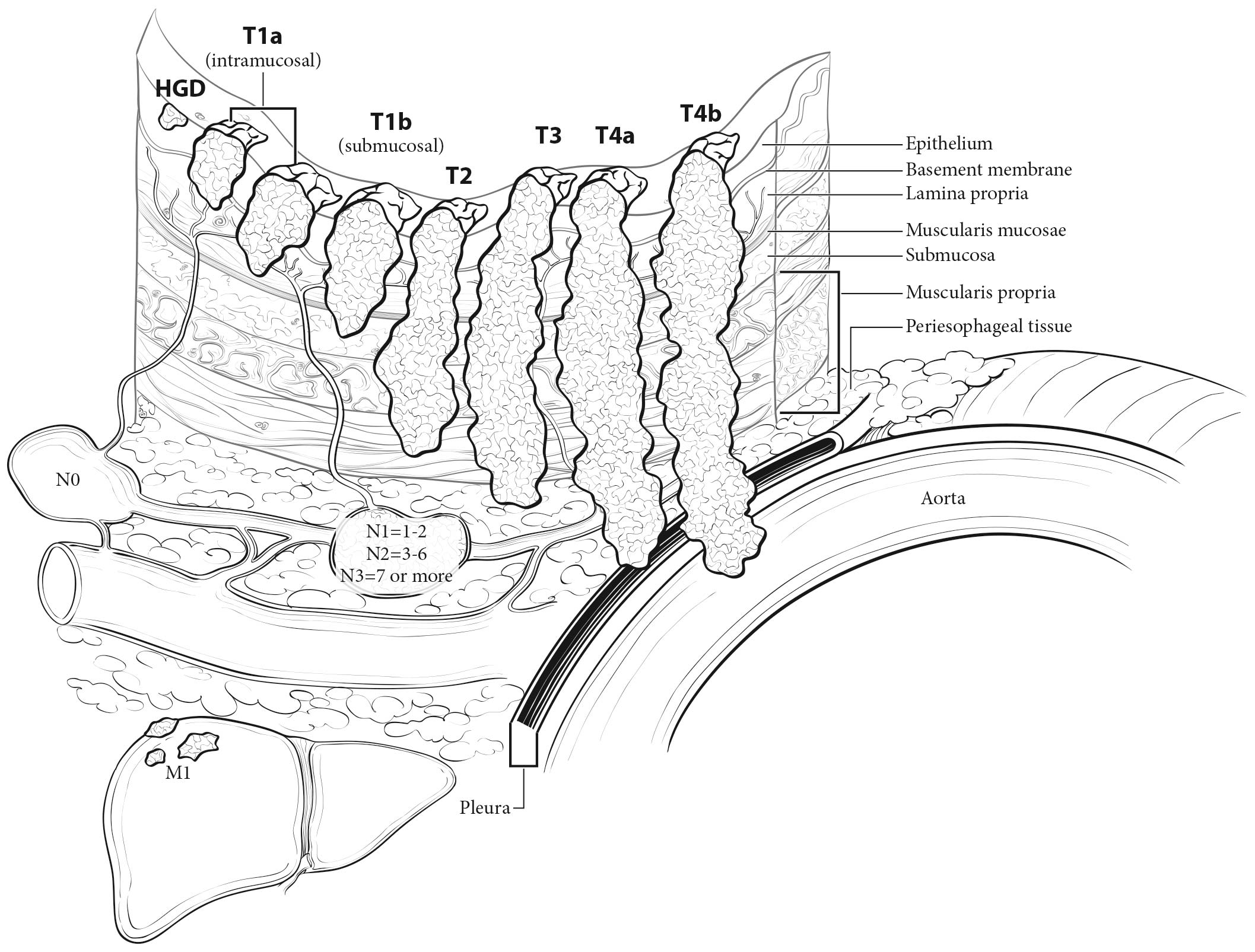
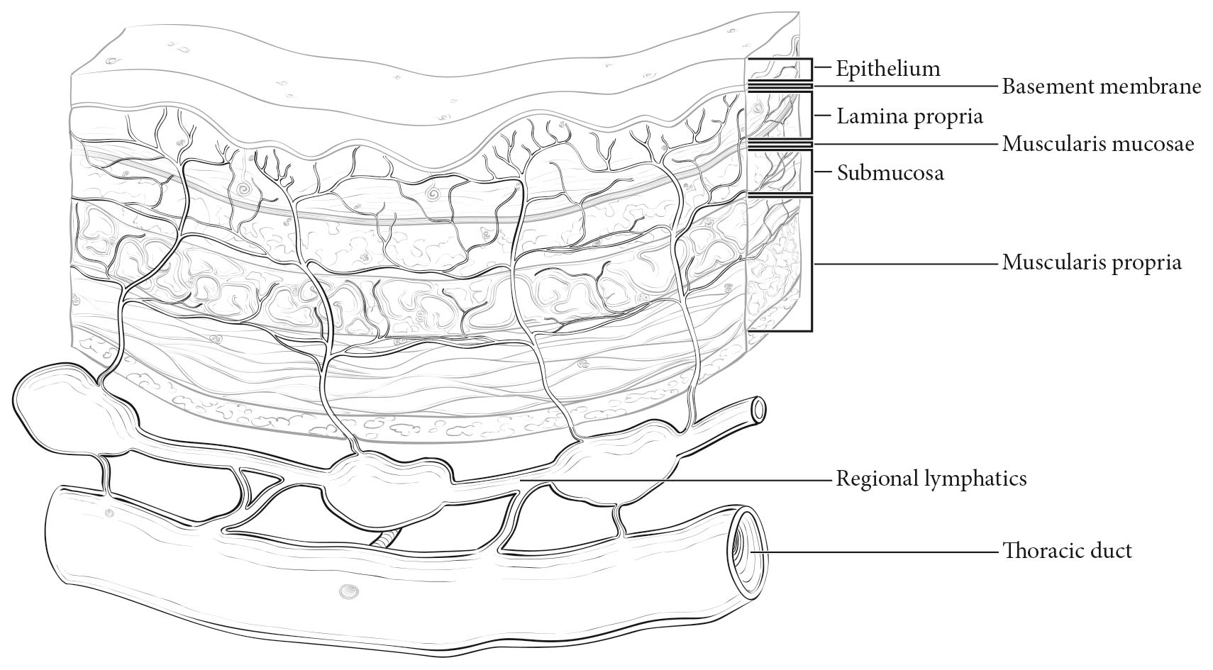
The esophageal wall has three layers: mucosa, submucosa, and muscularis propria (Figure 16.2). The mucosa is composed of epithelium, lamina propria, and muscularis mucosae. A basement membrane isolates the epithelium from the rest of the esophageal wall. In the columnar-lined esophagus, the muscularis mucosae may be a two-layered (duplicated) structure. The clinical importance of this duplicate layer is questionable.7,8 The outer layer is considered the true boundary. The mucosal division may be classified as m1 (epithelium), m2 (lamina propria), or m3 (muscularis mucosae).9 The submucosa has no land marks, but it may be divided into inner (sm1), middle (sm2), and outer (sm3) thirds.9 The muscularis propria has inner circular and outer longitudinal muscle layers. There is no serosa; rather, adventitia (periesophageal connective tissue) lies directly on the muscularis propria.

16.2 Esophageal wall.

Location

Cervical Esophagus

Cancers located in the cervical esophagus are staged as upper thoracic esophageal cancers, not as head and neck cancers.

Anatomically, the cervical esophagus lies in the neck, bordered superiorly by the hypopharynx and inferiorly by the thoracic inlet, which lies at the level of the sternal notch. It is subtended by the trachea, carotid sheaths, and vertebrae. Although the length of the esophagus differs somewhat with body habitus, gender, and age, typical endoscopic measurements for the cervical esophagus measured from the incisors are from 15 to less than 20 cm (Figure 16.1). If esophagoscopy is not available, location may be assessed by computed tomography (CT). If the epicenter of the tumor begins above the sternal notch, the location is defined as cervical esophagus.

Upper Thoracic Esophagus

The upper thoracic esophagus is bordered superiorly by the thoracic inlet and inferiorly by the lower border of the azygos vein. Anterolaterally, it is surrounded by the trachea, aortic arch, and great vessels, posteriorly by the vertebrae. Typical endoscopic measurements from the incisor teeth are from 20 to less than 25 cm (Figure 16.1). On CT, to determine the location, the epicenter of an upper thoracic cancer is visible between the sternal notch and the azygos vein.

Middle Thoracic Esophagus

The middle thoracic esophagus is bordered superiorly by the lower border of the azygos vein and inferiorly by the lower border of the inferior pulmonary vein. It is sand wiched between the pulmonary hilum anteriorly, descending thoracic aorta on the left, and vertebrae posteriorly; on the right, it lies freely on the pleura. Typical endoscopic measurements from the incisors are from 25 to less than 30 cm (Figure 16.1). On CT, to determine the location, the epicenter of a middle thoracic cancer is between the azygos vein and the inferior pulmonary vein.

Lower Thoracic Esophagus/Esophagogastic Junction (EGJ)

The lower thoracic esophagus is bordered superiorly by the lower border of the inferior pulmonary vein and inferiorly by the stomach. It is bordered anteriorly by the pericardium, posteriorly by vertebrae, and on the left by the descending thoracic aorta. It normally passes through the diaphragm to reach the stomach, but there is a variable intra-abdominal portion, and in the presence of a hiatal hernia, this portion may be absent. Typical endoscopic measurements from the incisors are from 30 to 40 cm (Figure 16.1). On CT to determine the location, the epicenter of a lower thoracic esophagus/EGJ cancer is below the inferior pulmonary vein. The abdominal esophagus is included in the lower thoracic esophagus. Cancers involving the EGJ that have their epicenter within the proximal 2 cm of the cardia (Siewert types I/II) are to be staged as esophageal cancers. Cancers whose epicenter is more than 2 cm distal from the EGJ, even if the EGJ is involved, will be staged using the stomach cancer TNM and stage groupings (see Chapter 17).

16.1 Anatomy of esophageal cancer primary site by ICD-O-3 topography codes

Esophageal location
Anatomic nameCompartment ICD-O-3ICD-O-3NameAnatomic boundariesTypical esophagectomy, cm
CervicalC15.0C15.3UpperHypopharynx to sternal notch15 to less than 20
ThoracicC15.1C15.3UpperSternal notch to azygos vein20 to less than 25
C15.4MiddleLower border of azygos vein to inferior pulmonary vein25 to less than 30
C15.5LowerLower border of inferior pulmonary vein to EGJ30 to less than 40
AbdominalC15.2C15.5LowerEGJ to 2 cm below EGJ40 to 45
C16.0EGJ/cardiaEGJ to 2 cm below EGJ40 to 45

Regional Lymph Nodes

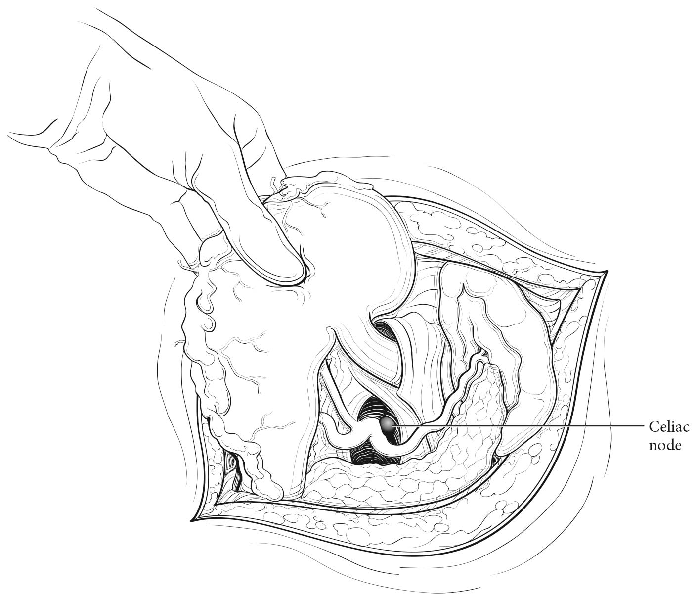
Esophageal lymphatic drainage is intramural and longitudinal. The lymphatic network within the esophagus is concentrated in the submucosa, although lymphatic channels also are present in the lamina propria. This arrangement may permit lymphatic metastases early in the course of the disease from otherwise superficial cancers.10 Lymphatic drainage of the muscularis propria is more limited, but lymphatic channels pierce this layer to drain into regional lymphatic channels and lymph nodes in the periesophageal fat. Up to 43% of autopsy dissections demonstrate direct drainage from the submucosal plexus into the thoracic duct, which facilitates systemic metastases.11-13 The longitudinal nature of the submucosal lymphatic plexus permits lymphatic metastases orthogonal to depth of tumor invasion.14 The implication of the longitudinal nature of lymphatic drainage is that the anatomic site of the cancer and the lymph nodes to which lymphatics drain from that site may not be the same (Figures 16.3_A, 16.3_B, and 16.3_C).

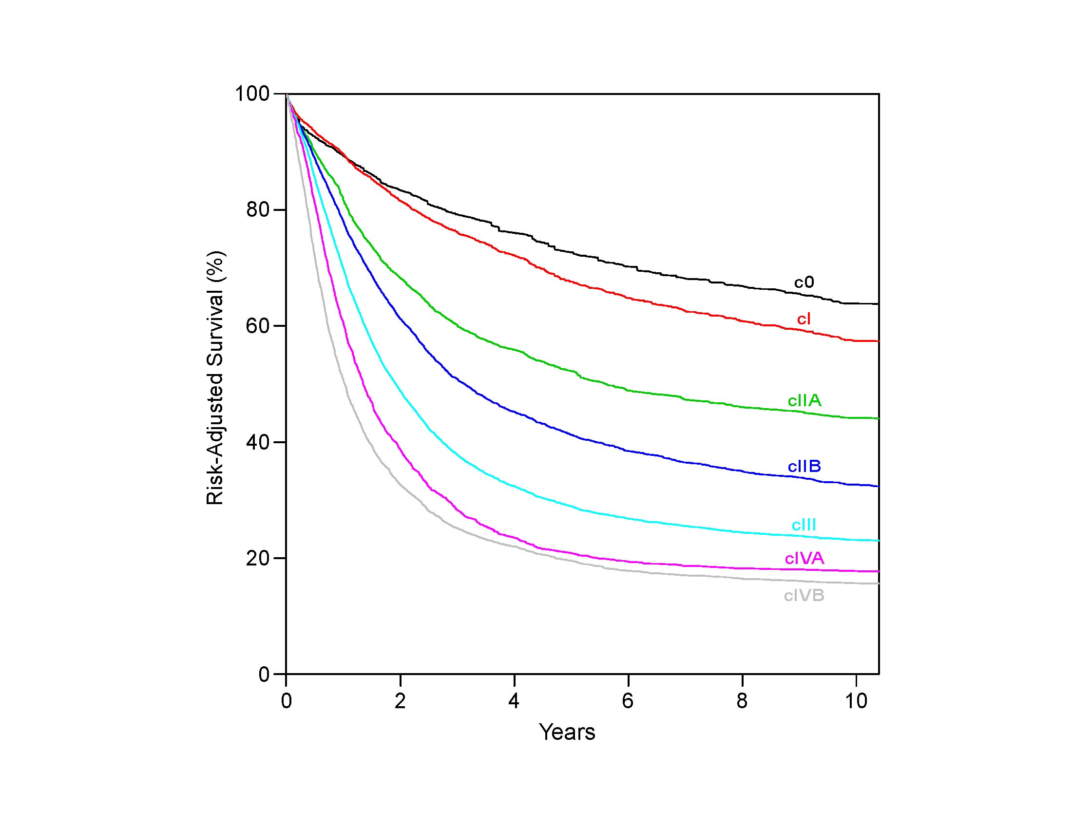
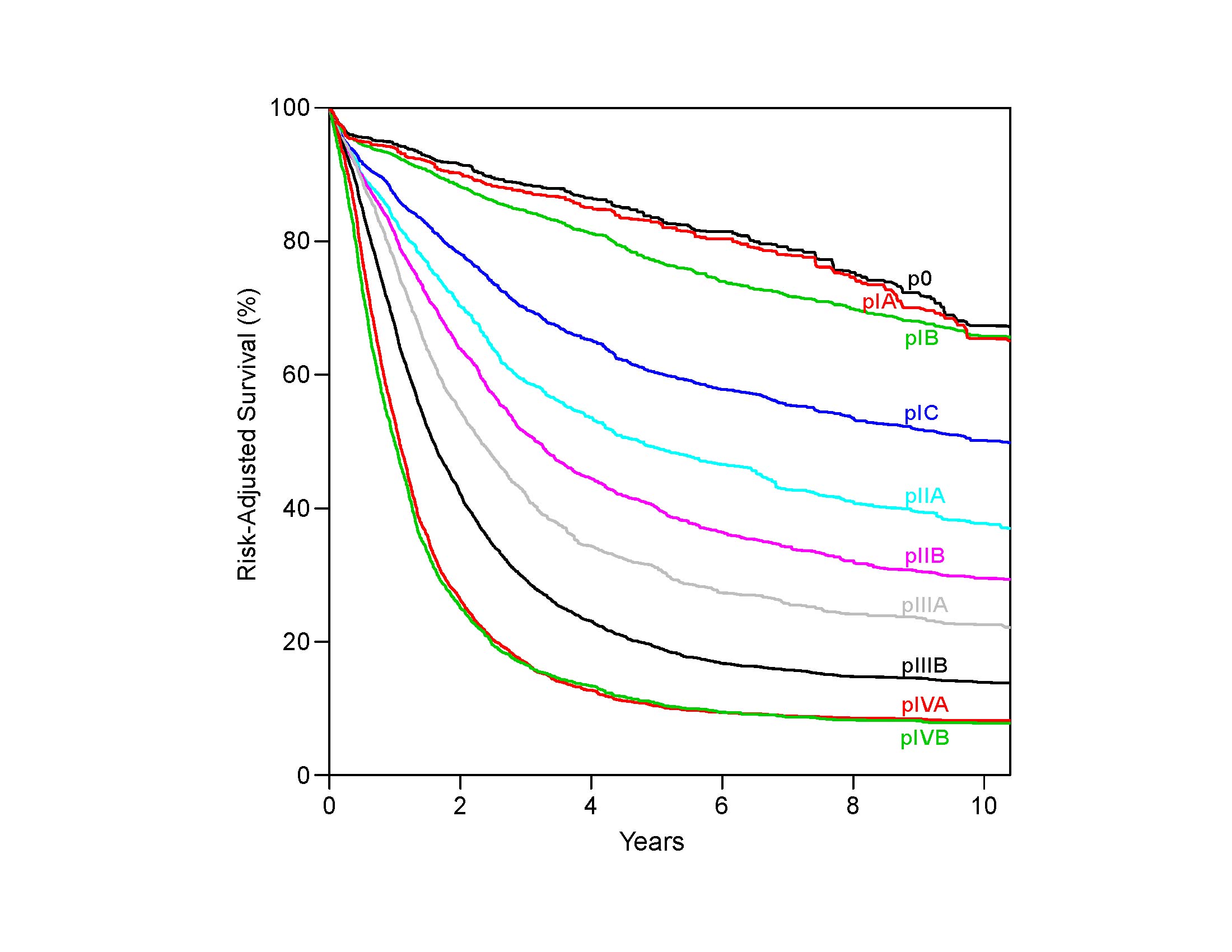
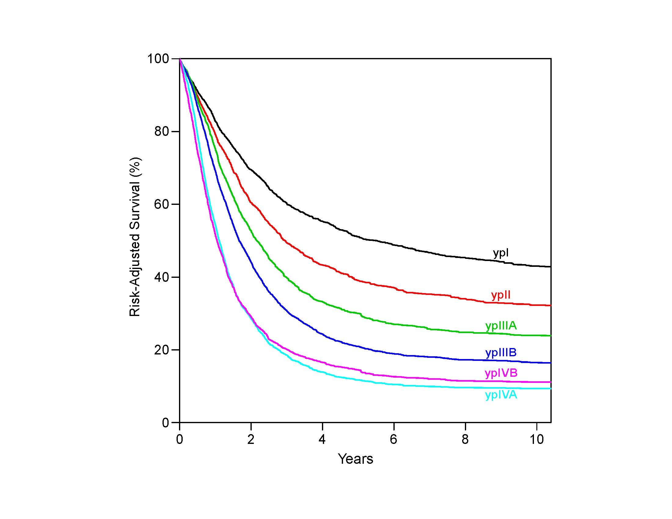
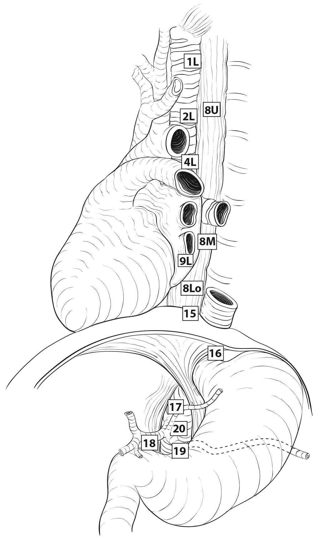
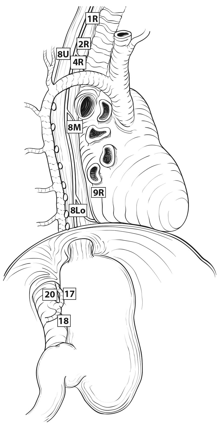
Therefore it follows, and analysis of data supports, that regional lymph nodes for all locations in the esophagus discussed in this chapter extend from periesophageal cervical nodes to celiac nodes (Figures 16.3_A 16.3_B 16.3_Cand 16.4). The nomenclature for thoracic and abdominal regional lymph nodes is listed in Figures 16.3_A, 16.3_B, and 16.3_C. The nomenclature for cervical regional lymph nodes follows that of head and neck chapters (see Chapter 6) and are located in periesophageal levels VI and VII. Lymph nodes in continuity with the esophagus would be considered regional.

The specific regional lymph nodes are as follows:

  • Right lower cervical paratracheal nodes: between the supraclavicular paratracheal space and apex of the lung
  • Left lower cervical paratracheal nodes: between the supraclavicular paratracheal space and apex of the lung
  • Right upper paratracheal nodes: between the intersection of the caudal margin of the brachiocephalic artery with the trachea and the apex of the lung
  • Left upper paratracheal nodes: between the top of the aortic arch and apex of the lung
  • Right lower paratracheal nodes: between the intersection of the caudal margin of the brachiocephalic artery with the trachea and cephalic border of the azygos vein
  • Left lower paratracheal nodes: between the top of the aortic arch and the carina •Subcarinal nodes: caudal to the carina of the trachea
  • Subcarinal nodes: caudal to the carina of the trachea
  • Upper thoracic paraesophageal lymph nodes: from the apex of the lung to the tracheal bifurcation
  • Middle thoracic paraesophageal lymph nodes: from the tracheal bifurcation to the caudal margin of the inferior pulmonary vein
  • Lower thoracic paraesophageal lymph nodes: from the caudal margin of the inferior pulmonary vein to the EGJ
  • Pulmonary ligament nodes: within the right inferior pulmonary ligament
  • Pulmonary ligament nodes: within the left inferior pulmonary ligament
  • Diaphragmatic nodes: lying on the dome of the diaphragm and adjacent to or behind its crura
  • Paracardial nodes: immediately adjacent to the gastroesophageal junction
  • Left gastric nodes: along the course of the left gastric artery
  • Common hepatic nodes: immediately on the proximal common hepatic artery
  • Splenic nodes: immediately on the proximal splenic artery
  • Celiac nodes: at the base of the celiac artery
  • Cervical periesophageal level VI lymph nodes (see Chapter 6)
  • Cervical periesophageal level VII lymph nodes (see Chapter 6)

16.3_A (A) Lymph node maps for esophageal cancer. Regional lymph node stations for staging esophageal cancer from left. 1R, Right lower cervical paratracheal nodes, between the supraclavicular paratracheal space and apex of the lung. 1L, Left lower cervical paratracheal nodes, between the supraclavicular paratracheal space and apex of the lung. 2R, Right upper paratracheal nodes, between the intersection of the caudal margin of the brachiocephalic artery with the trachea and the apex of the lung. 2L, Left upper paratracheal nodes, between the top of the aortic arch and the apex of the lung. 4R, Right lower paratracheal nodes, between the intersection of the caudal margin of the brachiocephalic artery with the trachea and cephalic border of the azygos vein. 4L, Left lower paratracheal nodes, between the top of the aortic arch and the carina. 7, Subcarinal nodes, caudal to the carina of the trachea. 8U, Upper thoracic paraesophageal lymph nodes, from the apex of the lung to the tracheal bifurcation. 8M, Middle thoracic paraesophageal lymph nodes, from the tracheal bifurcation to the caudal margin of the inferior pulmonary vein. 8Lo, Lower thoracic paraesophageal lymph nodes, from the caudal margin of the inferior pulmonary vein to the EGJ. 9R, Pulmonary ligament nodes, within the right inferior pulmonary ligament. 9L, Pulmonary ligament nodes, within the left inferior pulmonary ligament. 15, Diaphragmatic nodes, lying on the dome of the diaphragm and adjacent to or behind its crura. 16, Paracardial nodes, immediately adjacent to the gastroesophageal junction. 17, Left gastric nodes, along the course of the left gastric artery. 18, Common hepatic nodes, immediately on the proximal common hepatic artery. 19, Splenic nodes, immediately on the proximal splenic artery. 20, Celiac nodes, at the base of the celiac artery.

16.3_B (B) Lymph node maps for esophageal cancer. Regional lymph node stations for staging esophageal cancer from right. 1R, Right lower cervical paratracheal nodes, between the supraclavicular paratracheal space and apex of the lung. 1L, Left lower cervical paratracheal nodes, between the supraclavicular paratracheal space and apex of the lung. 2R, Right upper paratracheal nodes, between the intersection of the caudal margin of the brachiocephalic artery with the trachea and the apex of the lung. 2L, Left upper paratracheal nodes, between the top of the aortic arch and the apex of the lung. 4R, Right lower paratracheal nodes, between the intersection of the caudal margin of the brachiocephalic artery with the trachea and cephalic border of the azygos vein. 4L, Left lower paratracheal nodes, between the top of the aortic arch and the carina. 7, Subcarinal nodes, caudal to the carina of the trachea. 8U, Upper thoracic paraesophageal lymph nodes, from the apex of the lung to the tracheal bifurcation. 8M, Middle thoracic paraesophageal lymph nodes, from the tracheal bifurcation to the caudal margin of the inferior pulmonary vein. 8Lo, Lower thoracic paraesophageal lymph nodes, from the caudal margin of the inferior pulmonary vein to the EGJ. 9R, Pulmonary ligament nodes, within the right inferior pulmonary ligament. 9L, Pulmonary ligament nodes, within the left inferior pulmonary ligament. 15, Diaphragmatic nodes, lying on the dome of the diaphragm and adjacent to or behind its crura. 16, Paracardial nodes, immediately adjacent to the gastroesophageal junction. 17, Left gastric nodes, along the course of the left gastric artery. 18, Common hepatic nodes, immediately on the proximal common hepatic artery. 19, Splenic nodes, immediately on the proximal splenic artery. 20, Celiac nodes, at the base of the celiac artery.

16.3_C C) Lymph node maps for esophageal cancer. Regional lymph node stations for staging esophageal cancer, anterior view. 1R, Right lower cervical paratracheal nodes, between the supraclavicular paratracheal space and apex of the lung. 1L, Left lower cervical paratracheal nodes, between the supraclavicular paratracheal space and apex of the lung. 2R, Right upper paratracheal nodes, between the intersection of the caudal margin of the brachiocephalic artery with the trachea and the apex of the lung. 2L, Left upper paratracheal nodes, between the top of the aortic arch and the apex of the lung. 4R, Right lower paratracheal nodes, between the intersection of the caudal margin of the brachiocephalic artery with the trachea and cephalic border of the azygos vein. 4L, Left lower paratracheal nodes, between the top of the aortic arch and the carina. 7, Subcarinal nodes, caudal to the carina of the trachea. 8U, Upper thoracic paraesophageal lymph nodes, from the apex of the lung to the tracheal bifurcation. 8M, Middle thoracic paraesophageal lymph nodes, from the tracheal bifurcation to the caudal margin of the inferior pulmonary vein. 8Lo, Lower thoracic paraesophageal lymph nodes, from the caudal margin of the inferior pulmonary vein to the EGJ. 9R, Pulmonary ligament nodes, within the right inferior pulmonary ligament. 9L, Pulmonary ligament nodes, within the left inferior pulmonary ligament. 15, Diaphragmatic nodes, lying on the dome of the diaphragm and adjacent to or behind its crura. 16, Paracardial nodes, immediately adjacent to the gastroesophageal junction. 17, Left gastric nodes, along the course of the left gastric artery. 18, Common hepatic nodes, immediately on the proximal common hepatic artery. 19, Splenic nodes, immediately on the proximal splenic artery. 20, Celiac nodes, at the base of the celiac artery.

16.4 Celiac lymph node.

Metastatic Sites

Sites of distant metastases are those not in direct continuity with the esophagus, and include nonregional lymph nodes (M1).

Classification Rules

T

Malignant cells confined to the esophageal epithelium are categorized as Tis (high-grade dysplasia). Cancers confined to the mucosa are T1a (intramucosal), and those that invade beyond, but are confined to the submucosa, are T1b (submucosal). Cancers confined to the muscularis propria are T2. Cancers invading the adventitia are T3. Cancers invading adjacent structures are T4, which are subcategorized into T4a and T4b. See Figure 16.5.

N

The data on which this chapter is based demonstrate that the total number of lymph nodes containing metastases (positive nodes) is an important prognostic factor. In classifying N, the data support convenient coarse groupings of number of positive nodes (zero, one to two, three to six, seven or more). These groups have been designated N1 (one to two), N2 (three to six), and N3 (seven or more) (Figure 16.5). Nevertheless, there are no sharp cut points; rather, each additional positive node reduces survival. Clinical determination of the number of positive lymph nodes is possible and correlates with survival.15-17

M

If there is no evidence of metastasis to distant sites, the category is M0. If metastases to distant sites are evident, these are categorized as M1 (Figure 16.5).

Classifications

Staging recommendations presented in this chapter for adenocarcinoma of the esophagus and EGJ apply to clinical staging (cTNM; newly diagnosed, not yet treated patients), pathological staging (pTNM) for patients directly undergoing resection without prior treatment, and patients who have received preoperative therapy (ypTNM).

Clinical Classification

Clinical assessment begins with a patient's history and physical examination. The recent onset of dysphagia and weight loss often heralds at least locally advanced disease. Abnormal physical findings suggesting distant metastasis, such as palpable lymphadenopathy or subcutaneous masses, should prompt immediate definition of the cause via imaging, aspiration cytology, biopsy, or other methods.

Imaging and endoscopy currently are critical components of clinical staging. This section describes current recommendations for studies to define T, N, and M. Blood-based assays and tumor genomics analysis so far have not identified validated biomarkers to inform staging.

Imaging

Given the disparity in outcomes when comparing esophageal cTNM with pTNM staging, there clearly is a need for more accurate and precise clinical staging modalities. It is important for physicians to clearly indicate in the medical record the modalities used to determine clinical stage (e.g., endoscopy with or without biopsy, endoscopic resection, CT, fluorine-18 fluoro-2-deoxy-D-glucose [FDG] positron emission tomography [PET]/CT, endoscopic ultrasound [EUS] with or without fine-needle aspiration [FNA]). These data will inform future clinical staging systems.

CT of the chest and abdomen with oral and intravenous contrast frequently is the initial imaging modality used to determine the proximity of the tumor to other structures, as well as the cN and cM categories. PET/CT with FDG is used to further refine cN category away from the primary tumor, and is more sensitive than CT for determining cM category.18-26 Some of these studies suggest that FDG PET/CT may also be useful in estimating the extent of gastric tumor extension for lower EGJ tumors, especially in obstructing tumors of the esophagus (Figure 16.1).

CT of the chest and abdomen with intravenous and oral contrast and FDG PET/CT imaging may be used to describe the primary cancer in terms of location in the cervical, upper thoracic, middle thoracic, lower thoracic, or abdominal esophagus, as well as its orientation to other structures. Determination of locoregional involvement with regard to adjacent structures is important in treatment planning. However, CT of the chest and abdomen and FDG PET/CT have a limited role in determining primary tumor category (cT). The inability to differentiate between cT1, cT2, and cT3 and invasion of adjacent structures (cT4) is a major limitation in the use of CT for the primary tumor category (cT). Additionally, although the intensity of FDG uptake and cT category are positively related, this association is weak.18,27,28

CT of the chest and abdomen with intravenous and oral contrast and FDG PET/CT imaging may be used to describe locoregional (cN) lymph nodes. Unfortunately, CT and FDG PET/CT imaging are not optimal for detecting locoregional nodal metastasis because of their low accuracy.18,19,21-23,26 In clinical practice, locoregional nodes generally are suspicious for tumor involvement when round and /or greater than 10 mm in short axis diameter. The portocaval lymph node, however, is an exception to these criteria. This lymph node has an elongated shape with a long transverse diameter and small anterior posterior diameter, and relying on measurement alone would result in frequent false positive interpretations. Additionally, the diagnostic benefit of FDG PET/CT is especially limited in patients with an early T category (pT1) because of the low prevalence of nodal and distant metastases and the high rate of false positive PET findings.27,29 Because the criteria for cN category have not been defined rigorously in peer-reviewed literature, the current cN category requires evaluation of the size, shape, and number of abnormal lymph nodes in determining the cN category by imaging. As we make an effort to make clinical stage more accurate, obtaining histologic samples through various endoscopic techniques (endobronchial ultrasound, EUS-FNA) also should be considered.

CT of the chest and abdomen with intravenous and oral contrast and FDG PET/CT imaging are useful in detecting distant metastasis (cM). The addition of FDG PET/CT imaging to conventional clinical staging improves the detection of distant metastases missed or not visualized on CT of the chest and abdomen. However, a potential pitfall is the poor detection of hepatic metastases when the CT component of the FDG PET/CT is performed without intravenous contrast. An additional pitfall is the high rate of false positive PET findings that may result in unnecessary additional investigations.23,25-27,29,30 Furthermore, the diagnostic benefit of performing FDG PET/CT may be limited if comprehensive conventional staging, including CT of the chest, abdomen and pelvis; EUS; and sonography of the neck, is performed.

Recent improvements in magnetic resonance (MR) imaging techniques have resulted in better imaging quality and improved determination of cT and cN categories.31-33 In addition, whole-body MR imaging with or without diffusion weighting may have a role in cM categorization. However, a current limitation is that because MR imaging is not commonly performed in the staging of patients with esophageal cancer, the studies indicating its utility in staging are small, and the ultimate role of MR imaging in staging is uncertain.

Endoscopy (cT, cN, c/pM, G, L)

Esophagoscopy with multiple biopsies provides information on cancer location (L) and tissue to determine the cell type and histologic grade (G) of the tumor. Location of the primary tumor in relation to the EGJ should always be documented for purposes of appropriate staging and therapy. The presence of skip lesions (multiple discrete lesions) should be recorded and included in the overall length of the tumor. This requires the suffix m: T(m).

The clinical assessment of depth of tumor invasion and nodal involvement, as well as some limited areas of distant disease, may be facilitated by the use of EUS or EUS-FNA. Esophageal staging is best performed with the use of commercially available ultrasound endoscopes with multifrequency (5-, 7.5-, 10-, and 12-MHz) radial transducers.34

Sonographic evaluation is performed as the instrument is withdrawn starting at the pylorus. Orienting the images in an anteroposterior axis enables careful assessment of anatomic land marks to permit correlation with the location of the tumor, lymph nodes, and surrounding organs. The individual layers of the gastrointestinal wall are visualized throughout the examination, to correlate the extent of the tumor relative to the alternating bright and dark layers seen on ultrasound. On the basis of in vitro studies, the first two layers (bright and dark starting at the lumen) correspond to the acoustical interface and mucosa, the third (bright) layer corresponds to the submucosa, the fourth (dark) layer to the muscularis propria, and the fifth (bright) layer to the adventitia.35 Alterations in thickness of individual layers are identified, permitting an estimate of depth of tumor invasion (cT).

The presence of a mass in the esophagus usually is diagnosed as a hypoechoic or dark thickening in one or more layers, or loss of the usual layer pattern.34,35 The first bright layer, which represents a transition echo layer, rarely is lost or thickened. Thickening of the second layer, or the inner dark layer, suggests a cT1 tumor. Although at higher EUS frequencies of 10 or 12 MHz one should be able to distinguish tumors limited to the mucosa (cT1a) from those extending into the submucosa (cT1b), most studies have shown poor accuracy.36-39 A dark thickening extending from the second to the third layer (mucosa and submucosa) but not reaching the fourth layer (muscularis propria) is evidence of a T1b tumor. A dark thickening extending to the fourth layer with a smooth outer border is associated with a cT2 tumor.

Suspicious nodules or lesions known to be malignant that are identified on endoscopy as potentially superficial should be excised by endoscopic resection to provide the best available determination of tumor depth in early carcinomas. Ultimately, a cancer that is completely removed by endoscopic resection (negative deep margin designated by a pathologist) should be designated as pT. The final stage designation of a patient who has undergone endoscopic resection followed by esophagectomy must take into account the conglomerate pathology results, using the deepest point of invasion for the final pT category.

Complete loss of all the layers, associated with an irregular outer surface, indicates penetration beyond the muscularis propria, consistent with a cT3 tumor in the esophagus. If the dark thickening extends to the pleura, pericardium, azygos vein, diaphragm, or peritoneum, the tumor is categorized as cT4a. Extension through the muscularis propria with loss of the echogenic stripe separating the esophagus from surrounding structures, such as the aorta, heart, lung parenchyma, or other adjacent structure, indicates a cT4b tumor.

The lymphatic drainage areas routinely investigated are both regional and nonregional (cN, cM), including the peritumoral, paratracheal, subcarinal, crural, celiac axis, splenic vein, portacaval, and gastrohepatic ligament areas. The presence of hypoechoic, rounded, sharply demarcated structures in these areas is considered diagnostic of malignant lymph nodes.34,36,37 Histologic confirmation of nodal disease (cN) by EUS-FNA is strongly encouraged.39,40 Since the 7th Edition of AJCC staging, clinical nodal staging in these areas has required documentation of the number and location of suspicious nodes. The appropriate nodal staging by EUS should include reporting of the number of suspicious nodes seen during the examination, followed by interpretation of the categorization according to AJCC N criteria: no suspicious nodes, N0; one or two suspicious nodes, N1; three to six suspicious nodes, N2; and seven or more suspicious nodes, N3.

Parts of the liver are readily seen with EUS with the endoscope positioned in the antrum and along the lesser curvature and cardia, permitting the identification of liver metastases (M1). Similarly, the presence of ascites adjacent to the stomach raises suspicion for peritoneal metastases, if other causes of ascites are ruled out.41,42 This, however, has not been shown to be a reliable indicator of M1 disease. If the site of distant metastases is seen on imaging or on EUS without histologic confirmation, the metastases should be considered clinically determined (cM1). If a biopsy is performed (strongly encouraged) and there is pathological confirmation of cancer, then it is assigned pM1 for the clinical classification.43

Pathological Classification

Comparing the survival of patients receiving surgery alone (pTNM) with that of patients receiving neoadjuvant therapy (ypTNM) with equivalent pathological classifications, it is evident that prognostic implications for neoadjuvant stage classifications differ from those of equivalent pathological stage classifications (pTNM).2,4-6 Survival of node-negative patients receiving neoadjuvant therapy (ypN0) is worse than that of equivalently pathologically staged patients undergoing esophagectomy alone (pN0); the prognosis of node-positive patients receiving neoadjuvant therapy (ypN+) is either worse or no better than that of equivalently staged patients receiving esophagectomy alone (pN+). Therefore, separate stage groupings for p and yp groupings are needed to stage patients more accurately within each treatment algorithm.

Accurate pathological staging requires careful examination of the gross specimen in terms of tumor size, shape, configuration, location, distance from margins (proximal, distal, and radial/circumferential), and nodal dissection. Amalgamation with clinical data is critical for pretreatment length or for final depth determination in patients who have undergone previous endoscopic resection. Pretreatment clinical M category (cM) would be included in the definition of ypTNM unless upstaged from cM0 to pM1 after resection (ypTypNcM).

Adjacent Structures

In close proximity to the esophagus lie the pleura, peritoneum, pericardium, azygos vein, and diaphragm. Cancers invading these structures are subcategorized as T4a. The aorta, arch vessels, airway, and vertebral body also are nearby, but cancers invading these structures are subcategorized as T4b.

Regional Lymph Node Assessment

Data demonstrate that in general, the more lymph nodes resected, the better the survival, which may be the result of either improved N categorization or a therapeutic effect of lymphadenectomy. Based on worldwide data, the adequacy of lymphadenectomy depends on T categorization. For pT1, approximately 10 nodes must be resected to maximize survival; for pT2, 20 nodes; and for pT3 or pT4, 30 nodes or more.44 Based on different data and analysis methods that focus on maximizing sensitivity, others have suggested that an adequate lymphadenectomy requires resecting 12 to 23 nodes.45,46 However, to determine pN category adequately, paradoxically more nodes must be resected for early-stage cancers than for advanced-stage cancers.47 Overall, it is desirable to resect as many regional lymph nodes as possible, balancing the extent of lymph node resection necessary to accurately determine pN and maximize survival without unnecessarily increasing the morbidity of radical lymphadenectomy.

Optimal lymph node yield and staging depend on the amount of nodal tissue resected by the surgeon as well as specimen hand ling by pathology personnel. The periesophageal soft tissue should be dissected thoroughly to maximize the lymph node yield. In cases in which lymph node tissue is submitted so that nodes may be individually counted, the number of lymph nodes should be documented in the pathology report. In cases in which the nodal specimens are received in multiple fragments, accurate lymph node count may not be possible, and this finding should be documented. However, in such cases, the surgeon should note the number of lymph nodes submitted in the fragmented specimen.

In patients who have received neoadjuvant therapy, lymph nodes may undergo atrophy and may be difficult to recognize macroscopically. Extent of lymphadenectomy may not be as related to survival as in pTNM.45 In these cases, histologic assessment of most of the periesophageal soft tissue is helpful to retrieve grossly impalpable lymph nodes.

Following neoadjuvant treatment, the lymph node parenchyma shows fibrosis, lymphoid depletion, and acellular mucin lakes. Lymph nodes with these changes, and without any viable tumor cells, should be considered negative for metastasis. Immunohistochemical stains, such as cytokeratin AE1/AE3, may be used to confirm the presence of rare residual tumor cells. However, as false positive results may occur, they should be interpreted in conjunction with morphologic findings.

Distant Metastasis

The categorization of distant metastasis for pathological staging may be cM0, cM1, or pM1. Extensive imaging is not required to assign cM0. Distant metastasis identified on imaging or during surgery but not biopsied is assigned cM1. Histologic evidence of distant metastasis is categorized as pM1.

In postneoadjuvant therapy staging (yp), the M category is identified during clinical staging and is not changed based on the response to therapy, unless upstaged from cM0 to pM1.

Prognostic Factors

Prognostic Factors Required for Stage Grouping

Histopathologic cell type is an important prognostic factor for all staging efforts in esophageal cancer. Recent genomic alteration analyses demonstrated that gastroesophageal adenocarcinomas may be classified molecularly into different subgroups, and that squamous cell and adenocarcinomas of the esophagus and EGJ are genomically distinct.48 Extensive data analysis also indicates that survival by stage is distinctly different for squamous cell carcinoma and adenocarcinoma, requiring a separate stage grouping system. Therefore, each major cell type is given its own section.

Histopathologic cell type and grade are required for staging esophageal adenocarcinoma.

Histopathologic Cell Type

Adenocarcinoma is defined as a neoplasm composed of atypical gland s in which the epithelial cells breach the basement membrane of the gland s and infiltrate the surrounding lamina propria or muscularis mucosae (intramucosal adenocarcinoma). Deeply invasive adenocarcinoma is defined as infiltration of neoplastic gland s into the submucosa or deeper layers of the esophageal wall.

AJCC Level of Evidence: I

Definition of Histologic Grade (G)

Grading of adenocarcinoma is based on the proportion of tumor that is composed of gland s.51

In biopsy specimens of well-differentiated tumors, the infiltrating component may be difficult to recognize as invasive. Grading of cancers on biopsy specimens follows the aforementioned guidelines that are applicable to resection specimens. The overall grade is assigned based on the foci with the highest grade within the specimen.

Note that every effort should be made to avoid signing out a histologic grade as “undifferentiated.” If this cannot be resolved, the cancer should be staged as a G3 squamous cell carcinoma.

AJCC Level of Evidence: II

Additional Factors Recommended for Clinical Care

Tumor Length

Tumor length may be a strong surrogate benchmark for the presence or absence of nodal disease in early- to intermediate-stage esophageal cancer. If skip lesions are present (multiple discrete lesions), these should be considered in overall length so that length is measured from the top of the highest lesion to the bottom of the lowest. The suffix m—T(m)—is required in this instance.

AJCC Level of Evidence: II

Lymphovascular Invasion

Lymphovascular invasion (LVI) refers to the presence of malignant cells within an endothelial-lined space, and correlates with the ability of the cancer to metastasize. It therefore is an important predictor of outcome. The presence or absence of LVI in preoperative biopsies, as well as resection specimens, should be documented. Whenever possible, invasion of lymphatic vessels should be reported separately from vascular invasion, as this may portend a difference in prognosis.

AJCC Level of Evidence: II

Histoviability

Neoadjuvant therapy induces a spectrum of changes within the tumor and nonneoplastic tissue of the esophagus. Residual cancer cells often are present only in the form of small nests or as single cells dispersed within the esophageal wall. The residual cancer is admixed with fibrosis and elastosis. Fibrosis causes significant obliteration of the histologic boundaries and hampers accurate assessment of depth of invasion.50

The tumor regression grading system described by Mand ard et al.52 appears to be the most widely used system to assess response to therapy.53

AJCC Level of Evidence: II

Surgical Margin: R Category

Assessment of the surgical margin (R category) applies only to a surgically resected specimen. In addition to proximal and distal margins of resection, the status of the radial or circumferential margin of resection determines whether the tumor has been excised completely. The surgical margin is based on a combination of intraoperative assessment by the surgeon and pathological evaluation of the resected specimen. R0 indicates no evidence of residual tumor. R1 indicates presence of microscopic tumor at margins, as defined by College of American Pathologists (CAP); however, the Royal College of Pathologists (RCP) R1 definition includes tumors within a 1-mm margin. Macroscopically visible tumor at margins is classified as R2. Presence of tumor cells at the inked radial margin constitutes a positive margin by CAP criteria.

Tumors undergoing endoscopic resection should be assessed at the deepest (vertical) margin. Lateral margins typically are not useful in piecemeal mucosal resection cases and should not be considered in R designation. Lateral margins may be considered important in cases in which endoscopic submucosal dissection has been performed, and there is one complete resection specimen.

AJCC Level of Evidence: I

Extranodal Extension

Extranodal extension, or extracapsular lymph node invasion, is the extension of tumor cells through the lymph node capsule into the perinodal soft tissue. It is encountered more frequently in patients with node-positive adenocarcinoma than in those with node-positive squamous cell carcinoma.54

AJCC Level of Evidence: II

HER2 (Adenocarcinoma Only)

Overexpression or amplification of HER2 in an adenocarcinoma tumor specimen directs the choice of systemic therapy for patients with advanced, incurable disease, but is not yet validated as a prognostic biomarker.

AJCC Level of Evidence: II

Genomic Studies

There is significant controversy regarding where the esophagus ends and the stomach begins. Cardialization of the esophagus due to metaplasia and effacement of the esophagus is a theory forwarded to describe the rapid increase in EGJ cancers related to reflux.55 The designation of Siewert type II/III tumors encompassing the distal esophagus and proximal 5 cm of the stomach (cardia) is an area of contention for staging.56 Cancers arising in this segment have been staged variably as esophageal or gastric tumors, often depending on the treating physician. In this edition, as a compromise to the treatment algorithms surrounding stomach cancers, which differ substantially from esophageal protocols, and to maintain harmony with an international delegation of gastric surgeons treating lower EGJ cancers, tumors of the cardia with an epicenter greater than 2 cm from the EGJ will be staged using the stomach cancer staging system (see Chapter 17). It is well recognized that the prognosis of all EGJ tumors depends on cancer categories (T, N, M, G) and not on EGJ location or Siewert type.57 It is our hope that data from major genomic studies eventually will be integrated into the staging system and remove the need to assign either esophageal or stomach cancer staging based solely on an assessment of tumor epicenter in relation to the distal end of the EGJ .

Presently, there are no validated biomarkers (in blood or somatic tumor specimens) that are part of the staging system. Although FDG PET/CT is part of the clinical evaluation for extent of disease, and is being studied as a measure of response to treatment, the intensity of FDG avidity (measured as subtraction uptake value) has not yet been validated as a surrogate for outcome so as to affect the staging system.

Data are emerging from extensive genomic analyses of squamous cell carcinoma and adenocarcinoma of the esophagus. It is anticipated that these data will be developed and that molecular analysis to evaluate specific somatic genomic alterations and expression of distinct genes will increasingly guide therapy for esophageal cancer, especially in relation to the use of emerging biologic or targeted therapies. Several studies documented that esophageal adenocarcinoma versus squamous cell carcinoma have distinct patterns of recurrent somatic alterations.58,59 Although there currently are no targeted therapies used routinely in esophageal squamous cell carcinoma, there are recurrent genomic alterations at the loci of potentially targetable oncogenes, including amplification of genes encoding cyclin D1 and epidermal growth factor receptor and mutations of the gene encoding key PI3-kinase subunits.60 Additional recurrent alterations, such as frequent amplification of chromosome 3q, are shared across squamous cell cancers of multiple histologies,61 demonstrating molecular features that link tumors of this histologic type.

Recurrent somatic alterations in esophageal adenocarcinoma resemble many of the features described for gastric adenocarcinoma,62 as they share similar patterns of structural genomic alterations. These structural alterations frequently lead to massive amplification of genes encoding key growth-promoting proteins, many of which may be targeted by existing or emerging therapeutics. Among these, metastatic esophgeal adenocarcinoma patients with amplification of the gene encoding receptor tyrosine kinase ERBB2 (HER2) are commonly treated with a combination of the therapeutic antibody trastuzumab and cytotoxic therapy, based on results from the ToGa (Trastuzumab for Gastric Cancer) trial in gastric/gastroesophageal junction cancer.63 At this time, evaluation for ERBB2 amplifcation in metastatic esophageal adenocarcinoma is the only stand ard genomic assay in the care of esophageal cancer. Beyond ERBB2, there are frequent amplifications at the loci of other receptor tyrosine kinases and cell cycle mediators,62,64 which may become targets for future therapies.

AJCC Level of Evidence: III

Risk Assessment

Risk Assesment Models

The AJCC recently established guidelines that will be used to evaluate published statistical prediction models for the purpose of granting endorsement for clinical use.65 Although this is a monumental step toward the goal of precision medicine, this work was published only very recently. Therefore, the existing models that have been published or may be in clinical use have not yet been evaluated for this cancer site by the Precision Medicine Core of the AJCC. In the future, the statistical prediction models for this cancer site will be evaluated, and those that meet all AJCC criteria will be endorsed.

Recommendations

Clinical trials should either stratify cancers by histology (squamous cell cancer vs. adenocarcinomas) or be limited to either squamous cell cancer or to adenocarcinoma of the esophagus. Epidemiologic and genomic data currently available strongly support the idea that squamous cell cancers and adenocarcinomas of the esophagus are totally different cancers arising in the same organ. Trials including patients of intermediate stage (lymph node negative vs. lymph node positive) should be powered to allow analysis of outcome by stage.

TNM Definitions

Definition of Primary Tumor (T)

T CategoryT Criteria
TXTumor cannot be assessed
T0No evidence of primary tumor
TisHigh-grade dysplasia, defined as malignant cells confined to the epithelium by the basement membrane
T1Tumor invades the lamina propria, muscularis mucosae, or submucosa
T1aTumor invades the lamina propria or muscularis mucosae
T1bTumor invades the submucosa
T2Tumor invades the muscularis propria
T3Tumor invades adventitia
T4Tumor invades adjacent structures
T4aTumor invades the pleura, pericardium, azygos vein, diaphragm, or peritoneum
T4bTumor invades other adjacent structures, such as the aorta, vertebral body, or airway

Definition of Regional Lymph Node (N)

N CategoryN Criteria
NXRegional lymph nodes cannot be assessed
N0No regional lymph node metastasis
N1Metastasis in one or two regional lymph nodes
N2Metastasis in three to six regional lymph nodes
N3Metastasis in seven or more regional lymph nodes

Definition of Distant Metastasis (M)

M CategoryM Criteria
M0No distant metastasis
M1Distant metastasis

Definition of Grade (G)

GG Definition
GXGrade cannot be assessed
G1Well-differentiated adenocarcinoma. In these tumors, greater than 95% of the tumor is composed of well-formed gland s.
G2Moderately differentiated adenocarcinoma. In these tumors, 50-95% of the tumor shows gland formation. Most adenocarcinomas are categorized as moderately differentiated tumors.
G3Poorly differentiated adenocarcinoma. These tumors are composed predominantly of nests and sheets of neoplastic cells. Only less than 50% of the tumor shows gland formation.

Stage Prognostic

!!Calculator!!

Clinical

When T is…and N is…and M is…Then the stage group is…
TisN0M00
T1N0M0I
T1N1M0IIA
T2N0M0IIB
T2N1M0III
T3N0-N1M0III
T4aN0-N1M0III
T1-T4aN2M0IVA
T4bN0-N2M0IVA
Any TN3M0IVA
Any TAny NM1IVB

Pathological

When T is…and N is…and M is…and Grade is...Then the stage group is…
TisN0M0N/A0
T1aN0M0G1IA
T1aN0M0GXIA
T1aN0M0G2IB
T1bN0M0G1-G2IB
T1bN0M0GXIB
T1N0M0G3IC
T2N0M0G1-G2IC
T2N0M0G3IIA
T2N0M0GXIIA
T1N1M0AnyIIB
T3N0M0AnyIIB
T1N2M0AnyIIIA
T2N1M0AnyIIIA
T2N2M0AnyIIIB
T3N1-N2M0AnyIIIB
T4aN0-N1M0AnyIIIB
T4aN2M0AnyIVA
T4bN0-N2M0AnyIVA
Any TN3M0AnyIVA
Any TAny NM1AnyIVB

Neoadjuvant Pathological

When T is…and N is…and M is…Then the stage group is…
T0-T2N0M0I
T3N0M0II
T0-T2N1M0IIIA
T3N1M0IIIB
T0-3N2M0IIIB
T4aN0M0IIIB
T4aN1-N2M0IVA
T4aNXM0IVA
T4bN0-N2M0IVA
Any TN3M0IVA
Any TAny NM1IVB

Registry Data

Registry Data Collection Variables

  1. Clinical staging modalities (endoscopy and biopsy, EUS, EUS-FNA, CT, PET/CT)
  2. Tumor length
  3. Depth of invasion
  4. Number of nodes involved, clinical
  5. Number of nodes involved, pathological
  6. Location of nodal disease, clinical
  7. Location of nodal disease, pathological
  8. Sites of metastasis, if applicable
  9. Presence of skip lesions: T(m)
  10. Perineural invasion
  11. LVI (lymphatic, vascular, both)
  12. Extranodal extension
  13. HER2 status (positive or negative)
  14. Type of surgery
  15. Chemotherapy
  16. Chemoradiation theraypy (for ypTNM)
  17. Surgical margin (negative, microscopic, macroscopic)

Survival

The stated pupose of cancer staging is to link clusters of cancer facts, particularly TNM, with prognosis. Survival data for staging recommendations in this chapter were collected by WECC institutions and included vital status on 22,654 esophageal and esophagogastric epithelial cancer patients from six continents and 33 centers.1,2,6 Risk-adjusted all-cause mortality was considered the hardest and most reliable end point after accounting for patient demographics, comorbidities, region of the world, and center by rand om survival forest analysis, attributing to cancer characteristics the residual mortality.3-5

Generally, the survival data indicated that stage groups could not be shared across clinical (cTNM), pathological (pTNM), and neoadjuvant pathological (ypTNM) cancer categories.3-5 Survival analysis also confirmed that separate groups were needed for squamous cell carcinoma and adenocarcinoma, except for yp classification.

For adenocarcinoma, clinical (Figure 16.9), pathological (Figure 16.10), and pathological after neoadjuvant therapy (Figure 16.11) stage groups revealed generally better survival than for squamous cell carcinoma. Pathological stage groups were generally distinctive except for p0 and pIA, which were separated by consensus. All IVA and IVB separations for adenocarcinoma were by consensus.

16.9 Risk-adjusted survival after treatment decision for clinically staged (c) adenocarcinoma of the esophagus based on WECC data.1,3

16.10 Risk-adjusted survival after treatment decision for pathologically staged (p) adenocarcinoma of the esophagus based on WECC data.2,4

16.11 Risk-adjusted survival after treatment decision for postneoadjuvant pathologically staged (yp) adenocarcinoma of the esophagus based on WECC data.5,6

Illustrations

16.5 T, N, and M categories. Primary tumor (T) is classified by depth of tumor invasion. Regional lymph node categories are determined by metastatic burden. Distant metastatic sites are designated M1.

Bibliography

  1. Rice TW, Apperson-Hansen C, DiPaola LM, et al. Worldwide Esophageal Cancer Collaboration: clinical staging data. Dis Esophagus (in press).
  2. Rice TW, Chen LQ, Hofstetter WL, et al. Worldwide Esophageal Cancer Collaboration: pathologic staging data. Dis Esophagus (in press).
  3. Rice TW, Ishwaran H, Blackstone EH, et al. Recommendations for clinical staging (cTNM) of cancer of the esophagus and esophagogastric junction for the 8th edition AJCC/UICC staging manuals. Dis Esophagus (in press).
  4. Rice TW, Ishwaran H, Hofstetter WL, et al. Recommendations for pathologic staging (pTNM) of cancer of the esophagus and esophagogastric junction for the 8th edition AJCC/UICC staging manuals. Dis Esophagus (in press).
  5. Rice TW, Ishwaran H, Kelsen DP, et al. Recommendations for neoadjuvant staging (ypTNM) of cancer of the esophagus and esophagogastric junction for the 8th edition AJCC/UICC staging manuals. Dis Esophagus (in press).
  6. Rice TW, Lerut TEMR, Orringer MB, et al. Worldwide Esophageal Cancer Collaboration: neoadjuvant staging data. Dis Esophagus (in press).
  7. Abraham SC, Krasinskas AM, Correa AM, et al. Duplication of the muscularis mucosae in Barrett esophagus: an underrecognized feature and its implication for staging of adenocarcinoma. The American journal of surgical pathology. 2007;31(11):1719-1725.
  8. Kaneshiro DK, Post JC, Rybicki L, Rice TW, Goldblum JR. Clinical significance of the duplicated muscularis mucosae in Barrett esophagus-related superficial adenocarcinoma. The American journal of surgical pathology. 2011;35(5):697-700.
  9. Kodama M, Kakegawa T. Treatment of superficial cancer of the esophagus: a summary of responses to a questionnaire on superficial cancer of the esophagus in Japan. Surgery. 1998;123(4):432-439.
  10. Rice TW, Blackstone EH, Goldblum JR, et al. Superficial adenocarcinoma of the esophagus. J Thorac Cardiovasc Surg. 2001;122(6):1077-1090.
  11. Riquet M, Saab M, Le Pimpec Barthes F, Hidden G. Lymphatic drainage of the esophagus in the adult. Surgical and radiologic anatomy : SRA. 1993;15(3):209-211.
  12. Murakami G, Sato I, Shimada K, Dong C, Kato Y, Imazeki T. Direct lymphatic drainage from the esophagus into the thoracic duct. Surgical and radiologic anatomy : SRA. 1994;16(4):399-407.
  13. Kuge K, Murakami G, Mizobuchi S, Hata Y, Aikou T, Sasaguri S. Submucosal territory of the direct lymphatic drainage system to the thoracic duct in the human esophagus. J Thorac Cardiovasc Surg. 2003;125(6):1343-1349.
  14. Akiyama H, Tsurumaru M, Kawamura T, Ono Y. Principles of surgical treatment for carcinoma of the esophagus: analysis of lymph node involvement. Annals of surgery. 1981;194(4):438-446.
  15. Natsugoe S, Yoshinaka H, Shimada M, et al. Number of lymph node metastases determined by presurgical ultrasound and endoscopic ultrasound is related to prognosis in patients with esophageal carcinoma. Annals of surgery. 2001;234(5):613-618.
  16. Chen J, Xu R, Hunt GC, Krinsky ML, Savides TJ. Influence of the number of malignant regional lymph nodes detected by endoscopic ultrasonography on survival stratification in esophageal adenocarcinoma. Clin Gastroenterol Hepatol. 2006;4(5):573-579.
  17. Twine CP, Roberts SA, Rawlinson CE, et al. Prognostic significance of the endoscopic ultrasound defined lymph node metastasis count in esophageal cancer. Dis Esophagus. 2010;23(8):652-659.
  18. Kato H, Kuwano H, Nakajima M, et al. Comparison between positron emission tomography and computed tomography in the use of the assessment of esophageal carcinoma. Cancer. 2002;94(4):921-928.
  19. Lowe VJ, Booya F, Fletcher JG, et al. Comparison of positron emission tomography, computed tomography, and endoscopic ultrasound in the initial staging of patients with esophageal cancer. Molecular imaging and biology : MIB : the official publication of the Academy of Molecular Imaging. 2005;7(6):422-430.
  20. van Westreenen HL, Heeren PA, van Dullemen HM, et al. Positron emission tomography with F-18-fluorodeoxyglucose in a combined staging strategy of esophageal cancer prevents unnecessary surgical explorations. Journal of gastrointestinal surgery : official journal of the Society for Surgery of the Alimentary Tract. 2005;9(1):54-61.
  21. Takizawa K, Matsuda T, Kozu T, et al. Lymph node staging in esophageal squamous cell carcinoma: a comparative study of endoscopic ultrasonography versus computed tomography. J Gastroenterol Hepatol. 2009;24(10):1687-1691.
  22. Choi J, Kim SG, Kim JS, Jung HC, Song IS. Comparison of endoscopic ultrasonography (EUS), positron emission tomography (PET), and computed tomography (CT) in the preoperative locoregional staging of resectable esophageal cancer. Surg Endosc. 2010;24(6):1380-1386.
  23. Walker AJ, Spier BJ, Perlman SB, et al. Integrated PET/CT fusion imaging and endoscopic ultrasound in the pre-operative staging and evaluation of esophageal cancer. Molecular imaging and biology : MIB : the official publication of the Academy of Molecular Imaging. 2011;13(1):166-171.
  24. You JJ, Wong RK, Darling G, Gulenchyn K, Urbain JL, Evans WK. Clinical utility of 18F-fluorodeoxyglucose positron emission tomography/computed tomography in the staging of patients with potentially resectable esophageal cancer. J Thorac Oncol. 2013;8(12):1563-1569.
  25. Purand are NC, Pramesh CS, Karimundackal G, et al. Incremental value of 18F-FDG PET/CT in therapeutic decision-making of potentially curable esophageal adenocarcinoma. Nuclear medicine communications. 2014;35(8):864-869.
  26. Findlay JM, Bradley KM, Maile EJ, et al. Pragmatic staging of oesophageal cancer using decision theory involving selective endoscopic ultrasonography, PET and laparoscopy. The British journal of surgery. 2015;102(12):1488-1499.
  27. Cuellar SL, Carter BW, Macapinlac HA, et al. Clinical staging of patients with early esophageal adenocarcinoma: does FDG-PET/CT have a role? J Thorac Oncol. 2014;9(8):1202-1206.
  28. Omloo JM, Sloof GW, Boellaard R, et al. Importance of fluorodeoxyglucose-positron emission tomography (FDG-PET) and endoscopic ultrasonography parameters in predicting survival following surgery for esophageal cancer. Endoscopy. 2008;40(6):464-471.
  29. Little SG, Rice TW, Bybel B, et al. Is FDG-PET indicated for superficial esophageal cancer? European journal of cardio-thoracic surgery : official journal of the European Association for Cardio-thoracic Surgery. 2007;31(5):791-796.
  30. Adams HL, Jaunoo SS. Clinical significance of incidental findings on staging positron emission tomography for oesophagogastric malignancies. Ann R Coll Surg Engl. 2014;96(3):207-210.
  31. Malik V, Harmon M, Johnston C, et al. Whole Body MRI in the Staging of Esophageal Cancer--A Prospective Comparison with Whole Body 18F-FDG PET-CT. Digestive surgery. 2015;32(5):397-408.
  32. Yamada I, Miyasaka N, Hikishima K, et al. Ultra-high-resolution MR imaging of esophageal carcinoma at ultra-high field strength (7.0T) ex vivo: correlation with histopathologic findings. Magn Reson Imaging. 2015;33(4):413-419.
  33. Yamada I, Hikishima K, Miyasaka N, et al. Esophageal carcinoma: Evaluation with q-space diffusion-weighted MR imaging ex vivo. Magn Reson Med. 2015;73(6):2262-2273.
  34. Botet JF, Lightdale CJ, Zauber AG, Gerdes H, Urmacher C, Brennan MF. Preoperative staging of esophageal cancer: comparison of endoscopic US and dynamic CT. Radiology. 1991;181(2):419-425.
  35. Kimmey MB, Martin RW, Haggitt RC, Wang KY, Franklin DW, Silverstein FE. Histologic correlates of gastrointestinal ultrasound images. Gastroenterology. 1989;96(2 Pt 1):433-441.
  36. Barbour AP, Rizk NP, Gerdes H, et al. Endoscopic ultrasound predicts outcomes for patients with adenocarcinoma of the gastroesophageal junction. Journal of the American College of Surgeons. 2007;205(4):593-601.
  37. Blackshaw G, Lewis WG, Hopper AN, et al. Prospective comparison of endosonography, computed tomography, and histopathological stage of junctional oesophagogastric cancer. Clin Radiol. 2008;63(10):1092-1098.
  38. Murata Y, Napoleon B, Odegaard S. High-frequency endoscopic ultrasonography in the evaluation of superficial esophageal cancer. Endoscopy. 2003;35(5):429-435; discussion 436.
  39. Puli SR, Reddy JB, Bechtold ML, Antillon D, Ibdah JA, Antillon MR. Staging accuracy of esophageal cancer by endoscopic ultrasound: a meta-analysis and systematic review. World journal of gastroenterology : WJG. 2008;14(10):1479-1490.
  40. Eloubeidi MA, Wallace MB, Reed CE, et al. The utility of EUS and EUS-guided fine needle aspiration in detecting celiac lymph node metastasis in patients with esophageal cancer: a single-center experience. Gastrointestinal endoscopy. 2001;54(6):714-719.
  41. Lee YT, Ng EK, Hung LC, et al. Accuracy of endoscopic ultrasonography in diagnosing ascites and predicting peritoneal metastases in gastric cancer patients. Gut. 2005;54(11):1541-1545.
  42. Sultan J, Robinson S, Hayes N, Griffin SM, Richardson DL, Preston SR. Endoscopic ultrasonography-detected low-volume ascites as a predictor of inoperability for oesophagogastric cancer. The British journal of surgery. 2008;95(9):1127-1130.
  43. tenBerge J, Hoffman BJ, Hawes RH, et al. EUS-guided fine needle aspiration of the liver: indications, yield, and safety based on an international survey of 167 cases. Gastrointestinal endoscopy. 2002;55(7):859-862.
  44. Rizk NP, Ishwaran H, Rice TW, et al. Optimum lymphadenectomy for esophageal cancer. Annals of surgery. 2010;251(1):46-50.
  45. Chen YJ, Schultheiss TE, Wong JY, Kernstine KH. Impact of the number of resected and involved lymph nodes on esophageal cancer survival. Journal of surgical oncology. 2009;100(2):127-132.
  46. Peyre CG, Hagen JA, DeMeester SR, et al. The number of lymph nodes removed predicts survival in esophageal cancer: an international study on the impact of extent of surgical resection. Annals of surgery. 2008;248(4):549-556.
  47. Rice TW, Ishwaran H, Hofstetter WL, al. E. Esophageal cancer: association with pN+. Ann Surg (in press).
  48. Cancer Genome Atlas Research N. Comprehensive molecular characterization of gastric adenocarcinoma. Nature. 2014;513(7517):202-209.
  49. Montgomery E FJ, Boffetta P, et al. . Squamous cell carcinoma of the oesophagus. In: Bosman FT, Carneiro F, Hruban RH, and Theise ND eds. WHO Classification of Tumors of the Digestive System. 4th ed. Lyon, France: International Agency for Research and Cancer (IARC. 2010:18-24.
  50. Chang F, Deere H, Mahadeva U, George S. Histopathologic examination and reporting of esophageal carcinomas following preoperative neoadjuvant therapy: practical guidelines and current issues. Am J Clin Pathol. 2008;129(2):252-262.
  51. Flejou J, Odze R, Montgomery E, al. E. Adenocarcinoma of the oesophagus. WHO Classification of Tumors of the Digestive System. 4th ed. . 2010;4th ed. Lyon, France, : International Agency for Research and Cancer (IARC).
  52. Mand ard AM, Dalibard F, Mand ard JC, et al. Pathologic assessment of tumor regression after preoperative chemoradiotherapy of esophageal carcinoma. Clinicopathologic correlations. Cancer. 1994;73(11):2680-2686.
  53. Ryan R, Gibbons D, Hyland JM, et al. Pathological response following long-course neoadjuvant chemoradiotherapy for locally advanced rectal cancer. Histopathology. 2005;47(2):141-146.
  54. Nafteux PR, Lerut AM, Moons J, et al. International Multicenter Study on the Impact of Extracapsular Lymph Node Involvement in Primary Surgery Adenocarcinoma of the Esophagus on Overall Survival and Staging Systems. Annals of surgery. 2015;262(5):809-815; discussion 815-806.
  55. Oh DS, DeMeester SR, Tanaka K, et al. The gene expression profile of cardia intestinal metaplasia is similar to that of Barrett's esophagus, not gastric intestinal metaplasia. Dis Esophagus. 2011;24(7):516-522.
  56. Siewert JR, Stein HJ. Classification of adenocarcinoma of the oesophagogastric junction. The British journal of surgery. 1998;85(11):1457-1459.
  57. Feith M, Stein HJ, Siewert JR. Adenocarcinoma of the esophagogastric junction: surgical therapy based on 1602 consecutive resected patients. Surg Oncol Clin N Am. 2006;15(4):751-764.
  58. Band la S, Pennathur A, Luketich JD, et al. Comparative genomics of esophageal adenocarcinoma and squamous cell carcinoma. The Annals of thoracic surgery. 2012;93(4):1101-1106.
  59. Wang K, Johnson A, Ali SM, et al. Comprehensive Genomic Profiling of Advanced Esophageal Squamous Cell Carcinomas and Esophageal Adenocarcinomas Reveals Similarities and Differences. The oncologist. 2015;20(10):1132-1139.
  60. Song Y, Li L, Ou Y, et al. Identification of genomic alterations in oesophageal squamous cell cancer. Nature. 2014;509(7498):91-95.
  61. Bass AJ, Watanabe H, Mermel CH, et al. SOX2 is an amplified lineage-survival oncogene in lung and esophageal squamous cell carcinomas. Nature genetics. 2009;41(11):1238-1242.
  62. Dulak AM, Schumacher SE, van Lieshout J, et al. Gastrointestinal adenocarcinomas of the esophagus, stomach, and colon exhibit distinct patterns of genome instability and oncogenesis. Cancer Res. Vol 72. 2012/07/04 ed:4383-4393.
  63. Bang YJ, Van Cutsem E, Feyereislova A, et al. Trastuzumab in combination with chemotherapy versus chemotherapy alone for treatment of HER2-positive advanced gastric or gastro-oesophageal junction cancer (ToGA): a phase 3, open-label, rand omised controlled trial. Lancet. 2010;376(9742):687-697.
  64. Dulak AM, Stojanov P, Peng S, et al. Exome and whole-genome sequencing of esophageal adenocarcinoma identifies recurrent driver events and mutational complexity. Nature genetics. 2013;45(5):478-486.
  65. Kattan MW, Hess KR, Amin MB, et al. American Joint Committee on Cancer acceptance criteria for inclusion of risk models for individualized prognosis in the practice of precision medicine. CA: a cancer journal for clinicians. 2016.