section name header

Disease Prologue

Summary

Cancers Staged Using This Staging System

Carcinomas arising in the kidney

Cancers Not Staged Using This Staging System

These histopathologic types of cancer…Are staged according to the classification for…and can be found in chapter…
Urothelial carcinomaRenal pelvis and ureter61
LymphomaHodgkin and Non-Hodgkin lymphoma79
SarcomaSoft tissue sarcoma of the abdomen and thoracic visceral organs42
Wilms tumorNo AJCC staging systemN/A

Summary of Changes

ChangeDetails of ChangeLevel of Evidence
Definition of Primary Tumor (T)For T3a disease: The word “grossly” was eliminated from the description of renal vein involvement, and “muscle containing” was changed to “segmental veins.”II
Definition of Primary Tumor (T)For T3a disease: Invasion of the pelvicalyceal system was added.II

ICD-O-3 Topography Codes

CodeDescription
C64.9Kidney, NOS

WHO Classification of Tumors

This list includes histology codes and preferred terms from the WHO Classification of Tumors and the International Classification of Diseases for Oncology (ICD-O). Most of the terms in this list represent malignant behavior. For cancer reporting purposes, behavior codes /3 (denoting malignant neoplasms), /2 (denoting in situ neoplasms), and in some cases /1 (denoting neoplasms with uncertain and unknown behavior) may be appended to the 4-digit histology codes to create a complete morphology code.

CodeDescription
8000Neoplasm, malignant
8010Carcinoma, NOS
8140Adenocarcinoma, NOS
8255Adenocarcinoma with mixed subtypes
8260Papillary renal cell carcinoma
8310Clear cell renal cell carcinoma
8311Hereditary leiomyomatosis renal cell carcinoma (HLRCC)-associated renal cell carcinoma
8311MiT family translocation renal cell carcinomas
8312Renal cell carcinoma, unclassified
8316Acquired cystic disease-associated renal cell carcinoma
8316Multilocular cystic renal neoplasm of low malignant potential
8316Tubulocystic renal cell carcinoma
8317Chromophobe renal cell carcinoma
8318Renal cell carcinoma, sarcomatoid
8319Collecting duct carcinoma
8323Clear cell papillary renal cell carcinoma
8480Mucinous tubular and spindle cell carcinoma
8510Renal medullary carcinoma

Histology is not ideal for clinical use in patient care, as it describes an unspecified or outdated diagnosis. Data collectors may use this code only if there is not enough information in the medical record to document a more specific diagnosis.

Moch H, Humphrey PA, Ulbright TM, Reuter VE, eds. World Health Organization Classification of Tumours of the Urinary System and Male Genital Organs. Lyon: IARC; 2016. Used with permission.

International Agency for Research on Cancer, World Health Organization. International Classification of Diseases for Oncology. ICD-O-3-Online.http://codes.iarc.fr/home. Accessed August 16, 2017. Used with permission.

Introduction

Cancers of the kidney account for 3% of all malignancies. Nearly all malignant tumors are carcinomas arising from the renal tubular epithelium. Tumors arising from the renal pelvis, sarcomas, lymphomas, and pediatric tumors, such as Wilms tumor, are covered in different chapters. Kidney cancers are more common in males by a 3:2 ratio. Most are sporadic, but 2-3% are hereditary. Pain and hematuria are potential presenting signs, and 3-5% of patients may present with evidence of vascular tumor thrombus. The majority of kidney tumors currently are detected incidentally in asymptomatic individuals. Staging depends on the size of the primary tumor, invasion of adjacent structures, and vascular extension, in addition to regional lymph node and distant spread.

Recent data also demonstrate that multiple adverse features act in a collaborative manner to further worsen the prognosis, and emerging algorithms are incorporating all of these parameters. These adverse features include perirenal fat invasion, tumor size as a continuous variable, size of the largest involved lymph node, and extranodal extension. It also is becoming clear that prognosis and outcome of different renal cancer histologic subtypes may differ. Finally, cancers of the kidney have a number of potential molecular prognostic factors, including genetic variables, proliferative markers, angiogenic parameters, growth factors and receptor, and adhesion molecules. Most of these factors have not been formally validated and are best still considered experimental. Ideally, future staging protocols would capture this information to facilitate individualized counseling and foster further progress in this field. Specific factors to be examined include degree of invasion, the presence/level of venous involvement, the presence and type of adrenal gland involvement, the type of grading system employed and grade determined, the presence/absence of sarcomatoid features, the presence/absence of lymphovascular invasion, the presence/absence of necrosis, and the molecular features of the primary tumor.

Anatomy

Primary Site(s)

Encased by a fibrous capsule and surrounded by perirenal fat, the kidney consists of the cortex (glomeruli, convoluted tubules) and the medulla (Henle's loops, collecting ducts, and pyramids of converging tubules). Each papilla opens into the minor calyces; these, in turn, unite into the major calices and drain into the renal pelvis. At the hilum are the pelvis, ureter, and renal artery and vein. Gerota's fascia overlies the psoas and quadratus lumborum muscles. Renal cancer primary tumors can arise in any part of the renal unit. The anatomic sites and subsites of the kidney are illustrated in Figure 58.1. One unique feature of renal cell carcinoma (RCC) is growth of the primary renal tumor into the draining renal vein, and sometimes into the inferior vena cava as high as the right atrium.

58.1 Anatomic sites and subsites of the kidney.

Regional Lymph Nodes

The regional lymph nodes, illustrated in Figures 58.2 and 58.3, are as follows:

  • Renal hilar
  • Caval (precaval, interaortocaval, paracaval, and retrocaval)
  • Aortic (preaortic, paraaortic, and retroaortic)

The primary land ing zone for right-sided tumors is the interaortocaval zone and for left-sided tumors the aortic region. The more extended land ing zones for RCC are analogous to those for right and left testicular tumors, respectively, although patterns of spread are somewhat more unpredictable. Lymph nodes outside of these templates should be considered distal (metastatic), rather than regional.

58.2 Regional lymph nodes of the kidney.

58.3 Regional lymph nodes of the kidney.

Metastatic Sites

Common metastatic sites include the bone, liver, lung, brain, ipsilateral and contralateral adrenal gland s, and distant lymph nodes. RCCs are known to metastasize to unusual sites (nasal sinuses, penis, skin, etc.) and for widespread dissemination.

Classification Rules

Clinical Classification

Clinical examination, abdominal/pelvic computed tomography (CT) scanning, and other appropriate imaging techniques, such as magnetic resonance (MR) imaging of the primary tumor, are required for assessment of the tumor and its extensions, both local and distant (see below). Although percutaneous biopsy may not be necessary if surgical resection is planned, it is necessary if a non-renal cell cancer is suspected (e.g., lymphoma), or if an ablative rather than surgical extirpative procedure is planned, and can be performed safely. Extensive laboratory-based workup is generally not necessary, but should include a complete blood count, basic chemistries to assess renal and liver function, and calcium and lactate dehydrogenase (LDH) levels, which may be important for prognostication, at least in metastatic disease.

Imaging

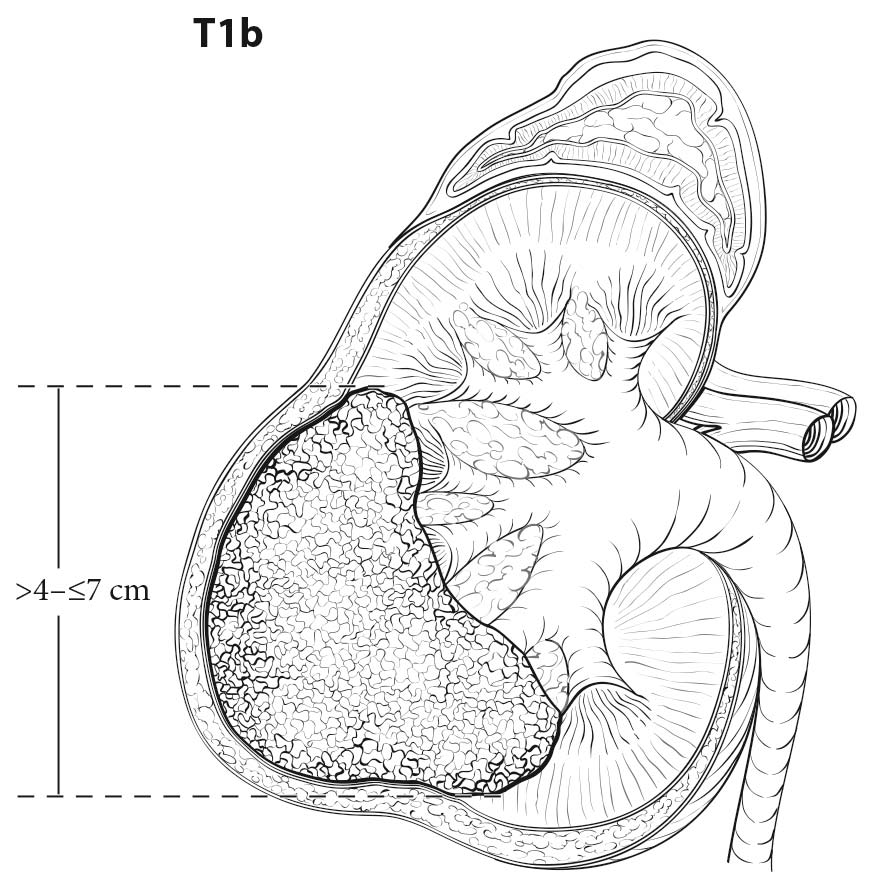
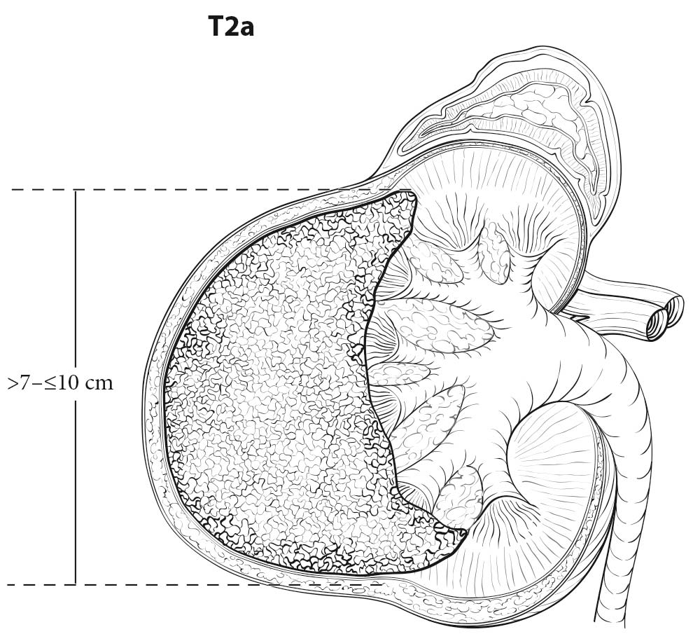
Both CT and MR imaging are equally useful in local staging of renal cell carcinoma and can be considered as first line tests. However, CT has an advantage over MR imaging as it shows calcification and allows better visualization of other body parts, such as the chest, which may be used for staging. RCC is usually a solid, contrast-enhancing mass or cystic with solid components. Most contrast-enhancing renal masses tend to be malignant, and the odds ratio of malignancy increases with increasing size. Multiplanar imaging in MR or multiplanar reconstruction of CT images allows accurate measurement of renal tumors. However, small differences between imaging size measurements and measurements made on resected tumors postoperatively are common, but may not be clinically significant.1,2 High-resolution CT using thin sections appears to improve detection of perinephric infiltration, although false positives are common.3,4

Presence of a pseudocapsule on MR imaging is useful in separating T1 or T2 tumors from T3a tumors in patients with RCC.5 Involvement of renal sinus fat is more difficult to detect on preoperative imaging. Although CT and MR imaging have a high negative predictive value, detection of renal sinus fat invasion is often difficult with relatively low positive predictive value.6

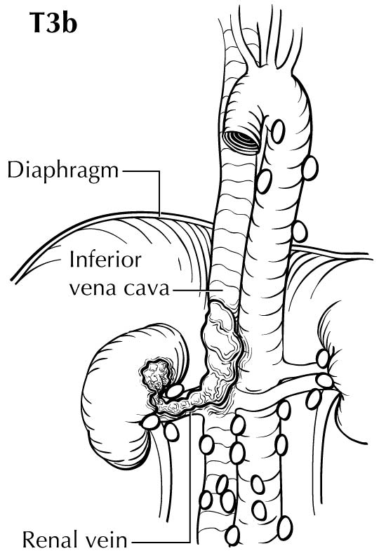
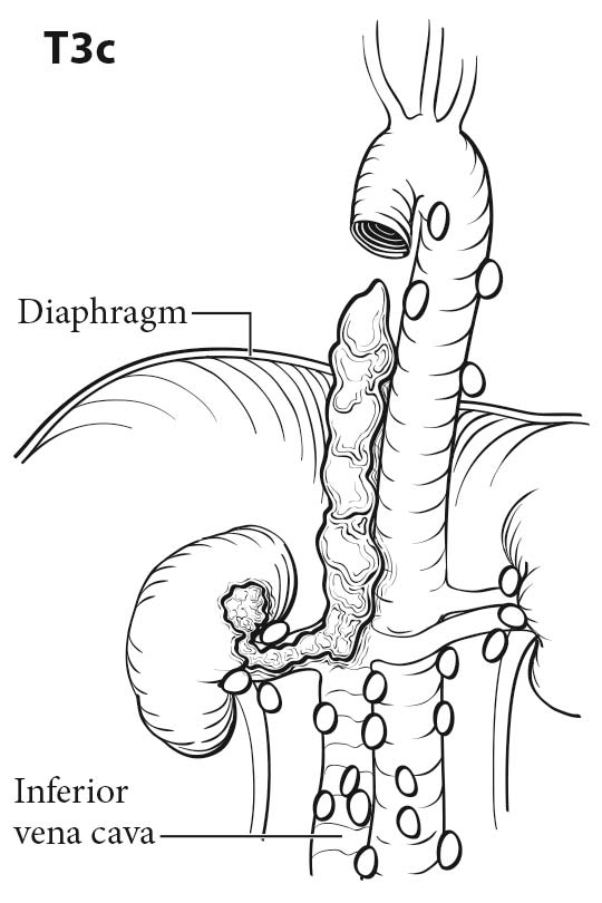
Tumor thrombus in the renal vein or inferior vena cava (IVC) can be identified on the venous or delayed phase of contrast-enhanced CT or MR imaging. Signs suggestive of renal vein or caval thrombus include filling defects, enlargement of the vessel, and rim enhancement. Tumor thrombus in the segmental branches of the renal vein may be more difficult to detect than thrombus in the main renal vein and IVC.7 Both CT and MR imaging have poor positive predictive value for distinguishing invasion from mere abutment with adjacent organs, such as adrenal gland s, liver, diaphragm, psoas muscles, pancreas, and bowel. CT and MR imaging have a high sensitivity and nearly a 100% negative predictive value in detecting direct contiguous spread to the ipsilateral adrenal gland .8,9

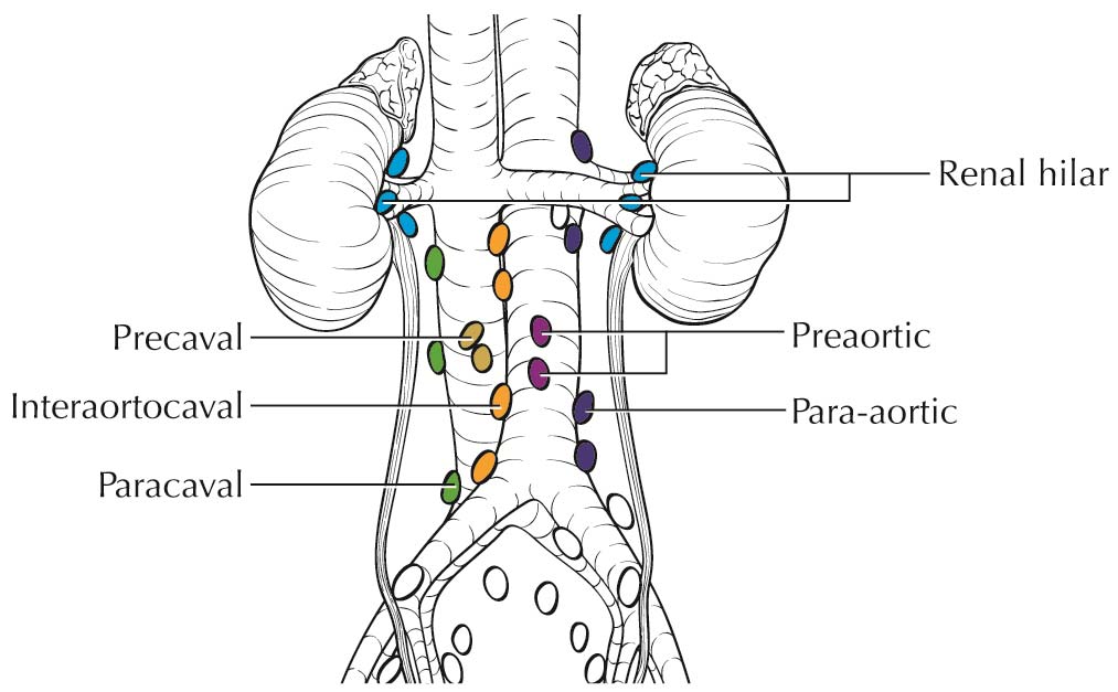
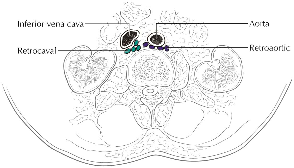
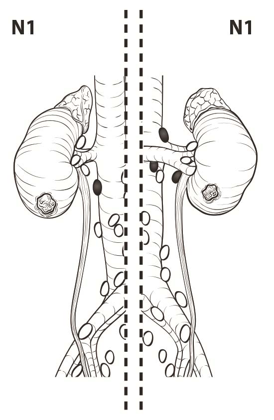
Lymph nodes larger than 1 cm in short axis diameter or nodes that appear to have distorted architecture on imaging should raise suspicion for nodal metastasis. Although 10% of nodes that harbor metastases may be smaller than 1 cm, reactive hyperplasia is common and can be seen in up to 58% of enlarged nodes.10

The risk of metastasis depends on the size of renal tumors and other factors, such as subtype and sarcomatoid differentiation.11-14 Distant metastases typically occur in lung, bone, liver, ipsilateral and contralateral adrenal gland s, and brain. Chest CT is the most sensitive test for detecting pulmonary metastasis, but a plain chest X-ray may be sufficient in low-risk patients.12,15 Patients with advanced primary tumors with symptoms attributable to bone metastasis or patients with abnormal laboratory findings, such as elevated alkaline phosphatase, can be investigated with bone scan to detect bone metastasis.16 Patients with localizing neurological signs should be investigated with MR imaging of the brain or a contrast-enhanced CT scan of the head to detect brain metastasis. Although no evidence justifies routine use of brain MR imaging, it can be used to detect asymptomatic occult brain metastasis in patients with advanced RCC.17 Combined 18-F-fluorodeoxyglucose positron emission tomography/CT (18F-FDG PET-CT or PETCT) does not have an established role in the initial staging of renal cancer, in part because RCC lesions typically have low avidity for FDG relative to high background uptake and excretion in the kidneys.18 Although low in sensitivity to detect distant metastasis, PET-CT has superior specificity and may have a complementary role as a problem-solving tool in cases that are equivocal by conventional imaging.19,20

Pathological Classification

Pathological staging requires surgical resection, which can be performed with either an open or minimally invasive approach. Resection of the primary tumor along with the overlying Gerota's fascia and perinephric fat is recommended. Partial nephrectomy is an acceptable treatment for localized tumors amenable to this approach and is the preferred form of management for clinical T1 tumors and when preservation of renal function is an issue.12,21 Formal retroperitoneal lymph node dissection improves nodal staging accuracy; however, the impact of lymphadenectomy on oncologic outcome remains uncertain.22-24 Adrenal gland involvement is categorized as M1 unless the mechanism is by direct extension from the renal tumor into the ipsilateral adrenal gland , which is category T4. En bloc resection of the ipsilateral adrenal gland is recommended if there is evidence of involvement by imaging or intraoperative findings, but is not necessary if the adrenal gland appears normal.25-27

For staging purposes, pathological tumor size is required. For large tumors, particularly those larger than 7 cm and those occurring in the region of the renal sinus, renal sinus invasion should be suspected and tumor sampling should be targeted to help make this determination. For tumor with extrarenal invasion, the greatest dimension of the tumor mass, including the extrarenal extension, should be measured. For tumor with intravascular extension, the tumor thrombus is excluded from the tumor size measurement. If a specimen contains multiple tumor nodules, a maximum of 5 nodules should be measured, provided all tumors have similar gross appearance and the largest is used to assign T-category with (m) used to indicate multiple tumors. Measurement should be taken for additional nodules if they have variable gross appearance. Tumors with differing histologies should have separate staging.

It is not uncommon that tumor involvement of the renal vein and , in particular, its branches is unrecognized at the time of gross examination of the specimen. This is even truer in partial nephrectomy specimens. Evaluation at microscopy not infrequently reveals such gross misses. Therefore, the word “grossly” has been excluded in the current pT3a staging. In addition, the diameter of a sinus vein or the quantity or the presence or absence of muscle in sinus veins is a poor indicator of the vein segment or its relationship to the main renal vein, and thus muscle does not need to be identified in a vein to classify it as a “renal vein segmental branch” and thus categorize the tumor as pT3a.28 Note that circumscribed tumor nodules in the renal sinus fat likely represent vascular invasion.28 Vascular invasion should be confirmed microscopically.

Perinephric/sinus fat invasion should be confirmed microscopically. Invasion into fat by tumor cells, with or without desmoplastic reaction, and vascular invasion in perinephric/sinus soft tissue are all evidence of perinephric/sinus invasion. It is reported as “present” or “absent.”

Specimen Hand ling

The pathological specimen should be processed in a stand ardized fashion to allow for full pathological assessment. The International Society of Urological Pathology (ISUP) and College of American Pathologists (www.cap.org) have established practical guidelines for specimen processing.29

Prognostic Factors

Prognostic Factors Required for Stage Grouping

Beyond the factors used to assign T, N, or M categories, no additional prognostic factors are required for stage grouping.

Additional Factors Recommended for Clinical Care

Histologic Subtype

Histologic subtype is a strong prognostic factor and an increasingly important factor for treatment decisions, especially in the metastatic state. Histologic subtype should be categorized as discussed below. For example, type 1 papillary renal cancers tend to have a good prognosis, whereas medullary and collecting duct carcinomas tend to have a poor prognosis.30

AJCC Level of Evidence: I
World Health Organization (WHO)/ISUP nucleolar grade

Grade is a strong prognostic factor, especially for clear cell RCC and should be described as discussed in the section titled Histologic Grade (G).

AJCC Level of Evidence: II
Sarcomatoid Features

Sarcomatoid differentiation in RCC consists of sheets and fascicles of malignant spindle cells, which can occur across all histologic subtypes. Occasionally, the sarcomatoid component resembles specific types of sarcoma, such as osteogenic sarcoma, chondrosarcoma, rhabdomyosarcoma, etc. Sarcomatoid differentiation can be seen in any of the RCC subtypes and is associated with a poor prognosis.

A minimum quantity of sarcomatoid component is not required to make the diagnosis; some experts require a low power field or a clear-cut area of sarcoma-like histology. The percentage of sarcomatoid component has been shown to correlate with cancer-specific mortality and an estimate of its quantity should be provided.31

AJCC Level of Evidence: II
Rhabdoid Features

Rhabdoid differentiation is characterized by the presence of cells with abundant eosinophilic cytoplasm expand ed by an eccentric granular eosinophilic inclusion and a large eccentrically placed nucleus and prominent eosinophilic nucleolus. Rhabdoid differentiation, like sarcomatoid differentiation, is a de-differentiation pathway common to all RCC subtypes. Both components may co-exist in the same tumor. The presence of rhabdoid differentiation in RCC is associated with a poor prognosis independent of histological subtypes, grade, and stage.32 The presence of rhabdoid differentiation should be noted in the pathology report.30

AJCC Level of Evidence: II
Histologic tumor necrosis

Coagulative necrosis is correlated with prognosis and , on microscopic examination, characterized by homogeneous clusters and sheets of degenerating and dead cells, or granular pink coagulum admixed with nuclear and cytoplasmic debris. Degenerative changes—such as hemorrhage, hyalinization and scar—and ischemic necrosis should not be mistaken for necrosis. Any amount of coagulative necrosis should be reported.33

AJCC Level of Evidence: II
Microscopic angiolymphatic invasion

Microscopic lymphovascular invasion (LVI) is defined as the presence of tumor in the small vascular spaces within the host kidney. Its reported incidence is quite variable due to variable detection methods used (by immunohistochemical or stand ard hematoxylin and eosin stains). In the majority of the published studies, LVI has been shown to correlate with other prognostic parameters, including tumor size, grade, pT category, and the presence of lymph node and distant metastases. LVI also has been significantly associated with outcome determined as disease-free survival and cancer-specific survival. It is recommended to report LVI when identified on hematoxylin and eosin stains.30

AJCC Level of Evidence: II

Gene Expression of the Primary Tumor

Over the past several years, techniques to incorporate tumor gene expression have been applied to patients with localized RCC who undergo nephrectomy. These gene expression results have then been incorporated into known prognostic variables and have been shown to add value in terms of prognostication. Although none are in routine clinical use presently, with broader availability and application of gene expression techniques, further refinement of prognostic schema may occur.

One approach examined 48 clear cell RCC samples by unsupervised clustering of gene expression data to reveal two distinct subsets, designated ccA and ccB.34 These independent tumor types were confirmed in an independent cohort of 177 clear cell RCC tumors and shown to be significantly associated with different cancer-specific and overall survival. These associations with survival were of borderline significance (p = 0.089) when accounting for stage, grade, and performance status.

Another dataset looked at gene expression in 942 resected Stage I-III clear cell RCC primary tumors and developed a score based on gene expression (in 11 cancer-related and 5 reference genes), which was validated in 626 separate primary renal tumors.35 In multivariable analysis accounting for known prognostic factors, this recurrence score was significantly associated with risk of tumor recurrence and overall survival (p < 0.0001)

AJCC Level of Evidence: Not identified

Risk Assessment

Risk Assesment Models

The American Joint Committee on Cancer (AJCC) recently has established guidelines that will be used to evaluate published statistical prediction models for the purpose of granting endorsement for clinical use.36 Although this is a monumental step toward the goal of precision medicine, this work was published only very recently. For this reason, existing models that have been published or may be in clinical use have not yet been evaluated for this cancer site by the Precision Medicine core of the AJCC. In the future, the statistical prediction models for this cancer site will be evaluated, and those that meet all AJCC criteria will be endorsed.

Recommendations

Clinical trials tend to be focused on patients with high-risk localized disease for which the TNM staging system and the prognostic variables, especially histologic subtype, become important clinical trial stratification or selection features. In patients with metastatic disease, prognosis remains poor. Important additional considerations for patient selection and stratification in the design of clinical trials for these patients include prior therapy, performance status, metastatic burden, distribution of metastatic disease, and presence of anemia, thrombocytosis, neutrophilia, hypercalcemia, and elevated LDH.

TNM Definitions

Clinical T (cT)

cT CategorycT Criteria
cTXPrimary tumor cannot be assessed
cT0No evidence of primary tumor
cT1Tumor less than or equal to 7 cm in greatest dimension, limited to the kidney
cT1aTumor less than or equal to 4 cm in greatest dimension, limited to the kidney
cT1bTumor greater than 4 cm but less than or equal to 7 cm in greatest dimension limited to the kidney
cT2Tumor greater than 7 cm in greatest dimension, limited to the kidney
cT2aTumor greater than 7 cm but less than or equal to 10 cm in greatest dimension, limited to the kidney
cT2bTumor greater than 10 cm, limited to the kidney
cT3Tumor extends into major veins or perinephric tissues, but not into the ipsilateral adrenal gland and not beyond Gerota's fascia
cT3aTumor extends into the renal vein or its segmental branches, or invades the pelvicalyceal system, or invades perirenal and /or renal sinus fat but not beyond Gerota's fascia
cT3bTumor extends into the vena cava below the diaphragm
cT3cTumor extends into the vena cava above the diaphragm or invades the wall of the vena cava
cT4Tumor invades beyond Gerota's fascia (including contiguous extension into the ipsilateral adrenal gland )

Pathological T (pT)

pT CategorypT Criteria
pTXPrimary tumor cannot be assessed
pT0No evidence of primary tumor
pT1Tumor less than or equal to 7 cm in greatest dimension, limited to the kidney
pT1aTumor less than or equal to 4 cm in greatest dimension, limited to the kidney
pT1bTumor greater than 4 cm but less than or equal to 7 cm in greatest dimension limited to the kidney
pT2Tumor greater than 7 cm in greatest dimension, limited to the kidney
pT2aTumor greater than 7 cm but less than or equal to 10 cm in greatest dimension, limited to the kidney
pT2bTumor greater than 10 cm, limited to the kidney
pT3Tumor extends into major veins or perinephric tissues, but not into the ipsilateral adrenal gland and not beyond Gerota's fascia
pT3aTumor extends into the renal vein or its segmental branches, or invades the pelvicalyceal system, or invades perirenal and /or renal sinus fat but not beyond Gerota's fascia
pT3bTumor extends into the vena cava below the diaphragm
pT3cTumor extends into the vena cava above the diaphragm or invades the wall of the vena cava
pT4Tumor invades beyond Gerota's fascia (including contiguous extension into the ipsilateral adrenal gland )
cTXPrimary tumor cannot be assessed
cT0No evidence of primary tumor
cT1Tumor less than or equal to 7 cm in greatest dimension, limited to the kidney
cT1aTumor less than or equal to 4 cm in greatest dimension, limited to the kidney
cT1bTumor greater than 4 cm but less than or equal to 7 cm in greatest dimension limited to the kidney
cT2Tumor greater than 7 cm in greatest dimension, limited to the kidney
cT2aTumor greater than 7 cm but less than or equal to 10 cm in greatest dimension, limited to the kidney
cT2bTumor greater than 10 cm, limited to the kidney
cT3Tumor extends into major veins or perinephric tissues, but not into the ipsilateral adrenal gland and not beyond Gerota's fascia
cT3aTumor extends into the renal vein or its segmental branches, or invades the pelvicalyceal system, or invades perirenal and /or renal sinus fat but not beyond Gerota's fascia
cT3bTumor extends into the vena cava below the diaphragm
cT3cTumor extends into the vena cava above the diaphragm or invades the wall of the vena cava
cT4Tumor invades beyond Gerota's fascia (including contiguous extension into the ipsilateral adrenal gland )

Neoadjuvant Clinical T (yT)

ycT CategoryycT Criteria
ycTXPrimary tumor cannot be assessed
ycT0No evidence of primary tumor
ycT1Tumor less than or equal to 7 cm in greatest dimension, limited to the kidney
ycT1aTumor less than or equal to 4 cm in greatest dimension, limited to the kidney
ycT1bTumor greater than 4 cm but less than or equal to 7 cm in greatest dimension limited to the kidney
ycT2Tumor greater than 7 cm in greatest dimension, limited to the kidney
ycT2aTumor greater than 7 cm but less than or equal to 10 cm in greatest dimension, limited to the kidney
ycT2bTumor greater than 10 cm, limited to the kidney
ycT3Tumor extends into major veins or perinephric tissues, but not into the ipsilateral adrenal gland and not beyond Gerota's fascia
ycT3aTumor extends into the renal vein or its segmental branches, or invades the pelvicalyceal system, or invades perirenal and /or renal sinus fat but not beyond Gerota's fascia
ycT3bTumor extends into the vena cava below the diaphragm
ycT3cTumor extends into the vena cava above the diaphragm or invades the wall of the vena cava
ycT4Tumor invades beyond Gerota's fascia (including contiguous extension into the ipsilateral adrenal gland )

Neoadjuvant Pathological T (yT)

ypT CategoryypT Criteria
ypTXPrimary tumor cannot be assessed
ypT0No evidence of primary tumor
ypT1Tumor less than or equal to 7 cm in greatest dimension, limited to the kidney
ypT1aTumor less than or equal to 4 cm in greatest dimension, limited to the kidney
ypT1bTumor greater than 4 cm but less than or equal to 7 cm in greatest dimension limited to the kidney
ypT2Tumor greater than 7 cm in greatest dimension, limited to the kidney
ypT2aTumor greater than 7 cm but less than or equal to 10 cm in greatest dimension, limited to the kidney
ypT2bTumor greater than 10 cm, limited to the kidney
ypT3Tumor extends into major veins or perinephric tissues, but not into the ipsilateral adrenal gland and not beyond Gerota's fascia
ypT3aTumor extends into the renal vein or its segmental branches, or invades the pelvicalyceal system, or invades perirenal and /or renal sinus fat but not beyond Gerota's fascia
ypT3bTumor extends into the vena cava below the diaphragm
ypT3cTumor extends into the vena cava above the diaphragm or invades the wall of the vena cava
ypT4Tumor invades beyond Gerota's fascia (including contiguous extension into the ipsilateral adrenal gland )

Definition of Regional Lymph Node (N)

Clinical N (cN)
cN CategorycN Criteria
cNXRegional lymph nodes cannot be assessed
cN0No regional lymph node metastasis
cN1Metastasis in regional lymph node(s)
Pathological N (pN)
pN CategorypN Criteria
pNXRegional lymph nodes cannot be assessed
pN0No regional lymph node metastasis
pN1Metastasis in regional lymph node(s)
cNXRegional lymph nodes cannot be assessed
cN0No regional lymph node metastasis
cN1Metastasis in regional lymph node(s)
Neoadjuvant Clinical N (pY)
ycN CategoryycN Criteria
ycNXRegional lymph nodes cannot be assessed
ycN0No regional lymph node metastasis
ycN1Metastasis in regional lymph node(s)
Neoadjuvant Pathological N (pY)
ypN CategoryypN Criteria
ypNXRegional lymph nodes cannot be assessed
ypN0No regional lymph node metastasis
ypN1Metastasis in regional lymph node(s)

Definition of Distant Metastasis (M)- Clinical M (cN)

cM CategorycM Criteria
cM0No distant metastasis
cM1Distant metastasis
pM1Microscopic evidence of distant metastasis

Definition of Distant Metastasis (M)- Pathological M (pN)

pM CategorypM Criteria
cM0No distant metastasis
cM1Distant metastasis
pM1Microscopic evidence of distant metastasis

Definition of Distant Metastasis (M)- Neoadjuvant Clinical M (pY)

ycM CategoryycM Criteria
cM0No distant metastasis
cM1Distant metastasis
pM1Microscopic evidence of distant metastasis

Definition of Distant Metastasis (M)- Neoadjuvant Pathological M (pY)

ypM CategoryypM Criteria
cM0No distant metastasis
cM1Distant metastasis
pM1Microscopic evidence of distant metastasis

Stage Prognostic

Clinical

When T is…and N is…and M is…Then the Clinical Prognostic Stage Group is…
cT1cN0cM0I
cT1cN0cM0I
cT1acN0cM0I
cT1bcN0cM0I
cT1cN1cM0III
cT1cN1cM0III
cT1acN1cM0III
cT1bcN1cM0III
cT2cN0cM0II
cT2cN0cM0II
cT2acN0cM0II
cT2bcN0cM0II
cT2cN1cM0III
cT2cN1cM0III
cT2acN1cM0III
cT2bcN1cM0III
cT3cNXcM0III
cT3cN0cM0III
cT3acNXcM0III
cT3acN0cM0III
cT3bcNXcM0III
cT3bcN0cM0III
cT3ccNXcM0III
cT3ccN0cM0III
cT3cN1cM0III
cT3cN1cM0III
cT3acN1cM0III
cT3bcN1cM0III
cT3ccN1cM0III
cT4cNXcM0IV
cT4cN0cM0IV
cT4cN1cM0IV
cTXcNXcM1IV
cTXcN0cM1IV
cTXcN1cM1IV
cT0cNXcM1IV
cT0cN0cM1IV
cT0cN1cM1IV
cT1cNXcM1IV
cT1cN0cM1IV
cT1cN1cM1IV
cT1acNXcM1IV
cT1acN0cM1IV
cT1acN1cM1IV
cT1bcNXcM1IV
cT1bcN0cM1IV
cT1bcN1cM1IV
cT2cNXcM1IV
cT2cN0cM1IV
cT2cN1cM1IV
cT2acNXcM1IV
cT2acN0cM1IV
cT2acN1cM1IV
cT2bcNXcM1IV
cT2bcN0cM1IV
cT2bcN1cM1IV
cT3cNXcM1IV
cT3cN0cM1IV
cT3cN1cM1IV
cT3acNXcM1IV
cT3acN0cM1IV
cT3acN1cM1IV
cT3bcNXcM1IV
cT3bcN0cM1IV
cT3bcN1cM1IV
cT3ccNXcM1IV
cT3ccN0cM1IV
cT3ccN1cM1IV
cT4cNXcM1IV
cT4cN0cM1IV
cT4cN1cM1IV
cTXcNXpM1IV
cTXcN0pM1IV
cTXcN1pM1IV
cT0cNXpM1IV
cT0cN0pM1IV
cT0cN1pM1IV
cT1cNXpM1IV
cT1cN0pM1IV
cT1cN1pM1IV
cT1acNXpM1IV
cT1acN0pM1IV
cT1acN1pM1IV
cT1bcNXpM1IV
cT1bcN0pM1IV
cT1bcN1pM1IV
cT2cNXpM1IV
cT2cN0pM1IV
cT2cN1pM1IV
cT2acNXpM1IV
cT2acN0pM1IV
cT2acN1pM1IV
cT2bcNXpM1IV
cT2bcN0pM1IV
cT2bcN1pM1IV
cT3cNXpM1IV
cT3cN0pM1IV
cT3cN1pM1IV
cT3acNXpM1IV
cT3acN0pM1IV
cT3acN1pM1IV
cT3bcNXpM1IV
cT3bcN0pM1IV
cT3bcN1pM1IV
cT3ccNXpM1IV
cT3ccN0pM1IV
cT3ccN1pM1IV
cT4cNXpM1IV
cT4cN0pM1IV
cT4cN1pM1IV

Pathological

When T is…and N is…and M is…Then the Pathological Prognostic Stage Group is…
pT1pN0cM0I
pT1pN0cM0I
pT1apN0cM0I
pT1bpN0cM0I
pT1pN1cM0III
pT1pN1cM0III
pT1apN1cM0III
pT1bpN1cM0III
pT2pN0cM0II
pT2pN0cM0II
pT2apN0cM0II
pT2bpN0cM0II
pT2pN1cM0III
pT2pN1cM0III
pT2apN1cM0III
pT2bpN1cM0III
pT3pNXcM0III
pT3pN0cM0III
pT3apNXcM0III
pT3apN0cM0III
pT3bpNXcM0III
pT3bpN0cM0III
pT3cpNXcM0III
pT3cpN0cM0III
pT3pN1cM0III
pT3pN1cM0III
pT3apN1cM0III
pT3bpN1cM0III
pT3cpN1cM0III
pT4pNXcM0IV
pT4pN0cM0IV
pT4pN1cM0IV
pTXpNXpM1IV
pTXpN0pM1IV
pTXpN1pM1IV
pT0pNXpM1IV
pT0pN0pM1IV
pT0pN1pM1IV
pT1pNXpM1IV
pT1pN0pM1IV
pT1pN1pM1IV
pT1apNXpM1IV
pT1apN0pM1IV
pT1apN1pM1IV
pT1bpNXpM1IV
pT1bpN0pM1IV
pT1bpN1pM1IV
pT2pNXpM1IV
pT2pN0pM1IV
pT2pN1pM1IV
pT2apNXpM1IV
pT2apN0pM1IV
pT2apN1pM1IV
pT2bpNXpM1IV
pT2bpN0pM1IV
pT2bpN1pM1IV
pT3pNXpM1IV
pT3pN0pM1IV
pT3pN1pM1IV
pT3apNXpM1IV
pT3apN0pM1IV
pT3apN1pM1IV
pT3bpNXpM1IV
pT3bpN0pM1IV
pT3bpN1pM1IV
pT3cpNXpM1IV
pT3cpN0pM1IV
pT3cpN1pM1IV
pT4pNXpM1IV
pT4pN0pM1IV
pT4pN1pM1IV
pTXpNXcM1IV
pTXpN0cM1IV
pTXpN1cM1IV
pT0pNXcM1IV
pT0pN0cM1IV
pT0pN1cM1IV
pT1pNXcM1IV
pT1pN0cM1IV
pT1pN1cM1IV
pT1apNXcM1IV
pT1apN0cM1IV
pT1apN1cM1IV
pT1bpNXcM1IV
pT1bpN0cM1IV
pT1bpN1cM1IV
pT2pNXcM1IV
pT2pN0cM1IV
pT2pN1cM1IV
pT2apNXcM1IV
pT2apN0cM1IV
pT2apN1cM1IV
pT2bpNXcM1IV
pT2bpN0cM1IV
pT2bpN1cM1IV
pT3pNXcM1IV
pT3pN0cM1IV
pT3pN1cM1IV
pT3apNXcM1IV
pT3apN0cM1IV
pT3apN1cM1IV
pT3bpNXcM1IV
pT3bpN0cM1IV
pT3bpN1cM1IV
pT3cpNXcM1IV
pT3cpN0cM1IV
pT3cpN1cM1IV
pT4pNXcM1IV
pT4pN0cM1IV
pT4pN1cM1IV
cTXcNXpM1IV
cTXcN0pM1IV
cTXcN1pM1IV
cT0cNXpM1IV
cT0cN0pM1IV
cT0cN1pM1IV
cT1cNXpM1IV
cT1cN0pM1IV
cT1cN1pM1IV
cT1acNXpM1IV
cT1acN0pM1IV
cT1acN1pM1IV
cT1bcNXpM1IV
cT1bcN0pM1IV
cT1bcN1pM1IV
cT2cNXpM1IV
cT2cN0pM1IV
cT2cN1pM1IV
cT2acNXpM1IV
cT2acN0pM1IV
cT2acN1pM1IV
cT2bcNXpM1IV
cT2bcN0pM1IV
cT2bcN1pM1IV
cT3cNXpM1IV
cT3cN0pM1IV
cT3cN1pM1IV
cT3acNXpM1IV
cT3acN0pM1IV
cT3acN1pM1IV
cT3bcNXpM1IV
cT3bcN0pM1IV
cT3bcN1pM1IV
cT3ccNXpM1IV
cT3ccN0pM1IV
cT3ccN1pM1IV
cT4cNXpM1IV
cT4cN0pM1IV
cT4cN1pM1IV

Neoadjuvant Clinical

There is no Postneoadjuvant Clinical Therapy (ycTNM) stage group available at this time.

Neoadjuvant Pathological

When T is…and N is…and M is…Then the Postneoadjuvant Pathological Stage Group is…
ypT1ypN0cM0I
ypT1ypN0cM0I
ypT1aypN0cM0I
ypT1bypN0cM0I
ypT1ypN1cM0III
ypT1ypN1cM0III
ypT1aypN1cM0III
ypT1bypN1cM0III
ypT2ypN0cM0II
ypT2ypN0cM0II
ypT2aypN0cM0II
ypT2bypN0cM0II
ypT2ypN1cM0III
ypT2ypN1cM0III
ypT2aypN1cM0III
ypT2bypN1cM0III
ypT3ypNXcM0III
ypT3ypN0cM0III
ypT3aypNXcM0III
ypT3aypN0cM0III
ypT3bypNXcM0III
ypT3bypN0cM0III
ypT3cypNXcM0III
ypT3cypN0cM0III
ypT3ypN1cM0III
ypT3ypN1cM0III
ypT3aypN1cM0III
ypT3bypN1cM0III
ypT3cypN1cM0III
ypT4ypNXcM0IV
ypT4ypN0cM0IV
ypT4ypN1cM0IV
ypTXypNXpM1IV
ypTXypN0pM1IV
ypTXypN1pM1IV
ypT0ypNXpM1IV
ypT0ypN0pM1IV
ypT0ypN1pM1IV
ypT1ypNXpM1IV
ypT1ypN0pM1IV
ypT1ypN1pM1IV
ypT1aypNXpM1IV
ypT1aypN0pM1IV
ypT1aypN1pM1IV
ypT1bypNXpM1IV
ypT1bypN0pM1IV
ypT1bypN1pM1IV
ypT2ypNXpM1IV
ypT2ypN0pM1IV
ypT2ypN1pM1IV
ypT2aypNXpM1IV
ypT2aypN0pM1IV
ypT2aypN1pM1IV
ypT2bypNXpM1IV
ypT2bypN0pM1IV
ypT2bypN1pM1IV
ypT3ypNXpM1IV
ypT3ypN0pM1IV
ypT3ypN1pM1IV
ypT3aypNXpM1IV
ypT3aypN0pM1IV
ypT3aypN1pM1IV
ypT3bypNXpM1IV
ypT3bypN0pM1IV
ypT3bypN1pM1IV
ypT3cypNXpM1IV
ypT3cypN0pM1IV
ypT3cypN1pM1IV
ypT4ypNXpM1IV
ypT4ypN0pM1IV
ypT4ypN1pM1IV
ypTXypNXcM1IV
ypTXypN0cM1IV
ypTXypN1cM1IV
ypT0ypNXcM1IV
ypT0ypN0cM1IV
ypT0ypN1cM1IV
ypT1ypNXcM1IV
ypT1ypN0cM1IV
ypT1ypN1cM1IV
ypT1aypNXcM1IV
ypT1aypN0cM1IV
ypT1aypN1cM1IV
ypT1bypNXcM1IV
ypT1bypN0cM1IV
ypT1bypN1cM1IV
ypT2ypNXcM1IV
ypT2ypN0cM1IV
ypT2ypN1cM1IV
ypT2aypNXcM1IV
ypT2aypN0cM1IV
ypT2aypN1cM1IV
ypT2bypNXcM1IV
ypT2bypN0cM1IV
ypT2bypN1cM1IV
ypT3ypNXcM1IV
ypT3ypN0cM1IV
ypT3ypN1cM1IV
ypT3aypNXcM1IV
ypT3aypN0cM1IV
ypT3aypN1cM1IV
ypT3bypNXcM1IV
ypT3bypN0cM1IV
ypT3bypN1cM1IV
ypT3cypNXcM1IV
ypT3cypN0cM1IV
ypT3cypN1cM1IV
ypT4ypNXcM1IV
ypT4ypN0cM1IV
ypT4ypN1cM1IV

Registry Data

Registry Data Collection Variables

  1. Histologic subtype
  2. WHO/ISUP grade
  3. Tumor size
  4. Invasion into perinephric fat or sinus tissues
  5. Venous involvement with specific mention of intra-renal lymphovascular invasion, branches of renal vein in the renal sinus invasion, renal vein involvement, or IVC involvement
  6. Lymphovascular invasion (LVI)
  7. Adrenal gland involvement by direct extension (T4) or as a seperate nodule (M1)
  8. Sarcomatoid features; present or absent and percentage
  9. Rhabdoid differentiation; present or absent
  10. Histologic tumor necrosis

Histopathologic type

Histologic grade

HISTOLOGIC GRADE (G)

The Fuhrman grading system, published in 1982, has been widely utilized. It is a four-tier system based on nuclear size, nuclear shape, and nucleolar prominence. Despite the widespread usage of Fuhrman grading, serious problems are associated with its implementation, reproducibility, and outcome prediction. As a result, a modified grading system has been proposed to be based on nucleolar prominence for the first three grading categories, while grade 4 is based on the presence of marked nuclear pleomorphism, which may include tumor giant cells or sarcomatoid and /or rhabdoid differentiation. Known as the WHO/ISUP grade, this grading system was validated for clear cell and papillary RCC, but was shown not to be useful for chromophobe RCC and has not been validated in other RCC histologic subtypes.30

GG Definition
GXGrade cannot be assessed
G1Nucleoli absent or inconspicuous and basophilic at 400x magnification
G2Nucleoli conspicuous and eosinophilic at 400x magnification, visible but not prominent at 100x magnification
G3Nucleoli conspicuous and eosinophilic at 100x magnification
G4Marked nuclear pleomorphism and /or multinucleate giant cells and /or rhabdoid and /or sarcomatoid differentiation

Illustrations

58.4 T1a: Tumor 4 cm or smaller in greatest dimension, limited to the kidney.

58.5 T1b: Tumor larger than 4 cm but not larger than 7 cm in greatest dimension, limited to the kidney.

58.6 T2a: Tumor larger than 7 cm in greatest dimension, but not larger than 10 cm, limited to the kidney.

58.7 T2b: Tumor larger than 10 cm in greatest dimension.

58.8 (Left) T3a: Invasion into perirenal and /or renal sinus fat but not beyond Gerota's fascia. (Right) T3a: In addition to perirenal and /or renal sinus fat, tumor invades into the renal vein or segmental branches of renal vein in the renal sinus.

58.9 T3b: Tumor extends into vena cava below the diaphragm.

58.10 T3c: Tumor extends into vena cava above diaphragm or invades the wall of the vena cava.

58.11 T4: Invasion beyond Gerota's fascia.

58.12 T4: Invasion into ipsilateral adrenal gland .

58.13 N1 disease is defined as a single or multiple regional lymph node involvement.

Bibliography

  1. Jeffery NN, Douek N, Guo DY, Patel MI. Discrepancy between radiological and pathological size of renal masses. BMC urology. 2011;11(1):2.
  2. Roberts WW, Bhayani SB, Allaf ME, Chan TY, Kavoussi LR, Jarrett TW. Pathological stage does not alter the prognosis for renal lesions determined to be stage T1 by computerized tomography. The Journal of urology. 2005;173(3):713-715.
  3. Catalano C, Fraioli F, Laghi A, et al. High-resolution multidetector CT in the preoperative evaluation of patients with renal cell carcinoma. AJR. American journal of roentgenology. May 2003;180(5):1271-1277.
  4. Hallscheidt PJ, Bock M, Riedasch G, et al. Diagnostic accuracy of staging renal cell carcinomas using multidetector-row computed tomography and magnetic resonance imaging: a prospective study with histopathologic correlation. Journal of computer assisted tomography. May-Jun 2004;28(3):333-339.
  5. Roy C, Sr., El Ghali S, Buy X, et al. Significance of the pseudocapsule on MRI of renal neoplasms and its potential application for local staging: a retrospective study. AJR. American journal of roentgenology. Jan 2005;184(1):113-120.
  6. Kim C, Choi HJ, Cho KS. Diagnostic value of multidetector computed tomography for renal sinus fat invasion in renal cell carcinoma patients. Eur J Radiol. Jun 2014;83(6):914-918.
  7. Hallscheidt P, Wagener N, Gholipour F, et al. Multislice computed tomography in planning nephron-sparing surgery in a prospective study with 76 patients: comparison of radiological and histopathological findings in the infiltration of renal structures. Journal of computer assisted tomography. Nov-Dec 2006;30(6):869-874.
  8. Tsui KH, Shvarts O, Smith RB, Figlin RA, deKernion JB, Belldegrun A. Prognostic indicators for renal cell carcinoma: a multivariate analysis of 643 patients using the revised 1997 TNM staging criteria. J Urol. Apr 2000;163(4):1090-1095; quiz 1295.
  9. Sawai Y, Kinouchi T, Mano M, et al. Ipsilateral adrenal involvement from renal cell carcinoma: retrospective study of the predictive value of computed tomography. Urology. Jan 2002;59(1):28-31.
  10. Studer UE, Scherz S, Scheidegger J, et al. Enlargement of regional lymph nodes in renal cell carcinoma is often not due to metastases. J Urol. Aug 1990;144(2 Pt 1):243-245.
  11. Lee H, Lee JK, Kim K, et al. Risk of metastasis for T1a renal cell carcinoma. World journal of urology. Apr 2016;34(4):553-559.
  12. Ljungberg B, Bensalah K, Canfield S, et al. EAU guidelines on renal cell carcinoma: 2014 update. Eur Urol. May 2015;67(5):913-924.
  13. Thompson RH, Hill JR, Babayev Y, et al. Metastatic renal cell carcinoma risk according to tumor size. J Urol. Jul 2009;182(1):41-45.
  14. Wunderlich H, Reichelt O, Schumann S, et al. Nephron sparing surgery for renal cell carcinoma 4 cm. or less in diameter: indicated or under treated? J Urol. May 1998;159(5):1465-1469.
  15. Lim DJ, Carter MF. Computerized tomography in the preoperative staging for pulmonary metastases in patients with renal cell carcinoma. J Urol. Oct 1993;150(4):1112-1114.
  16. Santini D, Procopio G, Porta C, et al. Natural history of malignant bone disease in renal cancer: final results of an Italian bone metastasis survey. PloS one. 2013;8(12):e83026.
  17. Shuch B, La Rochelle JC, Klatte T, et al. Brain metastasis from renal cell carcinoma: presentation, recurrence, and survival. Cancer. Oct 1 2008;113(7):1641-1648.
  18. Ozulker T, Ozulker F, Ozbek E, Ozpacaci T. A prospective diagnostic accuracy study of F-18 fluorodeoxyglucose-positron emission tomography/computed tomography in the evaluation of indeterminate renal masses. Nuclear medicine communications. Apr 2011;32(4):265-272.
  19. Kang DE, White RL, Zuger JH, Sasser HC, Teigland CM. Clinical use of fluorodeoxyglucose F 18 positron emission tomography for detection of renal cell carcinoma. The Journal of urology. 2004;171(5):1806-1809.
  20. Majhail NS, Urbain JL, Albani JM, et al. F-18 fluorodeoxyglucose positron emission tomography in the evaluation of distant metastases from renal cell carcinoma. J Clin Oncol. Nov 1 2003;21(21):3995-4000.
  21. Campbell SC, Novick AC, Belldegrun A, et al. Guideline for management of the clinical T1 renal mass. J Urol. Oct 2009;182(4):1271-1279.
  22. Blom JH, van Poppel H, Marechal JM, et al. Radical nephrectomy with and without lymph-node dissection: final results of European Organization for Research and Treatment of Cancer (EORTC) rand omized phase 3 trial 30881. European urology. 2009;55(1):28-34.
  23. Terrone C, Guercio S, De Luca S, et al. The number of lymph nodes examined and staging accuracy in renal cell carcinoma. BJU Int. Jan 2003;91(1):37-40.
  24. Whitson JM, Harris CR, Reese AC, Meng MV. Lymphadenectomy improves survival of patients with renal cell carcinoma and nodal metastases. J Urol. May 2011;185(5):1615-1620.
  25. Kutikov A, Piotrowski ZJ, Canter DJ, et al. Routine adrenalectomy is unnecessary during surgery for large and /or upper pole renal tumors when the adrenal gland is radiographically normal. The Journal of urology. 2011;185(4):1198-1203.
  26. TSUI K-H, SHVARTS O, BARBARIC Z, FIGLIN R, de KERNION JB, BELLDEGRUN A. Is adrenalectomy a necessary component of radical nephrectomy? UCLA experience with 511 radical nephrectomies. The Journal of urology. 2000;163(2):437-441.
  27. Weight CJ, Kim SP, Lohse CM, et al. Routine adrenalectomy in patients with locally advanced renal cell cancer does not offer oncologic benefit and places a significant portion of patients at risk for an asynchronous metastasis in a solitary adrenal gland . European urology. 2011;60(3):458-464.
  28. Bonsib SM. Renal veins and venous extension in clear cell renal cell carcinoma. Modern pathology : an official journal of the United States and Canadian Academy of Pathology, Inc. Jan 2007;20(1):44-53.
  29. Trpkov K, Grignon DJ, Bonsib SM, et al. Hand ling and staging of renal cell carcinoma: the International Society of Urological Pathology Consensus (ISUP) conference recommendations. The American journal of surgical pathology. Oct 2013;37(10):1505-1517.
  30. Delahunt B, Cheville JC, Martignoni G, et al. The International Society of Urological Pathology (ISUP) grading system for renal cell carcinoma and other prognostic parameters. The American journal of surgical pathology. Oct 2013;37(10):1490-1504.
  31. Zhang BY, Thompson RH, Lohse CM, et al. A novel prognostic model for patients with sarcomatoid renal cell carcinoma. BJU Int. Mar 2015;115(3):405-411.
  32. Przybycin CG, McKenney JK, Reynolds JP, et al. Rhabdoid differentiation is associated with aggressive behavior in renal cell carcinoma: a clinicopathologic analysis of 76 cases with clinical follow-up. The American journal of surgical pathology. Sep 2014;38(9):1260-1265.
  33. Leibovich BC, Blute ML, Cheville JC, et al. Prediction of progression after radical nephrectomy for patients with clear cell renal cell carcinoma: a stratification tool for prospective clinical trials. Cancer. Apr 1 2003;97(7):1663-1671.
  34. Brannon AR, Reddy A, Seiler M, et al. Molecular Stratification of Clear Cell Renal Cell Carcinoma by Consensus Clustering Reveals Distinct Subtypes and Survival Patterns. Genes & cancer. Feb 1 2010;1(2):152-163.
  35. Rini B, Goddard A, Knezevic D, et al. A 16-gene assay to predict recurrence after surgery in localised renal cell carcinoma: development and validation studies. The lancet oncology. Jun 2015;16(6):676-685.
  36. Kattan MW, Hess KR, Amin MB, et al. American Joint Committee on Cancer acceptance criteria for inclusion of risk models for individualized prognosis in the practice of precision medicine. CA: a cancer journal for clinicians. Jan 19 2016.