Cancers Staged Using This Staging System
Pheochromocytoma and paraganglioma are staged in this section.
Cancers Not Staged Using This Staging System
These histopathologic types of cancer | Are staged according to the classification for | and can be found in chapter |
---|---|---|
Neuroendocrine tumor of the pancreas | Neuroendocrine tumors of the pancreas | 34 |
Carotid body tumors | Not staged | N/A |
Summary of Changes
This is a new chapter for the AJCC Cancer Staging Manual.
ICD-O-3 Topography Codes
WHO Classification of Tumors
This list includes histology codes and preferred terms from the WHO Classification of Tumors and the International Classification of Diseases for Oncology (ICD-O). Most of the terms in this list represent malignant behavior. For cancer reporting purposes, behavior codes /3 (denoting malignant neoplasms), /2 (denoting in situ neoplasms), and in some cases /1 (denoting neoplasms with uncertain and unknown behavior) may be appended to the 4-digit histology codes to create a complete morphology code.
Code | Description |
---|---|
8680 | Paraganglioma, malignant |
8690 | Jugulotympanic paraganglioma |
8692 | Carotid body paraganglioma |
8693 | Composite paraganglioma |
8693 | Laryngeal paraganglioma |
8693 | Sympathetic paragangliomas |
8693 | Vagal paraganglioma |
8700 | Composite pheochromocytoma |
8700 | Pheochromocytoma |
Lloyd RV, Osamura RY, Klöppel G, Rosai J, eds. World Health Organization Classification of Tumours of Endocrine Organs. Lyon: IARC; 2017. Used with permission.
International Agency for Research on Cancer, World Health Organization. International Classification of Diseases for Oncology. ICD-O-3-Online.http://codes.iarc.fr/home. Accessed August 16, 2017. Used with permission.
Pheochromocytomas (PHs) and paragangliomas (PGs) are rare neuroendocrine tumors originating in the paraganglia. The paraganglia is a group of neuroendocrine cells that during embryonic life migrate to give origin to the different components of the autonomous nervous system. PHs originate in the adrenal medulla and are sympathetic tumors. PGs may originate either in parasympathetic or sympathetic autonomous nervous system ganglia.
Sympathetic PGs (SPGs) and PHs frequently secrete catecholamines, such as noradrenaline and /or adrenaline, predisposing to cardiovascular disease, gastrointestinal complications, and other endocrine problems. Unlike SPGs, many PHs secrete adrenaline. Approximately 30% of PH/PGs have a hereditary predisposition. At the end of the 20th century, several hereditary disorders with a relatively obvious phenotype were identified as predisposing diseases for PH/PGs, including von Hippel-Lindau (VHL) disease, multiple endocrine neoplasia type 2 syndrome (MEN2A and B), and neurofibromatosis type 1 (NF1).1 At the beginning of the 21st century, new germline mutations were identified in the genes that code for the different subunits of the mitochondrial enzymatic complex 2 or succinate dehydrogenase enzyme. These mutations predispose to PH/PGs, kidney cancer, and gastrointestinal stromal tumors. These syndromes are called paraganglioma syndromes 1 through 4. Most recently, mutations in the fumarase, malate dehydrogenase, MAX, and TMEM127 genes also were associated with the development of rare hereditary PH/PGs.2-4
Malignant PH/PGs account for 14-17% of PH/PGs.5 The incidence of malignant PH/PGs is less than 1% per million people per year, and malignant PH/PGs account for less than 1% of all endocrine tumors. Unlike most other tumors, no molecular or histologic markers exist for determining whether a PH/PG is malignant. Vascular invasion, mitotic activity, cellular atypia, and even local recurrence without soft tissue or lymph node tumor involvement cannot be used to definitively identify and differentiate tumors with metastatic potential.6
The current TNM means of staging patients with PH/PG is challenging because currently it is impossible to differentiate benign from malignant tumors from a histologic perspective, and because there are no molecular, biochemical, or genetic markers that can absolutely predict risk of distant spread. Nevertheless, TNM staging may help in the follow-up and treatment of patients with PH/PG; therefore, this staging system should be based on the recognition of clinical predictors of metastases and survival in the context of metastatic disease.
The adrenal gland s are in a retroperitoneal position above the kidneys and are surrounded by connective and adipose tissues. These gland s are enclosed within the renal (Gerota's) fascia. Each gland has an outer cortex that is lipid rich and an inner component, known as the adrenal medulla, composed of chromaffin cells. These gland s are very vascular organs; their vascular supply is derived from the aorta and the renal and inferior phrenic arteries. Veins emerge from the hilus of the gland s. The shorter right central vein drains into the inferior vena cava, and the left central vein drains into the renal vein.
The name pheochromocytoma is given to tumors arising from the adrenal medulla. Paragangliomas are tumors arising from the autonomic nervous system ganglia (paraganglia outside the adrenal medulla). PGs may occur in the head, neck, chest, abdomen, or pelvis.
PGs of the head and neck areas are classified as parasympathetic PGs (PPGs); PGs located in the thorax, abdomen, and pelvis usually are SPGs. PPGs almost never secrete catecholamines. These tumors may be locally invasive; however, they rarely develop metastases. SPGs frequently secrete catecholamines, such as dopamine and /or noradrenaline, leading to hormonal syndromes characterized by cardiovascular, gastrointestinal, and constitutional symptoms. They do not secrete adrenaline. Occasionally, these tumors do not secrete catecholamines, and their clinical manifestations are derived from location and tumor burden. Currently, malignancy is defined only by the presence of metastasis.
PG/PHs can metastasize to locoregional lymph nodes. For abdominal and pelvic PGs, the regional lymph nodes are aortic (para-aortic, periaortic) and retroperitoneal. For thoracic PGs, regional lymph nodes are usually located in the posterior mediastinum.
Patients who present with a PH larger than 5 cm, a SPG, and mutations in the succinate dehydrogenase subunit B gene (SDHB) should be suspected of harboring a malignant PH/PG.5 From a clinical perspective, the hormonal manifestations are similar to those observed in patients with benign tumors (e.g., hypertension, throbbing headaches, palpitations, diaphoresis). Constipation has been observed more often in patients with malignant disease.14 Some patients with malignant PH/PGs associated with an excessive secretion of catecholamines have minimal or no symptoms related to the excessive hormonal secretion.
Preoperative biopsy of patients with suspected PH/PGs is not recommended because of the increased risk of a catecholamine crisis, tumor rupture, and seeding.15
Evaluation of biochemical function is critical if there is clinical suspicion of the disease. In fact, the presence of plasma or urinary fractionated metanephrine concentrations higher than three times the upper limit of normal suggests the diagnosis of these tumors before they are removed.16 The final diagnosis usually is assigned once the primary tumor has been removed.
The prognostic significance of regional lymph node involvement is unclear. There are no current studies addressing this issue. Until more is known about the prognostic significance of regional lymph node metastases, data pertaining to the extent of lymph node dissection, number of lymph nodes removed, and number of lymph nodes with metastatic disease should be recorded. Malignant tumors may not be associated with locoregional lymph node metastases.7
Both computed tomography (CT) and magnetic resonance (MR) imaging provide useful information for assessing PH and PG, and either may be an appropriate first imaging test. MR provides T2-weighted imaging information on adrenal and extra-adrenal masses and does not involve the use of iodinated contrast media. Abdominal CT scanning has an accuracy of 85-95% for detecting adrenal masses with a spatial resolution of 1 cm or greater but is less accurate for lesions smaller than 1 cm. Differentiating an adenoma from a PH is difficult using CT scanning. Although most PHs have CT attenuation of greater than 10 Hounsfield units (HU), they very rarely contain sufficient intracellular fat to have an attenuation less than 10 HU.17 Although it has been thought that the use of intravenous contrast poses a risk of inducing hypertensive crisis in patients with PH, a controlled prospective study in patients receiving low-osmolar CT scan contrast18 and a retrospective review in patients who received nonionic contrast19 concluded that the use of intravenous contrast is safe, even in patients not receiving α- or β-blockers. When contrast is given, PHs usually are more hypervascular than adenomas on early postcontrast imaging. Approximately one third show washout-like adenomas on delayed-washout analysis.
MR imaging is preferred for detection of PH in children and in pregnant or lactating women. MR imaging has a reported sensitivity of up to 100% in detecting adrenal PHs, does not require contrast, and does not expose the patient to ionizing radiation. MR imaging also is superior to CT scanning for detecting PGs. In approximately 70% of cases, PHs appear hyperintense on T2-weighted images because of their high water content.20 Similar to what is observed with CT, the tumor is hypervascular post contrast, unless it has undergone mass degeneration. Even in such cases, the more peripheral nondegenerated regions of the mass usually continue to enhance.
Imaging with iodine-123 (I)-labeled metaiodobenzylguanidine (MIBG) is reserved for cases in which a PH is confirmed biochemically but CT scanning or MR imaging does not demonstrate a tumor. MIBG is a substrate for the norepinephrine transporter and concentrates within adrenal or extra-adrenal PHs. MIBG scanning frequently is used in cases of familial PH syndromes, recurrent PH, or malignant PH. Estimates of the sensitivity and specificity of I-MIBG vary widely; the reported sensitivity ranges from 53-94% and the specificity from 82-92%.21-23
Positron emission tomography (PET) scanning with 18-fluoro-2-deoxyglucose (18F-FDG) has been demonstrated to detect occult PHs. 18F-FDG is selectively concentrated as part of the abnormal metabolism of many neoplasms. PHs usually show increased uptake on FDG-PET scanning.24
Usually, CT or MR is the initial imaging modality of choice. MIBG imaging is considered if there is concern regarding metastases/multifocality or if the diagnosis of PH/PG is unclear. MR angiography/MR venography is performed if there is concern regarding vascular invasion.
Until now, there was no stand ard staging system for PH and PG. Patients traditionally were classified into one of three categories: localized (apparently benign) disease, regional disease, and metastatic disease.
The Radiology Society of North America (RSNA) advocates the use of structured reporting templates for describing adrenal masses (http://www.radreport.org). In terms of the primary tumor, the recommendation includes reporting the site and size of the mass, the unenhanced appearance, the unenhanced attenuation, the parenchymal phase attenuation, and the delayed phase attenuation. Absolute washout also should be documented.
CT and MR are useful in assessing for local extent of the tumor, as well as nodal involvement and distant spread. As previously mentioned, MR imaging is superior to CT in assessing for extra-adrenal PH. Dedicated adrenal CT is excellent for adrenal medulla tumors; MIBG imaging may also be of benefit for this indication. 18F-FDG PET is useful in assessing for nodal involvement and distant spread.
In terms of emerging imaging modalities, there are several promising options. Regarding scintigraphy, impressive results to date have been observed with 6-[18F]-fluorodopamine (FDOPA) PET scanning.22 Studies suggest that scans performed with this radioisotope are extremely useful in the detection and localization of PHs. Further results from studies with this agent are eagerly awaited.
Recent studies demonstrated high diagnostic accuracy with gallium-68 DOTA,1-Nal(3)-octreotide (Ga-DOTANOC) PET/CT in patients in whom PH is suspected.26,27 A study of 62 patients found that Ga-DOTANOC PET/CT was superior to I-MIBG imaging for this purpose. The best results of Ga-DOTANOC PET/CT were seen in patients with MEN2-associated and malignant PH.26
Initial studies suggest that MR spectroscopy may be used to distinguish PHs from other adrenal masses.28,29 Specifically, a resonance signature of 6.8 ppm appears to be unique to PHs; the signature apparently is attributable to the catecholamines and catecholamine metabolites present in PHs.29
Pathological classification of PH and extra-adrenal PG requires total tumor removal. If adjacent tissue is involved, extirpation should be complete. Tumors may vary in color from gray-white to pink-tan, with foci of congestion.30 Larger tumors may contain areas of fibrosis or may show focal cystic degeneration. Familial PHs are usually bilateral and multicentric, and the adjacent medulla may appear hyperplastic. Tumors are composed of medium-sized to large polygonal cells, which may be arranged in various trabecular or solid patterns. The nuclei are usually round to ovoid. Large tumors may show hemorrhage and necrosis, and the stroma may show myxoid change. In some cases, amyloid may be present in the stroma. Foci of capsular and vascular invasion may be observed, but these features do not correlate with malignant behavior.
Immunohistochemical stains are usually positive for chromogranin A and synaptophysin. S-100 protein usually stains the sustentacular cells, although these cells may be decreased or lost in malignant tumors. The diagnosis of malignant PH or PG is confirmed in the presence of metastatic disease. Recent studies used morphologic and biochemical features to assist in the diagnosis.31,32 These systems include the Pheochromocytoma of the Adrenal Gland Scaled Score (PASS)31 and the Grading System for Adrenal Pheochromocytomas and Paragangliomas (GAPP system) from Japan,32 which incorporates Ki-67 labeling index and epinephrine and norepinephrine secretion into the factors needed to make a diagnosis. However, these systems are difficult to reproduce by different pathologists who study the same tumor sample. Therefore, they cannot be used to confirm the diagnosis of malignancy.
Prognostic Factors Required for Stage Grouping
Beyond the factors used to assign T, N, or M categories, no additional prognostic factors are required for stage grouping.
Additional Factors Recommended for Clinical Care
Clinical Predictors of Malignancy and Overall Survival
Many patients with malignant PH/SPGs present with apparently benign tumors (no evidence of metastasis at the time of diagnosis); however, some of these patients will later develop metastatic disease (metachronous metastasis).5 Historically speaking, most of these patients have not had adequate follow-up; consequently, by the time malignancy is recognized most have extensive, unresectable disease. It is then important to recognize clinical predictors of metastasis to determine which patients need long-term follow-up so that metastatic recurrence could be identified early and treated, preventing further spread and /or complications. Age, gender, and histologic or biochemical characteristics do not predict the risk of metastases in patients with PH/PGS.5 Currently, there are only three well-recognized clinical predictors of metastasis: 1) primary tumor size, 2) primary tumor location (adrenal vs. extra-adrenal), and 3) germline mutations of SDHB.5,13
The size of the primary tumor is defined as the measurement of the longest axis of the primary tumor in millimeters. Retrospective studies evaluated size as a prognostic factor. A PH larger than 5 cm is associated with an increased risk of metastasis and shorter overall survival (OS). Metastatic disease in PHs smaller than 5 cm, although possible, is uncommon (<5% of cases).5,33
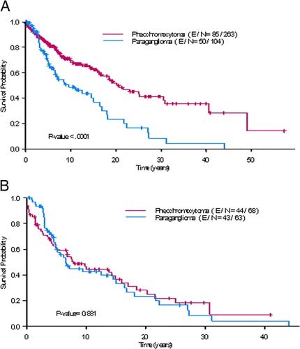
A PG located in the abdominal (e.g., Zuckerkand l organ, para-aortic, and perirenal), pelvic (bladder), and thoracic cavities are frequently malignant, and metastatic disease may be observed in 40-70% of cases. Although in most metastatic SPGs, the primary tumor is larger than 5 cm, in up to 20% of cases, the primary tumor is smaller than 5 cm, and distant metastases have been described in patients with tumors as small as 1 cm. An extra-adrenal location is associated with twice the risk of death from disease compared with a primary tumor larger than 5 cm, making an extra-adrenal location a stronger predictor of aggressiveness, metastasis, and decreased survival than primary tumor size. Nevertheless, metastatic PHs and SPGs have similar OS, suggesting that their oncopathogenesis overlaps.5 (Figure 75.1).
Metastatic disease and decreased OS are also observed in approximately 50% of patients with PH/SPG associated with SDHB mutations.13 Although most SDHB tumors are SPGs, several metastatic SPGs are not associated with SDHB mutations. Therefore, the higher prevalence of malignancy in SPG cannot be explained in every case by an association between the genetic background and tumor site alone.8
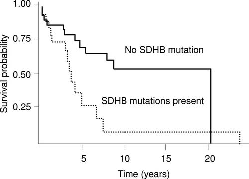
A retrospective study with a limited population of patients compared the OS of patients with metastatic SDHB PH/PGs with that of patients with sporadic metastic tumors. The study suggested that SDHB carriers may have a lower OS34 (Figure 75.2).
Prognostic Factors at the Time of Diagnosis of Metastases
The natural history of metastatic PH/SPG has not been described yet. As for most well-differentiated neuroendocrine tumors, the prognosis of metastatic tumors is characterized by their heterogeneity. The consequences of excess catecholamine on blood pressure increase, cardiovascular function, and other organ physiology (e.g., gastrointestinal tract) as well as the tumor extent should be actively characterized. Indeed, reported causes of death in these patients include hypertensive crisis, cardiac dysfunction, colonic obstruction, and /or tumor burden and progression. Only four studies looked for prognostic parameters in patients with metastatic disease.
Timmers et al.35 evaluated the impact of excessive catecholamine secretion and the tumor extent on clinical outcome in patients with PH/SPG. Although the excessive secretion of catecholamines was associated with substantial morbidity, the tumor burden was the main determinant of survival. This study, however, did not clearly define tumor burden.
No studies have addressed whether the extent of invasion of the primary tumor affects overall prognosis.
No studies have evaluated whether the presence of lymph node metastases might predict outcome.
Distant metastasis is defined as evidence of disease in organs in which chromaffin cells are not supposed to exist, including the liver, lungs, and bones. Several studies demonstrated that the presence of distant metastases portends worse survival. In fact, only 60% of patients with distant metastases are alive 5 years after initial diagnosis.8
Ayala-Ramirez et al.5 found that approximately 50% of patients with malignant PH/SPG present with metastasis >=6 months after the initial time of diagnosis and /or resection of the primary tumor (metachronous metastasis). As expected, timing of metastasis is an important determinant of prognosis, as patients with metachronous metastasis exhibit better OS than those with synchronous metastasis (diagnosis of metastases at the time of or <6 months after diagnosis of the primary tumor).
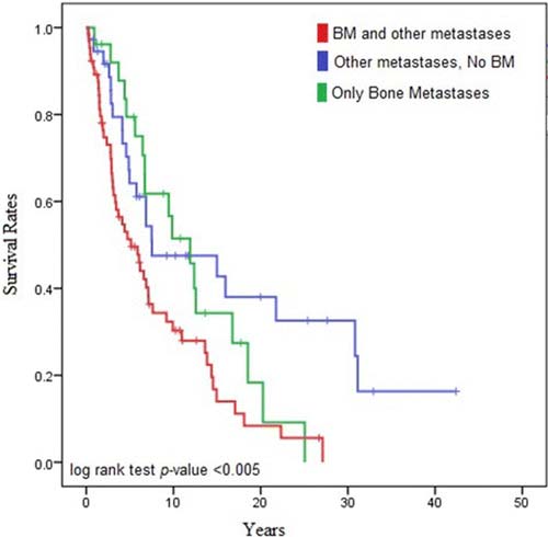
In another study by Ayala-Ramirez et al.,7 patients who manifested malignancy exclusively with skeletal metastasis (20%) exhibited a significantly longer OS that those with or without skeletal metastasis but with metastasis to other organs, such as the liver and lungs (12 years vs. 5 years vs. 7.5 years, respectively; log-rank test p value = 0.005; Figure 75.3).
The authors have not noted any emerging factors for clinical care.
The AJCC recently established guidelines that will be used to evaluate published statistical prediction models for the purpose of granting endorsement for clinical use.36 Although this is a monumental step toward the goal of precision medicine, this work was published only very recently. Therefore, the existing models that have been published or may be in clinical use have not yet been evaluated for this cancer site by the Precision Medicine Core of the AJCC. In the future, the statistical prediction models for this cancer site will be evaluated, and those that meet all AJCC criteria will be endorsed.
Clinical T (cT)
cT Category | cT Criteria |
---|---|
cTX | Primary tumor cannot be assessed |
cT1 | PH less than 5 cm in greatest dimension, no extra-adrenal invasion |
cT2 | PH greater than or equal to 5 cm or PG-sympathetic of any size, no extra-adrenal invasion |
cT3 | Tumor of any size with invasion into surrounding tissues (e.g., liver, pancreas, spleen, kidneys) |
PG Parasympathetic: nonfunctional, usually in the head and neck region. Parasympathetic paraganglioma are not staged because they are largely benign.
Pathological T (pT)
pT Category | pT Criteria |
---|---|
pTX | Primary tumor cannot be assessed |
pT1 | PH less than 5 cm in greatest dimension, no extra-adrenal invasion |
pT2 | PH greater than or equal to 5 cm or PG-sympathetic of any size, no extra-adrenal invasion |
pT3 | Tumor of any size with invasion into surrounding tissues (e.g., liver, pancreas, spleen, kidneys) |
cTX | Primary tumor cannot be assessed |
cT1 | PH less than 5 cm in greatest dimension, no extra-adrenal invasion |
cT2 | PH greater than or equal to 5 cm or PG-sympathetic of any size, no extra-adrenal invasion |
cT3 | Tumor of any size with invasion into surrounding tissues (e.g., liver, pancreas, spleen, kidneys) |
PG Parasympathetic: nonfunctional, usually in the head and neck region. Parasympathetic Paraganglioma are not staged because they are largely benign.
Neoadjuvant Clinical T (yT)
ycT Category | ycT Criteria |
---|---|
ycTX | Primary tumor cannot be assessed |
ycT1 | PH less than 5 cm in greatest dimension, no extra-adrenal invasion |
ycT2 | PH greater than or equal to 5 cm or PG-sympathetic of any size, no extra-adrenal invasion |
ycT3 | Tumor of any size with invasion into surrounding tissues (e.g., liver, pancreas, spleen, kidneys) |
PG Parasympathetic: nonfunctional, usually in the head and neck region. Parasympathetic Paraganglioma are not staged because they are largely benign.
Neoadjuvant Pathological T (yT)
ypT Category | ypT Criteria |
---|---|
ypTX | Primary tumor cannot be assessed |
ypT1 | PH less than 5 cm in greatest dimension, no extra-adrenal invasion |
ypT2 | PH greater than or equal to 5 cm or PG-sympathetic of any size, no extra-adrenal invasion |
ypT3 | Tumor of any size with invasion into surrounding tissues (e.g., liver, pancreas, spleen, kidneys) |
PG Parasympathetic: nonfunctional, usually in the head and neck region. Parasympathetic Paraganglioma are not staged because they are largely benign.
Definition of Regional Lymph Node (N)
Clinical N (cN)cN Category | cN Criteria |
---|---|
cNX | Regional lymph nodes cannot be assessed |
cN0 | No lymph node metastasis |
cN1 | Regional lymph node metastasis |
pN Category | pN Criteria |
---|---|
pNX | Regional lymph nodes cannot be assessed |
pN0 | No lymph node metastasis |
pN1 | Regional lymph node metastasis |
cNX | Regional lymph nodes cannot be assessed |
cN0 | No lymph node metastasis |
cN1 | Regional lymph node metastasis |
ycN Category | ycN Criteria |
---|---|
ycNX | Regional lymph nodes cannot be assessed |
ycN0 | No lymph node metastasis |
ycN1 | Regional lymph node metastasis |
ypN Category | ypN Criteria |
---|---|
ypNX | Regional lymph nodes cannot be assessed |
ypN0 | No lymph node metastasis |
ypN1 | Regional lymph node metastasis |
Definition of Distant Metastasis (M)- Clinical M (cN)
cM Category | cM Criteria |
---|---|
cM0 | No distant metastasis |
cM1 | Distant metastasis |
cM1a | Distant metastasis to only bone |
cM1b | Distant metastasis to only distant lymph nodes/liver or lung |
cM1c | Distant metastasis to bone plus multiple other sites |
pM1 | Microscopic evidence of distant metastasis |
pM1a | Microscopic evidence of distant metastasis to only bone |
pM1b | Microscopic evidence of distant metastasis to only distant lymph nodes/liver or lung |
pM1c | Microscopic evidence of distant metastasis to bone plus multiple other sites |
Definition of Distant Metastasis (M)- Pathological M (pN)
pM Category | pM Criteria |
---|---|
cM0 | No distant metastasis |
cM1 | Distant metastasis |
cM1a | Distant metastasis to only bone |
cM1b | Distant metastasis to only distant lymph nodes/liver or lung |
cM1c | Distant metastasis to bone plus multiple other sites |
pM1 | Microscopic evidence of distant metastasis |
pM1a | Microscopic evidence of distant metastasis to only bone |
pM1b | Microscopic evidence of distant metastasis to only distant lymph nodes/liver or lung |
pM1c | Microscopic evidence of distant metastasis to bone plus multiple other sites |
Definition of Distant Metastasis (M)- Neoadjuvant Clinical M (pY)
ycM Category | ycM Criteria |
---|---|
cM0 | No distant metastasis |
cM1 | Distant metastasis |
cM1a | Distant metastasis to only bone |
cM1b | Distant metastasis to only distant lymph nodes/liver or lung |
cM1c | Distant metastasis to bone plus multiple other sites |
pM1 | Microscopic evidence of distant metastasis |
pM1a | Microscopic evidence of distant metastasis to only bone |
pM1b | Microscopic evidence of distant metastasis to only distant lymph nodes/liver or lung |
pM1c | Microscopic evidence of distant metastasis to bone plus multiple other sites |
Definition of Distant Metastasis (M)- Neoadjuvant Pathological M (pY)
ypM Category | ypM Criteria |
---|---|
cM0 | No distant metastasis |
cM1 | Distant metastasis |
cM1a | Distant metastasis to only bone |
cM1b | Distant metastasis to only distant lymph nodes/liver or lung |
cM1c | Distant metastasis to bone plus multiple other sites |
pM1 | Microscopic evidence of distant metastasis |
pM1a | Microscopic evidence of distant metastasis to only bone |
pM1b | Microscopic evidence of distant metastasis to only distant lymph nodes/liver or lung |
pM1c | Microscopic evidence of distant metastasis to bone plus multiple other sites |
Clinical
When T is | and N is | and M is | Then the Clinical Prognostic Stage Group is |
---|---|---|---|
cT1 | cN0 | cM0 | I |
cT2 | cN0 | cM0 | II |
cT1 | cN1 | cM0 | III |
cT2 | cN1 | cM0 | III |
cT3 | cNX | cM0 | III |
cT3 | cN0 | cM0 | III |
cT3 | cN1 | cM0 | III |
cTX | cNX | cM1 | IV |
cTX | cNX | cM1a | IV |
cTX | cNX | cM1b | IV |
cTX | cNX | cM1c | IV |
cTX | cN0 | cM1 | IV |
cTX | cN0 | cM1a | IV |
cTX | cN0 | cM1b | IV |
cTX | cN0 | cM1c | IV |
cTX | cN1 | cM1 | IV |
cTX | cN1 | cM1a | IV |
cTX | cN1 | cM1b | IV |
cTX | cN1 | cM1c | IV |
cT1 | cNX | cM1 | IV |
cT1 | cNX | cM1a | IV |
cT1 | cNX | cM1b | IV |
cT1 | cNX | cM1c | IV |
cT1 | cN0 | cM1 | IV |
cT1 | cN0 | cM1a | IV |
cT1 | cN0 | cM1b | IV |
cT1 | cN0 | cM1c | IV |
cT1 | cN1 | cM1 | IV |
cT1 | cN1 | cM1a | IV |
cT1 | cN1 | cM1b | IV |
cT1 | cN1 | cM1c | IV |
cT2 | cNX | cM1 | IV |
cT2 | cNX | cM1a | IV |
cT2 | cNX | cM1b | IV |
cT2 | cNX | cM1c | IV |
cT2 | cN0 | cM1 | IV |
cT2 | cN0 | cM1a | IV |
cT2 | cN0 | cM1b | IV |
cT2 | cN0 | cM1c | IV |
cT2 | cN1 | cM1 | IV |
cT2 | cN1 | cM1a | IV |
cT2 | cN1 | cM1b | IV |
cT2 | cN1 | cM1c | IV |
cT3 | cNX | cM1 | IV |
cT3 | cNX | cM1a | IV |
cT3 | cNX | cM1b | IV |
cT3 | cNX | cM1c | IV |
cT3 | cN0 | cM1 | IV |
cT3 | cN0 | cM1a | IV |
cT3 | cN0 | cM1b | IV |
cT3 | cN0 | cM1c | IV |
cT3 | cN1 | cM1 | IV |
cT3 | cN1 | cM1a | IV |
cT3 | cN1 | cM1b | IV |
cT3 | cN1 | cM1c | IV |
cTX | cNX | pM1 | IV |
cTX | cNX | pM1a | IV |
cTX | cNX | pM1b | IV |
cTX | cNX | pM1c | IV |
cTX | cN0 | pM1 | IV |
cTX | cN0 | pM1a | IV |
cTX | cN0 | pM1b | IV |
cTX | cN0 | pM1c | IV |
cTX | cN1 | pM1 | IV |
cTX | cN1 | pM1a | IV |
cTX | cN1 | pM1b | IV |
cTX | cN1 | pM1c | IV |
cT1 | cNX | pM1 | IV |
cT1 | cNX | pM1a | IV |
cT1 | cNX | pM1b | IV |
cT1 | cNX | pM1c | IV |
cT1 | cN0 | pM1 | IV |
cT1 | cN0 | pM1a | IV |
cT1 | cN0 | pM1b | IV |
cT1 | cN0 | pM1c | IV |
cT1 | cN1 | pM1 | IV |
cT1 | cN1 | pM1a | IV |
cT1 | cN1 | pM1b | IV |
cT1 | cN1 | pM1c | IV |
cT2 | cNX | pM1 | IV |
cT2 | cNX | pM1a | IV |
cT2 | cNX | pM1b | IV |
cT2 | cNX | pM1c | IV |
cT2 | cN0 | pM1 | IV |
cT2 | cN0 | pM1a | IV |
cT2 | cN0 | pM1b | IV |
cT2 | cN0 | pM1c | IV |
cT2 | cN1 | pM1 | IV |
cT2 | cN1 | pM1a | IV |
cT2 | cN1 | pM1b | IV |
cT2 | cN1 | pM1c | IV |
cT3 | cNX | pM1 | IV |
cT3 | cNX | pM1a | IV |
cT3 | cNX | pM1b | IV |
cT3 | cNX | pM1c | IV |
cT3 | cN0 | pM1 | IV |
cT3 | cN0 | pM1a | IV |
cT3 | cN0 | pM1b | IV |
cT3 | cN0 | pM1c | IV |
cT3 | cN1 | pM1 | IV |
cT3 | cN1 | pM1a | IV |
cT3 | cN1 | pM1b | IV |
cT3 | cN1 | pM1c | IV |
Staging applies to Pheochromocytoma/Sympathetic Paraganglioma.
Pathological
When T is | and N is | and M is | Then the Pathological Prognostic Stage Group is |
---|---|---|---|
pT1 | pN0 | cM0 | I |
pT2 | pN0 | cM0 | II |
pT1 | pN1 | cM0 | III |
pT2 | pN1 | cM0 | III |
pT3 | pNX | cM0 | III |
pT3 | pN0 | cM0 | III |
pT3 | pN1 | cM0 | III |
pTX | pNX | cM1 | IV |
pTX | pNX | cM1a | IV |
pTX | pNX | cM1b | IV |
pTX | pNX | cM1c | IV |
pTX | pN0 | cM1 | IV |
pTX | pN0 | cM1a | IV |
pTX | pN0 | cM1b | IV |
pTX | pN0 | cM1c | IV |
pTX | pN1 | cM1 | IV |
pTX | pN1 | cM1a | IV |
pTX | pN1 | cM1b | IV |
pTX | pN1 | cM1c | IV |
pT1 | pNX | cM1 | IV |
pT1 | pNX | cM1a | IV |
pT1 | pNX | cM1b | IV |
pT1 | pNX | cM1c | IV |
pT1 | pN0 | cM1 | IV |
pT1 | pN0 | cM1a | IV |
pT1 | pN0 | cM1b | IV |
pT1 | pN0 | cM1c | IV |
pT1 | pN1 | cM1 | IV |
pT1 | pN1 | cM1a | IV |
pT1 | pN1 | cM1b | IV |
pT1 | pN1 | cM1c | IV |
pT2 | pNX | cM1 | IV |
pT2 | pNX | cM1a | IV |
pT2 | pNX | cM1b | IV |
pT2 | pNX | cM1c | IV |
pT2 | pN0 | cM1 | IV |
pT2 | pN0 | cM1a | IV |
pT2 | pN0 | cM1b | IV |
pT2 | pN0 | cM1c | IV |
pT2 | pN1 | cM1 | IV |
pT2 | pN1 | cM1a | IV |
pT2 | pN1 | cM1b | IV |
pT2 | pN1 | cM1c | IV |
pT3 | pNX | cM1 | IV |
pT3 | pNX | cM1a | IV |
pT3 | pNX | cM1b | IV |
pT3 | pNX | cM1c | IV |
pT3 | pN0 | cM1 | IV |
pT3 | pN0 | cM1a | IV |
pT3 | pN0 | cM1b | IV |
pT3 | pN0 | cM1c | IV |
pT3 | pN1 | cM1 | IV |
pT3 | pN1 | cM1a | IV |
pT3 | pN1 | cM1b | IV |
pT3 | pN1 | cM1c | IV |
pTX | pNX | pM1 | IV |
pTX | pNX | pM1a | IV |
pTX | pNX | pM1b | IV |
pTX | pNX | pM1c | IV |
pTX | pN0 | pM1 | IV |
pTX | pN0 | pM1a | IV |
pTX | pN0 | pM1b | IV |
pTX | pN0 | pM1c | IV |
pTX | pN1 | pM1 | IV |
pTX | pN1 | pM1a | IV |
pTX | pN1 | pM1b | IV |
pTX | pN1 | pM1c | IV |
pT1 | pNX | pM1 | IV |
pT1 | pNX | pM1a | IV |
pT1 | pNX | pM1b | IV |
pT1 | pNX | pM1c | IV |
pT1 | pN0 | pM1 | IV |
pT1 | pN0 | pM1a | IV |
pT1 | pN0 | pM1b | IV |
pT1 | pN0 | pM1c | IV |
pT1 | pN1 | pM1 | IV |
pT1 | pN1 | pM1a | IV |
pT1 | pN1 | pM1b | IV |
pT1 | pN1 | pM1c | IV |
pT2 | pNX | pM1 | IV |
pT2 | pNX | pM1a | IV |
pT2 | pNX | pM1b | IV |
pT2 | pNX | pM1c | IV |
pT2 | pN0 | pM1 | IV |
pT2 | pN0 | pM1a | IV |
pT2 | pN0 | pM1b | IV |
pT2 | pN0 | pM1c | IV |
pT2 | pN1 | pM1 | IV |
pT2 | pN1 | pM1a | IV |
pT2 | pN1 | pM1b | IV |
pT2 | pN1 | pM1c | IV |
pT3 | pNX | pM1 | IV |
pT3 | pNX | pM1a | IV |
pT3 | pNX | pM1b | IV |
pT3 | pNX | pM1c | IV |
pT3 | pN0 | pM1 | IV |
pT3 | pN0 | pM1a | IV |
pT3 | pN0 | pM1b | IV |
pT3 | pN0 | pM1c | IV |
pT3 | pN1 | pM1 | IV |
pT3 | pN1 | pM1a | IV |
pT3 | pN1 | pM1b | IV |
pT3 | pN1 | pM1c | IV |
cTX | cNX | pM1 | IV |
cTX | cNX | pM1a | IV |
cTX | cNX | pM1b | IV |
cTX | cNX | pM1c | IV |
cTX | cN0 | pM1 | IV |
cTX | cN0 | pM1a | IV |
cTX | cN0 | pM1b | IV |
cTX | cN0 | pM1c | IV |
cTX | cN1 | pM1 | IV |
cTX | cN1 | pM1a | IV |
cTX | cN1 | pM1b | IV |
cTX | cN1 | pM1c | IV |
cT1 | cNX | pM1 | IV |
cT1 | cNX | pM1a | IV |
cT1 | cNX | pM1b | IV |
cT1 | cNX | pM1c | IV |
cT1 | cN0 | pM1 | IV |
cT1 | cN0 | pM1a | IV |
cT1 | cN0 | pM1b | IV |
cT1 | cN0 | pM1c | IV |
cT1 | cN1 | pM1 | IV |
cT1 | cN1 | pM1a | IV |
cT1 | cN1 | pM1b | IV |
cT1 | cN1 | pM1c | IV |
cT2 | cNX | pM1 | IV |
cT2 | cNX | pM1a | IV |
cT2 | cNX | pM1b | IV |
cT2 | cNX | pM1c | IV |
cT2 | cN0 | pM1 | IV |
cT2 | cN0 | pM1a | IV |
cT2 | cN0 | pM1b | IV |
cT2 | cN0 | pM1c | IV |
cT2 | cN1 | pM1 | IV |
cT2 | cN1 | pM1a | IV |
cT2 | cN1 | pM1b | IV |
cT2 | cN1 | pM1c | IV |
cT3 | cNX | pM1 | IV |
cT3 | cNX | pM1a | IV |
cT3 | cNX | pM1b | IV |
cT3 | cNX | pM1c | IV |
cT3 | cN0 | pM1 | IV |
cT3 | cN0 | pM1a | IV |
cT3 | cN0 | pM1b | IV |
cT3 | cN0 | pM1c | IV |
cT3 | cN1 | pM1 | IV |
cT3 | cN1 | pM1a | IV |
cT3 | cN1 | pM1b | IV |
cT3 | cN1 | pM1c | IV |
Staging applies to Pheochromocytoma/Sympathetic Paraganglioma.
Neoadjuvant Clinical
There is no Postneoadjuvant Clinical Therapy (ycTNM) stage group available at this time.
Neoadjuvant Pathological
When T is | and N is | and M is | Then the Postneoadjuvant Pathological Stage Group is |
---|---|---|---|
ypT1 | ypN0 | cM0 | I |
ypT2 | ypN0 | cM0 | II |
ypT1 | ypN1 | cM0 | III |
ypT2 | ypN1 | cM0 | III |
ypT3 | ypNX | cM0 | III |
ypT3 | ypN0 | cM0 | III |
ypT3 | ypN1 | cM0 | III |
ypTX | ypNX | cM1 | IV |
ypTX | ypNX | cM1a | IV |
ypTX | ypNX | cM1b | IV |
ypTX | ypNX | cM1c | IV |
ypTX | ypN0 | cM1 | IV |
ypTX | ypN0 | cM1a | IV |
ypTX | ypN0 | cM1b | IV |
ypTX | ypN0 | cM1c | IV |
ypTX | ypN1 | cM1 | IV |
ypTX | ypN1 | cM1a | IV |
ypTX | ypN1 | cM1b | IV |
ypTX | ypN1 | cM1c | IV |
ypT1 | ypNX | cM1 | IV |
ypT1 | ypNX | cM1a | IV |
ypT1 | ypNX | cM1b | IV |
ypT1 | ypNX | cM1c | IV |
ypT1 | ypN0 | cM1 | IV |
ypT1 | ypN0 | cM1a | IV |
ypT1 | ypN0 | cM1b | IV |
ypT1 | ypN0 | cM1c | IV |
ypT1 | ypN1 | cM1 | IV |
ypT1 | ypN1 | cM1a | IV |
ypT1 | ypN1 | cM1b | IV |
ypT1 | ypN1 | cM1c | IV |
ypT2 | ypNX | cM1 | IV |
ypT2 | ypNX | cM1a | IV |
ypT2 | ypNX | cM1b | IV |
ypT2 | ypNX | cM1c | IV |
ypT2 | ypN0 | cM1 | IV |
ypT2 | ypN0 | cM1a | IV |
ypT2 | ypN0 | cM1b | IV |
ypT2 | ypN0 | cM1c | IV |
ypT2 | ypN1 | cM1 | IV |
ypT2 | ypN1 | cM1a | IV |
ypT2 | ypN1 | cM1b | IV |
ypT2 | ypN1 | cM1c | IV |
ypT3 | ypNX | cM1 | IV |
ypT3 | ypNX | cM1a | IV |
ypT3 | ypNX | cM1b | IV |
ypT3 | ypNX | cM1c | IV |
ypT3 | ypN0 | cM1 | IV |
ypT3 | ypN0 | cM1a | IV |
ypT3 | ypN0 | cM1b | IV |
ypT3 | ypN0 | cM1c | IV |
ypT3 | ypN1 | cM1 | IV |
ypT3 | ypN1 | cM1a | IV |
ypT3 | ypN1 | cM1b | IV |
ypT3 | ypN1 | cM1c | IV |
ypTX | ypNX | pM1 | IV |
ypTX | ypNX | pM1a | IV |
ypTX | ypNX | pM1b | IV |
ypTX | ypNX | pM1c | IV |
ypTX | ypN0 | pM1 | IV |
ypTX | ypN0 | pM1a | IV |
ypTX | ypN0 | pM1b | IV |
ypTX | ypN0 | pM1c | IV |
ypTX | ypN1 | pM1 | IV |
ypTX | ypN1 | pM1a | IV |
ypTX | ypN1 | pM1b | IV |
ypTX | ypN1 | pM1c | IV |
ypT1 | ypNX | pM1 | IV |
ypT1 | ypNX | pM1a | IV |
ypT1 | ypNX | pM1b | IV |
ypT1 | ypNX | pM1c | IV |
ypT1 | ypN0 | pM1 | IV |
ypT1 | ypN0 | pM1a | IV |
ypT1 | ypN0 | pM1b | IV |
ypT1 | ypN0 | pM1c | IV |
ypT1 | ypN1 | pM1 | IV |
ypT1 | ypN1 | pM1a | IV |
ypT1 | ypN1 | pM1b | IV |
ypT1 | ypN1 | pM1c | IV |
ypT2 | ypNX | pM1 | IV |
ypT2 | ypNX | pM1a | IV |
ypT2 | ypNX | pM1b | IV |
ypT2 | ypNX | pM1c | IV |
ypT2 | ypN0 | pM1 | IV |
ypT2 | ypN0 | pM1a | IV |
ypT2 | ypN0 | pM1b | IV |
ypT2 | ypN0 | pM1c | IV |
ypT2 | ypN1 | pM1 | IV |
ypT2 | ypN1 | pM1a | IV |
ypT2 | ypN1 | pM1b | IV |
ypT2 | ypN1 | pM1c | IV |
ypT3 | ypNX | pM1 | IV |
ypT3 | ypNX | pM1a | IV |
ypT3 | ypNX | pM1b | IV |
ypT3 | ypNX | pM1c | IV |
ypT3 | ypN0 | pM1 | IV |
ypT3 | ypN0 | pM1a | IV |
ypT3 | ypN0 | pM1b | IV |
ypT3 | ypN0 | pM1c | IV |
ypT3 | ypN1 | pM1 | IV |
ypT3 | ypN1 | pM1a | IV |
ypT3 | ypN1 | pM1b | IV |
ypT3 | ypN1 | pM1c | IV |
Staging applies to Pheochromocytoma/Sympathetic Paraganglioma.
Registry Data Collection Variables