section name header

Disease Prologue

Summary

Cancers Staged Using This Staging System

The classification applies only to carcinoma of the conjunctiva. Other tumors of the conjunctiva, including secondary conjunctival tumors (e.g., intraocular tumors extending through the conjunctiva, such as uveal melanoma or uveal non-Hodgkin lymphoma, and orbital tumors extending into the conjunctiva, such as rhabdomyosarcoma), are not classified using this schema.

Cancers Not Staged Using This Staging System

These histopathologic types of cancer…Are staged according to the classification for…and can be found in chapter…
Conjunctival lymphomaOcular adnexal lymphoma71
Conjunctival melanomaConjunctival melanoma66

Summary of Changes

ChangeDetails of ChangeLevel of Evidence
Definition of Primary Tumor (T)T1 and T2 definitions have been revised to include invasion of the conjunctival basement membrane.II
Prognostic FactorsExpand ed Emerging Prognostic Factors listII and III

ICD-O-3 Topography Codes

CodeDescription
C69.0Conjunctiva

WHO Classification of Tumors

This list includes histology codes and preferred terms from the WHO Classification of Tumors and the International Classification of Diseases for Oncology (ICD-O). Most of the terms in this list represent malignant behavior. For cancer reporting purposes, behavior codes /3 (denoting malignant neoplasms), /2 (denoting in situ neoplasms), and in some cases /1 (denoting neoplasms with uncertain and unknown behavior) may be appended to the 4-digit histology codes to create a complete morphology code.

CodeDescription
8010Carcinoma, NOS
8070Squamous cell carcinoma, NOS
8071Squamous cell carcinoma, keratinizing, NOS
8072Squamous cell carcinoma, large cell, nonkeratinizing, NOS
8073Squamous cell carcinoma, small cell, nonkeratinizing, NOS
8074Squamous cell carcinoma, spindle cell
8075Squamous cell carcinoma, adenoid
8076Squamous cell carcinoma, microinvasive
8090Basal cell carcinoma, NOS
8410Sebaceous adenocarcinoma
8430Mucoepidermoid carcinoma
8560Adenosquamous carcinoma

Histology is not ideal for clinical use in patient care, as it describes an unspecified or outdated diagnosis. Data collectors may use this code only if there is not enough information in the medical record to document a more specific diagnosis.

International Agency for Research on Cancer, World Health Organization. International Classification of Diseases for Oncology. ICD-O-3-Online.http://codes.iarc.fr/home. Accessed August 16, 2017. Used with permission.

Introduction

The AJCC staging system for conjunctival carcinoma remains largely unchanged from the AJCC Cancer Staging Manual, 7th Edition, apart from a more precisely defined disease extent for early invasive lesions (T1 and T2). This change has implications for management, as emerging studies support adjuvant treatment in some of these patients.

It is acknowledged that the TNM staging system for these tumors would benefit greatly from the establishment of multicenter registries, prospective rand omized trials, and prospective data mining to consolidate and expand the definitions of prognostic significance and to incorporate biomarkers.

Similarly, it is noted that data from a large registry would help provide significance to the currently applied subdivisions of intraepithelial disease (i.e., conjunctival squamous intraepithelial neoplasia, I-III). However, these data currently do not exist.

AJCC staging for conjunctival carcinoma is becoming more important, because the incidence of conjunctival squamous cell carcinoma (SCC) associated with HIV is increasing, especially in younger individuals in developing countries with high incidences of HIV. Such tumors behave more aggressively.

This chapter clearly defines the clinical term ocular squamous surface neoplasia and explains why a histopathologic tissue diagnosis is needed for TNM staging.

This staging system applies to conjunctival carcinoma comprising predominantly SCC and corneal squamous intraepithelial neoplasia, along with other histologic subtypes (see ICD-O-3 Histology Codes). Nonepithelial tumors of the conjunctiva are not staged using these criteria. Biopsy is required for tumor staging.

Risk factors for the disease are exposure to sun and ultraviolet B light, as well as light-colored skin. Other risk factors include radiation exposure, smoking, human papillomavirus (HPV) infection, chemical exposures, immunosuppression, and particular syndromes (e.g., xeroderma pigmentosum). In developed countries, this condition is more common in men, with a peak incidence in the seventh decade of life. At diagnosis, it is typically localized to the corneal limbus. Conjunctival carcinoma also is associated with HIV infection, and this association is particularly prevalent in developing countries, where it may be considered an AIDS-defining illness, especially in younger patients.1-10

Ocular surface squamous neoplasia is a clinical term encompassing the continuum of disease from mild epithelial dysplasia to SCC. Because this term includes overlapping histopathologic grades and entities, it is imprecise and should be avoided in histopathology reports. The precise morphologic changes should be documented using terminology applied in pathology. 11,12

After clinical and pathological diagnosis, excisional treatment typically is supplemented by adjunctive double freeze-thaw cryotherapy to the conjunctival margins and sclera base at the time of primary resection.13 Additional adjuvant and /or alternative treatments include topical chemotherapy (mitomycin C, 5-fluorouracil, or interferon alfa-2b). Radiation therapy (teletherapy or brachytherapy) may be used when complete resection is not possible or as salvage treatment to avoid orbital exenteration.14-20
Anatomy

Primary Site(s)

Anatomically, the conjunctiva consists of stratified epithelium that contains mucus-secreting goblet cells. These cells are most numerous in the fornices. Palpebral conjunctiva lines the eyelid; bulbar conjunctiva covers the eyeball. Conjunctival epithelium merges with that of the cornea at the limbus.2 Conjunctival squamous carcinomas are most likely to arise from the exposed bulbar limbal location.

Conjunctival squamous intraepithelial neoplasia embraces all forms of intraepithelial dysplasia, including SCC in situ. Spread occurs initially by direct local extension radially into the adjacent conjunctiva and cornea, and ultimately vertically into the conjunctival stroma, Tenon's capsule, and the sclera. Perineural invasion may occur at an earlier stage, typically in more aggressive histologic subtypes (e.g.,mucoepidermoid carcinoma), leading to orbital extension. Larger lesions may develop the capacity for metastatic spread to regional lymph nodes following invasion into lymphatic vessels in the conjunctiva. Intraocular spread may occur through the sclera, particularly if weakened by prior surgery.2,4,5,21-28

Advanced tumors may directly invade the eyelid, eye, nasolacrimal system, orbit, adjacent paranasal sinus structures, or brain.26,28,29

Regional Lymph Nodes

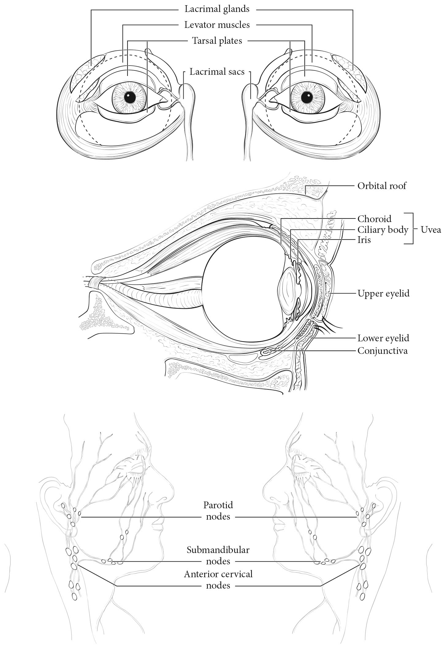
The regional lymph nodes are preauricular (parotid), submand ibular, and cervical nodes (Figure 63.1).

Metastatic Sites

In addition to spreading via regional lymphatics, tumors of the conjunctiva also may metastasize hematogenously, although this is rare. Sites of metastasis include the parotid and submand ibular gland , lungs, and bone.29

Classification Rules

Clinical Classification

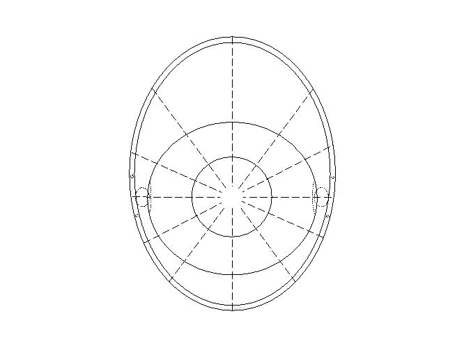
Initial and subsequent clinical assessments of conjunctival carcinoma are based on inspection, slit lamp examination, and palpation of the regional lymph nodes. All conjunctival surfaces should be inspected, measured, documented, and photographed (including eversion of the upper eyelid) (Figure 63.2). Tumor photography should pay particular attention to the lesion margins, evidence of pagetoid spread, corneal epithelial extension, and involvement of the punctum. Gonioscopy should be performed with photography (for limbal disease), particularly when intraocular extension is suspected. Examination of the ipsilateral sinuses is indicated (particularly if punctal involvement has been noted).

The diagnosis of conjunctival squamous carcinoma typically requires excisional biopsy because histolopathologic examination allows for the assessment of vertical tumor extent, which is needed to determine the levels required for TNM staging. In contrast, cytologic techniques have been found to be useful in determining cytologic atypia. Cytology has been particularly helpful as an adjunct to clinical diagnosis and in evaluation for recurrent disease.11

Imaging

Anterior segment ultrasound imaging and optical coherence tomography are useful for measuring tumor thickness and evaluating invasion of adjacent structures (e.g., the sclera, uvea, and anterior orbit). Ultrasound biomicroscopy (UBM) findings suspicious for intraocular invasion include angle blunting and uveal thickening.30 Low-frequency posterior segment ultrasonography also may be used to evaluate for choroidal and orbital invasion.31

Radiologic evaluation may be necessary to stage locally invasive and nodal disease and may include computed tomography (CT), magnetic resonance imaging, and positron emission tomography/CT. Metastatic survey typically includes a physical examination as well as hematologic screening and radiologic evaluations of the head, chest, and abdomen.

Pathological Classification

For pathological staging, complete resection of the primary site is indicated, if possible.

To obtain the best histopathologic information, it is important to send the conjunctival specimen to the laboratory, spread evenly on a piece of filter paper with orientation marks on the paper. These measures prevent the specimen from curling and enable orthogonal sections to be taken from the tissue, allowing assessment of the depth of tumor penetration. Histopathologic evaluation for negative peripheral or deep margins should be performed. If mapping biopsy samples are sent to the laboratory, they should be placed in separate containers labeled with the appropriate anatomic location. Sentinel lymph node biopsy is investigational.32

As stated earlier, the clinical term ocular squamous surface neoplasia is imprecise and may even include benign growths; thus, it should be avoided in surgical pathology reports.12

For pN, histologic examination of regional lymphadenectomy specimens, if performed, will include one or more regional lymph nodes.28,32

Prognostic Factors

Prognostic Factors Required for Stage Grouping

Beyond the factors used to assign T, N, or M categories, no additional prognostic factors are required for stage grouping.

Additional Factors Recommended for Clinical Care

For more information, see references 2-7, 10, and 33.

Presence or absence of subepithelial invasion

Presence or absence of subepithelial invasion, as determined by histopathologic examination

AJCC Level of Evidence: II
Tumor size

Tumor size as determined by clinical measurement, clock hour evaluation, and UBM

AJCC Level of Evidence: II
Local invasion

Local invasion as assessed by gonioscopy, ultrasound, and radiologic testing

AJCC Level of Evidence: II

The following factors are associated with higher recurrence rates after treatment. For more information, see references 2-7, 10, 14, 21-29, 34, and 35.

Caruncular location
AJCC Level of Evidence: IV
Scleral extension
AJCC Level of Evidence: III
Uveal extension
AJCC Level of Evidence: III
Initial treatment limited to excisional biopsy without cryotherapy
AJCC Level of Evidence: II
Histologic involvement of the margin of tumor resection
AJCC Level of Evidence: II
A history of xeroderma pigmentosum
AJCC Level of Evidence: II
Histologic subtype

SCC vs. spindle cell carcinoma and mucoepidermoid carcinoma, the latter being more aggressive variants of conjunctival carcinoma and associated with higher recurrence rates and worse prognosis due to a tendency for intraocular extension

AJCC Level of Evidence: Not identified
Lymphovascular Invasion

Presence or absence of lymphatic or vascular invasion

AJCC Level of Evidence: III
Perineural invasion
AJCC Level of Evidence: III
Ki-67 growth fraction

Ki-67 is reported as percentage of positive tumor cells

AJCC Level of Evidence: III

The following factors are prognostically significant potential therapeutic targets. For more information, see references 2-7,10,14,21-29,34, and 35.

Epidermal growth factor receptor (EGFR)
AJCC Level of Evidence: III
HPV high-risk genotypes
AJCC Level of Evidence: III
HIV infection
AJCC Level of Evidence: II

Risk Assessment

Risk Assesment Models

The AJCC recently established guidelines that will be used to evaluate published statistical prediction models for the purpose of granting endorsement for clinical use.36 Although this is a monumental step toward the goal of precision medicine, this work was published only very recently. Therefore, the existing models that have been published or may be in clinical use have not yet been evaluated for this cancer site by the Precision Medicine Core of the AJCC. In the future, the statistical prediction models for this cancer site will be evaluated, and those that meet all AJCC criteria will be endorsed.

Recommendations

The authors have not made any recommendations for clinical trial stratification.

TNM Definitions

Definition of Primary Tumor (T)

Adjacent structures include the cornea (3, 6, 9, or 12 clock hours), intraocular compartments, forniceal conjunctiva (lower and /or upper), palpebral conjunctiva (lower and /or upper), tarsal conjunctiva (lower and /or upper), lacrimal punctum and canaliculi (lower and /or upper), plica, caruncle, posterior eyelid lamella, anterior eyelid lamella, and /or eyelid margin (lower and /or upper).

T CategoryT Criteria
TXPrimary tumor cannot be assessed
T0No evidence of primary tumor
TisCarcinoma in situ
T1Tumor (less than or equal to 5 mm in greatest dimension) invades through the conjunctival basement membrane without invasion of adjacent structures
T2Tumor (greater than 5 mm in greatest dimension) invades through the conjunctival basement membrane without invasion of adjacent structures
T3Tumor invades adjacent structures (excluding the orbit)
T4Tumor invades the orbit with or without further extension
T4aTumor invades orbital soft tissues without bone invasion
T4bTumor invades bone
T4cTumor invades adjacent paranasal sinuses
T4dTumor invades brain

Definition of Regional Lymph Node (N)

N CategoryN Criteria
NXRegional lymph nodes cannot be assessed
N0No regional lymph node metastasis
N1Regional lymph node metastasis

Definition of Distant Metastasis (M)

M CategoryM Criteria
M0No distant metastasis
M1Distant metastasis

Stage Prognostic

AJCC PROGNOSTIC STAGE GROUPS

There is no proposal for anatomic stage and prognostic groups for conjunctival carcinoma.

Registry Data

Registry Data Collection Variables

  1. Ki-67 growth fraction, reported as percentage of positive tumor cells by immunohistochemistry

Histopathologic type

  • Conjunctival intraepithelial neoplasia, including SCC in situ
  • Squamous cell carcinoma
  • Mucoepidermoid carcinoma
  • Spindle cell carcinoma
  • Sebaceous gland carcinoma including pagetoid (conjunctival) spread
  • Basal cell carcinoma

Histologic grade

HISTOLOGIC GRADE (G)

GG Definition
GXGrade cannot be assessed
G1Well differentiated
G2Moderately differentiated
G3Poorly differentiated
G4Undifferentiated

Illustrations

63.1 Anatomic sites and regional lymph nodes for ophthalmic sites.

63.2 Clinical mapping system for conjunctival carcinoma. The map displays the entire conjunctiva as a flat surface, with the central point located at the center of the cornea and concentric regions, such as the limbus, bulbar conjunctiva, fornix, palpebral conjunctiva, and eyelid, considered progressively more peripheral. Radial lines represent clock hours (Modified from Damato and Coupland 37).

Bibliography

  1. Mehta M, Fay A. Squamous cell carcinoma of the eyelid and conjunctiva. International ophthalmology clinics. 2009;49(1):111-121.
  2. Eagle RC. Eye pathology: an atlas and text. Lippincott Williams & Wilkins; 2012.
  3. Ramberg I, Heegaard S, Prause JU, Sjo NC, Toft PB. Squamous cell dysplasia and carcinoma of the conjunctiva. A nationwide, retrospective, epidemiological study of Danish patients. Acta ophthalmologica. 2015;93(7):663-666.
  4. McKelvie PA, Daniell M, McNab A, Loughnan M, Santamaria JD. Squamous cell carcinoma of the conjunctiva: a series of 26 cases. The British journal of ophthalmology. 2002;86(2):168-173.
  5. Seitz B, Fischer M, Holbach LM, Naumann GO. [Differential diagnosis and prognosis of 112 excised epibulbar epithelial tumors]. Klin Monbl Augenheilkd. 1995;207(4):239-246.
  6. Lee GA, Hirst LW. Ocular surface squamous neoplasia. Survey of ophthalmology. 1995;39(6):429-450.
  7. Kenawy N, Garrick A, Heimann H, Coupland SE, Damato BE. Conjunctival squamous cell neoplasia: the Liverpool Ocular Oncology Centre experience. Graefe's archive for clinical and experimental ophthalmology = Albrecht von Graefes Archiv fur klinische und experimentelle Ophthalmologie. 2015;253(1):143-150.
  8. Kamal S, Kaliki S, Mishra DK, Batra J, Naik MN. Ocular Surface Squamous Neoplasia in 200 Patients: A Case-Control Study of Immunosuppression Resulting from Human Immunodeficiency Virus versus Immunocompetency. Ophthalmology. 2015;122(8):1688-1694.
  9. Rogena EA, Simbiri KO, De Falco G, Leoncini L, Ayers L, Nyagol J. A review of the pattern of AIDS defining, HIV associated neoplasms and premalignant lesions diagnosed from 2000-2011 at Kenyatta National Hospital, Kenya. Infectious agents and cancer. 2015;10(1):1-7.
  10. Yin VT, Merritt HA, Sniegowski M, Esmaeli B. Eyelid and ocular surface carcinoma: diagnosis and management. Clin Dermatol. 2015;33(2):159-169.
  11. Semenova EA, Milman T, Finger PT, et al. The diagnostic value of exfoliative cytology vs histopathology for ocular surface squamous neoplasia. American journal of ophthalmology. 2009;148(5):772-778 e771.
  12. Margo C, White A. Ocular surface squamous neoplasia: terminology that is conceptually friendly but clinically perilous. Eye. 2014;28(5):507.
  13. Finger PT. "Finger-tip" cryotherapy probes: treatment of squamous and melanocytic conjunctival neoplasia. The British journal of ophthalmology. 2005;89(8):942-945.
  14. Buuns DR, David TT, Folberg R. Microscopically controlled excision of conjunctival squamous cell carcinoma. American journal of ophthalmology. 1994;117(1):97-102.
  15. Li AS, Shih CY, Rosen L, Steiner A, Milman T, Udell IJ. Recurrence of Ocular Surface Squamous Neoplasia Treated With Excisional Biopsy and Cryotherapy. American journal of ophthalmology. 2015;160(2):213-219 e211.
  16. Wilson MW, Czechonska G, Finger PT, Rausen A, Hooper ME, Haik BG. Chemotherapy for eye cancer. Survey of ophthalmology. 2001;45(5):416-444.
  17. Arepalli S, Kaliki S, Shields CL, Emrich J, Komarnicky L, Shields JA. Plaque radiotherapy in the management of scleral-invasive conjunctival squamous cell carcinoma: an analysis of 15 eyes. JAMA ophthalmology. 2014;132(6):691-696.
  18. Graue GF, Tena LB, Finger PT. Electron beam radiation for conjunctival squamous carcinoma. Ophthalmic plastic and reconstructive surgery. 2011;27(4):277-281.
  19. Pe'er J. Ocular surface squamous neoplasia: evidence for topical chemotherapy. International ophthalmology clinics. 2015;55(1):9-21.
  20. Walsh-Conway N, Conway RM. Plaque brachytherapy for the management of ocular surface malignancies with corneoscleral invasion. Clinical & experimental ophthalmology. 2009;37(6):577-583.
  21. Brownstein S. Mucoepidermoid carcinoma of the conjunctiva with intraocular invasion. Ophthalmology. 1981;88(12):1226-1230.
  22. Cohen BH, Green WR, Iliff NT, Taxy JB, Schwab LT, de la Cruz Z. Spindle cell carcinoma of the conjunctiva. Archives of ophthalmology. 1980;98(10):1809-1813.
  23. Grossniklaus HE, Green WR, Luckenbach M, Chan CC. Conjunctival lesions in adults. A clinical and histopathologic review. Cornea. 1987;6(2):78-116.
  24. Grossniklaus HE, Martin DF, Solomon AR. Invasive conjunctival tumor with keratoacanthoma features. American journal of ophthalmology. 1990;109(6):736-738.
  25. Husain SE, Patrinely JR, Zimmerman LE, Font RL. Primary basal cell carcinoma of the limbal conjunctiva. Ophthalmology. 1993;100(11):17,20-1722.
  26. Rao NA, Font RL. Mucoepidermoid carcinoma of the conjunctiva: a clinicopathologic study of five cases. Cancer. 1976;38(4):1699-1709.
  27. Shields JA, Demirci H, Marr BP, Eagle RC, Jr., Stefanyszyn M, Shields CL. Conjunctival epithelial involvement by eyelid sebaceous carcinoma. The 2003 J. Howard Stokes lecture. Ophthalmic plastic and reconstructive surgery. 2005;21(2):92-96.
  28. Johnson TE, Tabbara KF, Weatherhead RG, Kersten RC, Rice C, Nasr AM. Secondary squamous cell carcinoma of the orbit. Archives of ophthalmology. 1997;115(1):75-78.
  29. Tabbara KF, Kersten R, Daouk N, Blodi FC. Metastatic squamous cell carcinoma of the conjunctiva. Ophthalmology. 1988;95(3):318-321.
  30. Garcia JP, Jr., Spielberg L, Finger PT. High-frequency ultrasound measurements of the normal ciliary body and iris. Ophthalmic Surg Lasers Imaging. 2011;42(4):321-327.
  31. Conway RM, Chew T, Golchet P, Desai K, Lin S, O'Brien J. Ultrasound biomicroscopy: role in diagnosis and management in 130 consecutive patients evaluated for anterior segment tumours. The British journal of ophthalmology. 2005;89(8):950-955.
  32. Mendoza PR, Grossniklaus HE. Sentinel Lymph Node Biopsy for Eyelid and Conjunctival Tumors: What is the Evidence? International ophthalmology clinics. 2015;55(1):123-136.
  33. Finger PT, Tran HV, Turbin RE, et al. High-frequency ultrasonographic evaluation of conjunctival intraepithelial neoplasia and squamous cell carcinoma. Archives of ophthalmology. 2003;121(2):168-172.
  34. Gichuhi S, Ohnuma S, Sagoo MS, Burton MJ. Pathophysiology of ocular surface squamous neoplasia. Exp Eye Res. 2014;129:172-182.
  35. Yousef YA, Finger PT. Squamous carcinoma and dysplasia of the conjunctiva and cornea: an analysis of 101 cases. Ophthalmology. 2012;119(2):233-240.
  36. Kattan MW, Hess KR, Amin MB, et al. American Joint Committee on Cancer acceptance criteria for inclusion of risk models for individualized prognosis in the practice of precision medicine. CA: a cancer journal for clinicians. 2016.
  37. Damato B, Coupland SE. Clinical mapping of conjunctival melanomas. The British journal of ophthalmology. 2008;92(11):1545-1549.