section name header

Disease Prologue

Summary

Cancers Staged Using This Staging System

Epithelial and minor salivary gland cancers of the lip and oral cavity

Cancers Not Staged Using This Staging System

These histopathologic types of cancer…Are staged according to the classification for…and can be found in chapter…
Nonepithelial tumors of lymphoid tissueHematologic malignancies78-83
Nonepithelial tumors of soft tissueSoft tissue sarcoma of the head and neck40
Nonepithelial tumors of bone and cartilageBone38
Mucosal melanomaof the lip and oral cavityMucosal melanoma of the head and neck14
Cutaneous squamous cell carcinoma of the vermilion lipCutaneous squamous cell carcinoma of the head and neck15

Summary of Changes

ChangeDetails of ChangeLevel of Evidence
Anatomy - Primary Site(s)Occult Primary Tumor: Staging of the patient who presents with EBV-unrelated and HPV-unrelated metastatic cervical lymphadenopathy is not included in this chapter.IV
Definition of Primary Tumor (T)Clinical and pathological depth of invasion (DOI) are now used to increase the T category.III
Definition of Primary Tumor (T)Extrinsic tongue muscle invasion is no longer used in T4 because this is a feature of DOI.III
Definition of Regional Lymph Node (N)Separate N staging approaches have been developed for HPV-related and HPV-unrelated cancers.II1,2
Definition of Regional Lymph Node (N)Separate N category approaches have been developed for patients treated without cervical lymph node dissection (clinical cN) and patients treated with cervical lymph neck dissection (pathological pN).II1,2
Definition of Regional Lymph Node (N)Extranodal extension (ENE) is introduced as a descriptor in all HPV-unrelated cancers.II2
Definition of Regional Lymph Node (N)ENE in HPV negative cancers: Only clinically and radiographically overt ENE—ENE(+)—should be used for cN.II2
Definition of Regional Lymph Node (N)ENE in HPV negative cancers: Any pathologically detected ENE is considered ENE(+) and is used for pN.II2
Definition of Regional Lymph Node (N)ENE in HPV-negative cancers: Presence of ENE is designated pN2a for a single ipsilateral node < 3 cm and pN3b for all other node(s).II2
Definition of Regional Lymph Node (N)Classification of ENE: Clinically overt ENE is classified as ENEc and is considered ENE(+) for cN.III3
Definition of Regional Lymph Node (N)Classification of ENE: Pathologically detected ENE is classified as either ENEmi ( 2 mm) or ENEma (> 2 mm) for data collection purposes only, but both are considered ENE(+) for definition of pN.III3

ICD-O-3 Topography Codes

CodeDescription
C00.0External upper lip (exclude vermilion border)
C00.1External lower lip (exclude vermilion border)
C00.2External lip NOS (exclude vermilion border)
C00.3Mucosa of upper lip
C00.4Mucosa of lower lip
C00.5Mucosa of lip, NOS
C00.6Commissure of lip
C00.8Overlapping lesion of lip
C00.9Lip, NOS
C02.0Dorsal surface of tongue, NOS
C02.1Border of tongue
C02.2Ventral surface of tongue, NOS
C02.3Anterior two-thirds of tongue, NOS
C02.8Overlapping lesion of tongue
C02.9Tongue, NOS
C03.0Upper gum
C03.1Lower gum
C03.9Gum, NOS
C04.0Anterior floor of mouth
C04.1Lateral floor of mouth
C04.8Overlapping lesion of floor of mouth
C04.9Floor of mouth, NOS
C05.0Hard palate
C05.8Overlapping lesion of palate
C05.9Palate, NOS
C06.0Cheek mucosa
C06.1Vestibule of mouth
C06.2Retromolar area
C06.8Overlapping lesion of other and unspecified parts of mouth
C06.9Mouth, NOS

WHO Classification of Tumors

Barnes L, Eveson JW, Reichart P, Sidransky D, eds. World Health Organization Classification of Tumours Pathology and Genetics of Head and Neck Tumours. Lyon: IARC; 2005.

CodeDescription
8070Squamous cell carcinoma, conventional
8075Acantholytic squamous cell carcinoma
8560Adenosquamous carcinoma
8083Basaloid squamous cell carcinoma
8051Carcinoma cuniculatum
8052Papillary squamous cell carcinoma
8074Spindle cell squamous carcinoma
8051Verrucous carcinoma
8082Lymphoepithelial carcinoma
8550Acinic cell carcinoma
8430Mucoepidermoid carcinoma
8200Adenoid cystic carcinoma
8525Polymorphous adenocarcinoma
8147Basal cell adenocarcinoma
8562Epithelial-myoepithelial carcinoma
8310Clear cell carcinoma
8480Mucinous adenocarcinoma
8290Oncocytic carcinoma
8500Salivary duct carcinoma
8982Myoepithelial carcinoma

Introduction

Cancers of the oral cavity continue to represent a major problem worldwide. The American Joint Committee on Cancer (AJCC) Cancer Staging Manual, 8th Edition (8th Edition), makes two significant changes based upon enhanced understand ing of the behavior of these malignancies.

The first modification is in T categorization incorporating depth of invasion (DOI). It is important to recognize the distinction between tumor thickness and true DOI. A detailed description of how this should be measured is included in this chapter. It has been recognized since the early work of Spiro and colleagues, in the mid-1980s, that prognosis of oral cancers worsens as the tumor grows thicker, similar to skin malignancies.4,5 The somewhat more sophisticated measure of DOI has been recorded for oral cavity cancers since the AJCC Cancer Staging Manual, 6th Edition. Extrinsic muscle infiltration is no longer a staging criterion for T4 designation because DOI supersedes it and extrinsic muscle invasion is difficult to assess (clinically and pathologically). Clinicians experienced with head and neck cancer will generally have few problems identifying a superficial and less invasive lesion ( 5 mm) from those of moderate depth (from > 5 to 10 mm ) or deeply invasive cancers (> 10 mm) through examination alone. Such experts have estimated the maximum dimensions for complicated lesions of the tonsil or palate for many years. The guiding principle, if there are doubts, is to select the less ominous attribute (a lesser depth) in a given case to avoid stage migration (according to the so-called uncertain rule of the AJCC/Union for International Cancer Control (UICC) TNM, as defined in Chapter 1).

A second significant change is in the use of extranodal extension (ENE) in categorizing metastatic cancer to neck nodes. The effect of ENE on prognosis in head and neck cancers is profound, except for those tumors associated with HPV.5 Including this important prognostic feature was considered critical in revising staging. Most of the data supporting ENE as an adverse prognostic factor is based on histopathological characterization of ENE, especially the distinction between microscopic and macroscopic ENE.6-8 Only unquestionable ENE is to be used for clinical staging (as in the uncertain rule noted above). For clinical ENE, the known limitations of current imaging modalities to define ENE accurately demand that stringent criteria be met prior to assigning a clinical diagnosis of ENE. However, unambiguous evidence of gross ENE on clinical examination (e.g., multiple matted nodes, invasion of skin, infiltration of musculature/dense tethering to adjacent structures, or cranial nerve, brachial plexus, sympathetic trunk, or phrenic nerve invasion with dysfunction) supported by strong radiographic evidence permits classification of disease as ENE(+). Pathological ENE also will be clearly defined as extension of metastatic tumor (present within the confines of the lymph node, through the lymph node capsule into the surrounding connective tissue, with or without associated stromal reaction). Again if there is doubt or uncertainty of the presence of ENE, the case should be categorized as ENE(-).

A staging system revision should address and respond to new information that influences patient outcome. An appropriate balance between complexity and utility (ease-of-use) is necessary for universal acceptance. The TNM system for oral cancers has strongly predicted prognosis and is applied worldwide. The introduction of two new parameters in oral cavity staging, DOI and ENE, better fits the prognostic modeling from large datasets. However, it must be balanced by the ability to derive accurate information from clinicians caring for patients with head and neck cancer in many different environments. Therefore, thorough descriptions of ENE and DOI are included in this chapter.

Effectively, DOI will increase the T category by 1 for each 5 mm of tumor depth (until >= 10 mm). Pathological ENE(+) will increase the nodal category by 1.

Anatomy

Primary Site(s)

The oral cavity extends from the skin-vermilion junction of the lips to the junction of the hard and soft palate above, to the line of circumvallate papillae below, and to the anterior tonsillar pillars laterally. It is additionally divided into multiple specific sites described in this section.

7.1 Anatomical subsites of the lip.

7.2 Anatomical sites and subsites of the oral cavity.

7.3 Anatomical sites and subsites of the oral cavity.

7.4 Anatomical sites and subsites of the oral cavity.

Mucosal Lip

The lip begins at the junction of the vermilion border with the skin and includes only the vermilion surface or that portion of the lip that comes into contact with the opposed lip. The remainder of the vermillion is staged using the chapter on cutaneous squamous cell carcinoma of the head and neck (Chapter 15). It is subdivided into an upper and lower lip, joined at the commissures of the mouth.

Buccal Mucosa

The buccal mucosa includes all the mucous membrane lining of the inner surface of the cheeks and lips from the line of contact of the opposing lips to the line of attachment of mucosa of the alveolar ridge (upper and lower) and pterygomand ibular raphe.

Lower Alveolar Ridge

The lower alveolar ridge refers to the mucosa overlying the alveolar process of the mand ible, which extends from the line of attachment of mucosa in the lower gingivobuccal sulcus to the line of attachment of free mucosa of the floor of the mouth. Posteriorly, it extends to the ascending ramus of the mand ible.

Upper Alveolar Ridge

The upper alveolar ridge refers to the mucosa overlying the alveolar process of the maxilla, which extends from the line of attachment of mucosa in the upper gingivobuccal sulcus to the junction of the hard palate. Its posterior margin is the upper end of the pterygopalatine arch.

Retromolar Gingiva (Retromolar Trigone)

The retromolar gingiva, or retromolar trigone, is the attached mucosa overlying the ascending ramus of the mand ible from the level of the posterior surface of the last lower molar tooth to the apex superiorly, adjacent to the tuberosity of the maxilla.

Floor of the Mouth

The floor of the mouth is a crescentic surface overlying the mylohyoid and hyoglossus muscles, extending from the inner surface of the lower alveolar ridge to the undersurface of the tongue. Its posterior boundary is the base of the anterior pillar of the tonsil. It is divided into two sides by the frenulum of the tongue and harbors the ostia of the submand ibular and sublingual salivary gland s.

Hard Palate

The hard palate is the semilunar area between the upper alveolar ridge and the mucous membrane covering the palatine process of the maxillary palatine bones. It extends from the inner surface of the superior alveolar ridge to the posterior edge of the palatine bone.

Anterior Two-Thirds of the Tongue (Oral Tongue)

The anterior two-thirds of the tongue is the freely mobile portion of the tongue that extends anteriorly from the line of circumvallate papillae to the undersurface of the tongue at the junction with the floor of the mouth. It is composed of four areas: the tip, the lateral borders, the dorsum, and the undersurface (nonvillous ventral surface of the tongue). The undersurface of the tongue is considered a separate category by the World Health Organization (WHO).

Regional Lymph Nodes

The risk of regional metastasis is generally related to the T category. In general, cervical lymph node involvement from oral cavity primary sites is predictable and orderly, spreading from the primary to upper, then middle, and subsequently lower cervical nodes. Any previous treatment of the neck, through surgery or radiation, may alter normal lymphatic drainage patterns and result in unusual dissemination of disease to the cervical lymph nodes. Cancer of the lip, with a low metastatic risk, initially involves adjacent submental and submand ibular nodes, then jugular nodes. Cancers of the hard palate likewise have a low metastatic potential and involve buccinator, pre-vascular facial and submand ibular, jugular, and , occasionally, retropharyngeal nodes. Other oral cancers spread primarily to submand ibular and jugular nodes and uncommonly to posterior triangle/supraclavicular nodes. Cancer of the anterior oral tongue may occasionally spread directly to lower jugular nodes. The closer the primary is to the midline, the greater the propensity for bilateral cervical nodal spread. Although patterns of regional lymph node metastases are typically predictable and sequential, disease in the anterior oral cavity also may spread directly to bilateral or mid-cervical lymph nodes.

Metastatic Sites

The lungs are the most common site of distant metastases; skeletal and hepatic metastases occur less often. Mediastinal lymph node metastases are considered distant metastases, except level VII lymph nodes (anterior superior mediastinal lymph nodes cephalad of the innominate artery).

Classification Rules

Clinical Classification

Clinical staging for Lip and Oral Cavity cancers is predicated most strongly upon the history and physical examination. Biopsy is necessary to confirm diagnosis and is typically done of the primary. Nodal biopsy is done by fine needle aspiration when indicated. Results from diagnostic biopsy of the primary tumor, regional nodes, and distant metastases can be included in clinical classification.

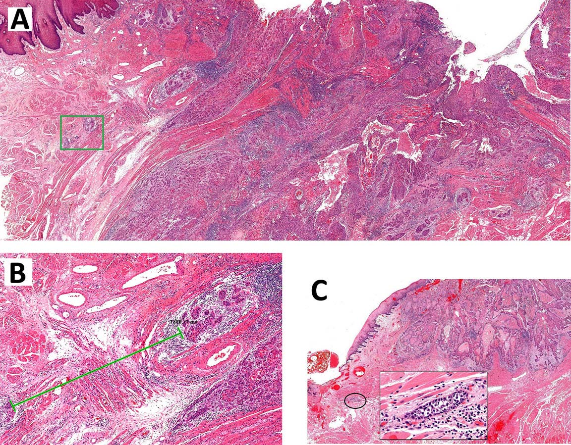
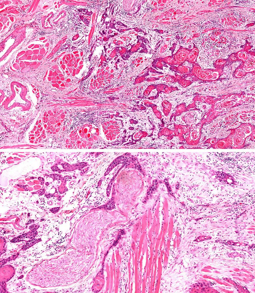
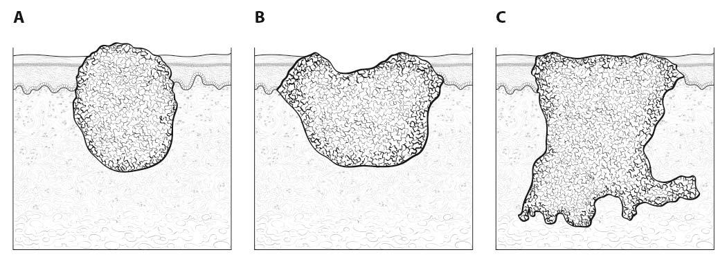
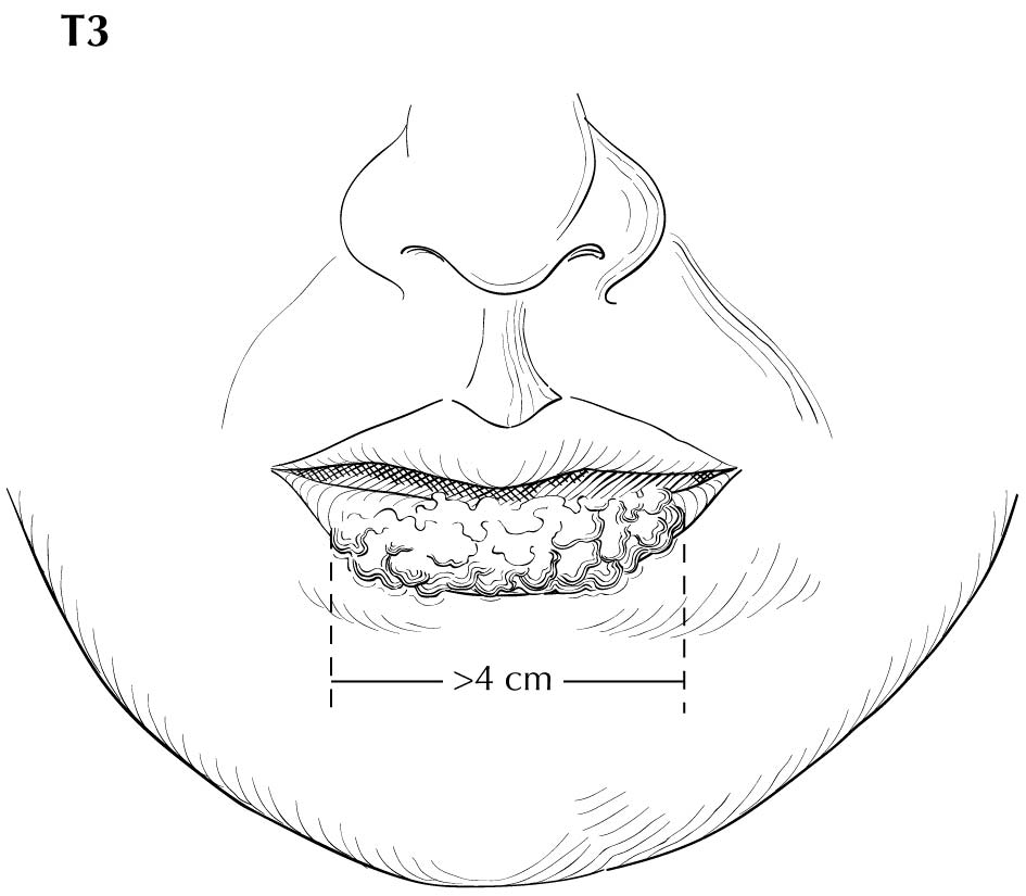
Inspection of the lip and oral cavity typically reveals the greatest diameter of a cancer, though palpation is essential to assess DOI and submucosal extension. The mucosal extent of the cancer usually reflects its true linear dimension. Induration surrounding a cancer typically is due to peritumoral inflammation. DOI should be distinguished from tumor thickness, and its determination is predicated on invasion beneath the plane defined by surrounding normal mucosa. Any exophytic character should be noted, but assignment of stage is determined by what transpires at or beneath the surface (defined by adjacent normal mucosa). Clinical evidence of bone destruction should be noted and its depth estimated (e.g., into bone versus through cortex into the marrow space). Thick lesions often are defined by computed tomography (CT) or magnetic resonance (MR) imaging, but the difference between thickness and DOI must be observed. Lesions located near the midline more often involve the contralateral side of the neck than well-lateralized cancers. Dysphagia is suggestive of a tumor with sufficient invasion of oral structures to engender dysfunction. It is seldom present when cancers have little DOI. Similarly, drooling or the inability to swallow liquids without difficulty suggests a tumor with substantial DOI. Trismus, when not caused by pain, is consistent with a deeply invasive lesion. Complaints of numbness of the lip and /or teeth are commonly associated with nerve invasion. The distinction between 4 mm DOI and 6 mm DOI (for example) may not be possible on clinical grounds. The stage should only be raised on the basis of DOI if the differences are clear.

Evidence of cranial nerve dysfunction should be sought (testing sensation and motion to command ) and skin should be examined for evidence of invasion by underlying nodes. Palpable neck nodes should be considered in terms of their location (level in the neck), size, number, character (smooth or irregular), attachment to other nodes, and mobility. Nodes that do not move in all directions may be invading nearby structures. Invasion of the sternomastoid muscle and /or cranial nerves is associated with lateral motion with restricted ability to move the node along the cranial-caudal axis. Inability to move the node at all (without moving the head) is worrisome for ENE, though the suspicion should be tempered for smaller nodes with limited mobility in level II. Assignment of ENE should be based almost entirely upon the physical examination, rather than upon imaging studies; gross ENE is required to raise the stage beyond the assignment based upon node size and number, and this may be overestimated with current imaging modalities.

Clinical or radiographic extranodal extension

ENE worsens the adverse outcome associated with nodal metastasis. The presence of ENE can be diagnosed clinically by the presence of a “matted” mass of nodes, involvement of overlying skin, adjacent soft tissue, or clinical signs of cranial nerve or brachial plexus, sympathetic chain or phrenic nerve invasion. Cross-sectional imaging (CT or MR) generally has low sensitivity (65-80%) but high specificity (86-93%) for the detection of ENE. The most reliable imaging signs are an indistinct nodal margin, irregular nodal capsular enhancement or infiltration into the adjacent fat or muscle, with the latter finding on CT and MR imaging as the most specific sign of ENE. Ultrasound appears to be less accurate than CT and MR imaging, but ENE is suggested by interrupted or undefined nodal contours with high-resolution ultrasound imaging. The absence or presence of clinical/radiologic ENE is designated ENE(-) or ENE(+), respectively.

Imaging

Cross-sectional imaging of the oral cavity may be performed with either CT or MR imaging, depending on availability, patient imaging tolerance, contrast allergies, and cost. With either modality, the coronal plane view—either as direct MR imaging or from reformats obtained from axially acquired thin-slice CT—allows excellent evaluation of the floor of the mouth.9 CT offers some advantage over MR imaging in the evaluation of cortical bone erosion, although MR imaging appears to be more sensitive but less specific for the detection of bone marrow invasion by tumor.10,11 MR imaging offers the additional advantage of evaluation of perineural tumor spread, which for oral cavity tumors is primarily along the inferior alveolar nerve (CNV3) of the mand ible and the greater and lesser palatine nerves (CNV2) of the maxilla. Gadolinium contrast is always recommended unless contraindicated by prior reaction or very poor renal function. Positron emission tomography (PET)/CT is primarily done for nodal staging of disease or when distant metastases are suspected, unless the CT component is performed as a post-contrast examination with dedicated neck imaging. Ultrasound does not allow adequate evaluation of the oral cavity primary tumor site, but it may be supplementary for nodal evaluation with otherwise equivocal nodal imaging findings.

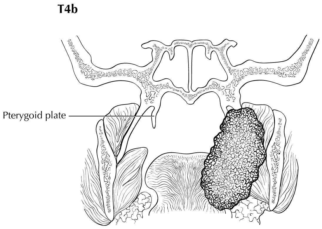
As small but clinically evident mucosal tumors may be subtle on imaging, it is important to review the imaging exam with knowledge of the tumor site. T1, T2, and T3 tumors are distinguished only by size and depth of invasion. The former is better determined by clinical examination, although a radiologic measurement should be given as part of the imaging report. The radiologist's more important role during tumor staging is to determine deep tissue involvement and assess for nodal and /or distant metastases. T4 disease entails deep tissue invasion, which varies according to the specific subsite of the oral cavity. For alveolar ridge, floor of mouth, retromolar triangle, hard palate, and large lip tumors, careful attention should be paid to the cortex and marrow space of the adjacent maxilla or mand ible, because such invasion denotes T4a disease. In the AJCC Cancer Staging Manual, 7th Edition, oral tongue tumors were designated T4a when there was deep invasion into the extrinsic muscles of the tongue and /or the floor of the mouth. DOI will supersede muscle invasion in the 8th Edition. Depth is frequently better evaluated in the coronal plane and /or sagittal plane. More posterior extensive spread of tumor—such as buccal tumors invading into the muscles of mastication, or spreading to the pterygoid plates or superiorly to the skull base—denotes T4b tumor. Additionally, posterolateral tumor spread to surround the internal carotid artery is also T4b disease.

Both CT and MR imaging allow evaluation of nodal morphology to determine possible tumor involvement. Levels IA, IB, and IIA are the most frequently involved sites, and these levels should be scrutinized specifically with concern for rounded contour, heterogeneous texture including cystic or necrotic change, enlargement, and ill-defined margins. It also is important to be cognizant that nodal spread may be bilateral, particularly with anterior and /or midline oral cavity tumors. Skip nodal metastases (level IV without level III involvement) while described with lateral tongue tumors, appear to be rare. As previously described, PET/CT may also be used to improve predictive yield for nodal metastases by the addition of physiologic information, and ultrasound may be an additive tool for evaluation of indeterminate nodes. PET/CT is the only modality to allow whole-body evaluation of distant metastatic spread, and the upper lungs and bone should always be reviewed as potential metastatic sites on any staging neck CT or MR imaging.

The risk of distant metastasis is more dependent on the N than on the T status of the head and neck cancer. In addition to the node size, number, and presence of ENE, regional lymph nodes also should be described according to the level of the neck that is involved. The level of involved nodes in the neck is prognostically significant for oral cavity (caudad nodal disease is worse), as is the presence of ENE metastatic tumor from individual nodes. Midline nodes are considered ipsilateral. Imaging studies showing amorphous spiculated margins of involved nodes or involvement of internodal fat resulting in loss of normal oval-to-round nodal shape strongly suggest extranodal extension; however, pathological examination is necessary to prove its presence. No imaging study can currently identify microscopic foci of cancer in regional nodes or distinguish between small reactive nodes and small nodes with metastatic deposits (in the absence of central radiographic inhomogeneity).

Pathological Classification

Complete resection of the primary site and /or regional lymph node dissections, followed by pathological examination of the resection specimen allows for the use of this designation for pT and /or pN, respectively. Resections after radiation or chemotherapy should be identified and considered in context. pT is derived from the actual measurement of the unfixed tumor in the surgical specimen. It should be noted, however, that up to 30% shrinkage of soft tissues may occur in resected specimen after formalin fixation. Pathological staging represents additional and important information and should be included as such in staging, but it does not supplant clinical staging as the primary staging scheme. Metastasis found on imaging is considered cM1. Biopsy-proven metastasis is considered pM1.

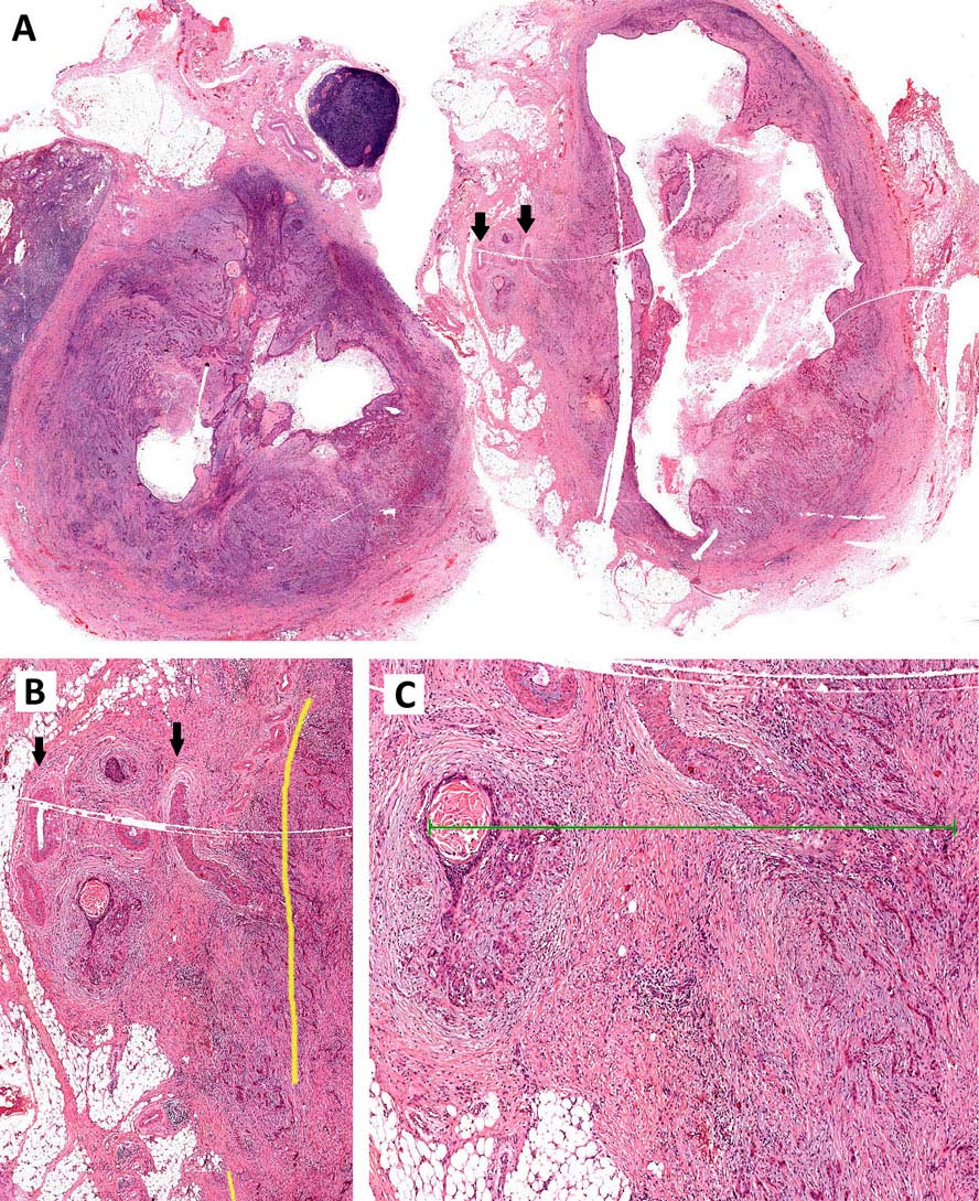
Pathological assessment of ENE

Resected positive lymph nodes require examination for the presence and extent of ENE. ENEmi is defined as microscopic ENE 2 mm. Macroscopic (ENEma) is defined as either extranodal extension apparent to the naked eye at the time of dissection or extension > 2 mm beyond the lymph node capsule microscopically. ENEmi and ENEma are used to define pathological ENE(+) nodal status.

For assessment of pN, a selective neck dissection will ordinarily include 10 or more lymph nodes, and a comprehensive neck dissection (radical or modified radical neck dissection) will ordinarily include 15 or more lymph nodes. Examination of fewer tumor-free nodes still mand ates a pN0 designation.

Prognostic Factors

Prognostic Factors Required for Stage Grouping

Beyond the factors used to assign T, N, or M categories, no additional prognostic factors are required for stage grouping.

Additional Factors Recommended for Clinical Care

Extranodal Extension

ENE is defined as extension of metastatic carcinoma within lymph node, through the capsule, and into the surrounding connective tissue, regardless of associated stromal reaction. Histopathologic designations for ENE are as follows:

  • ENEn (none)
  • ENEmi (microscopic ENE 2 mm)
  • ENEma (ENE > 2 mm or gross ENE)

ENEmi ENEma are used to define pathological ENE nodal status (Figure 7.5). ENEmi versus ENEn will affect current nodal staging, and data collection is recommended to allow stand ardization of data collection and future analysis.

7.5 A. Extranodal extension of metastatic carcinoma, low-power. The large vessels (black arrows) are extranodal in location. B. The direction of the collagen and the location of vessels guide the estimation of the natural lymph node boundary (yellow line). C. This carcinoma extends > 2mm from the estimated lymph node boundary (green line) and should be classified as ENEma.

AJCC Level of Evidence: III
Depth of Invasion

DOI assesses the invasiveness of a carcinoma, regardless of any exophytic component. It is measured by first finding the “horizon” of the basement membrane of the adjacent squamous mucosa (Figure 7.6). A perpendicular “plumb line” is established from this horizon to the deepest point of tumor invasion, which represents DOI. The DOI is recorded in millimeters. Measurements in millimeters can easily be accomplished by printing rulers on acetate sheets, which can be overlaid onto glass slides. Figure 7.7 demonstrates DOI of an ulcerated carcinoma.

7.6 Depth of invasion (DOI). The horizon is established at the level of the basement membrane relative to the closest intact squamous mucosa. The greatest DOI is measured by dropping a “plumb line” from the horizon.

7.7 Depth of invasion (DOI) in an ulcerated carcinoma. Notice how “tumor thickness” would be deceptively thinner than DOI.

AJCC Level of Evidence: III
Resection Margins

The ideal manner of intraoperative margin assessment is the “specimen driven approach.” 12,13 Direct discussion between surgeon and pathologist at specimen hand -off allows for correct anatomic orientation and identification of any intra-operative non-margin tissue tears or cuts. The pathologist maps the specimen, paints the different margin planes with unique colors, and documents the designations. In the event of non-margin tissue tears, these non-margins should be inked first using a unique color (e.g., yellow). This obviates the problem of ink running. The pathologist then makes multiple cuts into the margins at 5- to 10-mm intervals perpendicular to the resection plane. Initial gross assessment yields important preliminary information. This is followed by targeted microscopic examination of margins of interest. The margin sections should be taken perpendicular to the resection plane. The distance between carcinoma and resection margin should be reported in millimeters (Figure 7.8).

7.8 WPOI-5” describes a dispersed tumor pattern of invasion which is significantly predictive of worst outcome. Carcinomas are classifiable as WPOI-5 when satellite dispersion is >= 1 mm from neighboring satellites. A: Low-power overview demonstrating a context of generalized tumor dispersion. Tumor dispersion is measured at the advancing tumor edge. Carcinoma satellites in the green box are shown in panel B, lower edge. The green line measures dispersion of almost 2 mm. C. This carcinoma reveals only few dispersed satellites fulfilling this criteria, likely due to extratumoral lymphovascular emboli.

AJCC Level of Evidence: II
Worst Pattern of Invasion

Worst pattern of invasion (WPOI) is a validated outcome predictor for oral cavity squamous carcinoma patients in multivariate analysis.14-16 To simplify prognostication and enhance adaptation, the only cutpoint recommended for assessment is whether or not WPOI-5 is present. WPOI-5 is defined as tumor dispersion of >= 1 mm between tumor satellites. With respect to low-stage oral cavity squamous carcinomas > 4 mm DOI, the presence of WPOI-5 is significantly predictive of locoregional recurrence and disease-specific survival (p = 0.0008, HR 2.55, 95% CI 1.48, 4.41, and p = 0.0001, HR 6.34, 95% CI 2.50, 16.09, respectively) and the probability of developing locoregional recurrence is almost 42%. Figures 7.8 and 7.9 illustrate examples of WPOI-5. Tumor dispersion is assessed at the advancing tumor edge. The most common WPOI-5 phenotype is tumor dispersion through soft tissue. Dispersed extratumoral perineural invasion, or extratumoral lymphovascular invasion, also can qualify for classification as WPOI-5.

7.9 Top: A “strand y” pattern with intervening skeletal muscle observable at low-power is often classifiable as WPOI-5. Bottom: This strand pattern is also often associated with perineural invasion.

AJCC Level of Evidence: II
Perineural Invasion

Perineural invasion (PNI) should be subclassified as either intratumoral or extratumoral (Figure 7.10). Involvement of named nerves should be specifically reported.17 PNI should be subclassified as focal or multifocal. Extensive multifocal PNI is usually extratumoral and frequently associated with a “strand -like” tumor phenotype. The largest nerve diameter should be reported for multifocal, extratumoral PNI.

7.10 Carcinoma should demonstrate a specific relationship with nerve, such as wrapping around nerves, in order to be classified as perineural invasion (PNI). Merely “bumping” into a nerve does not constitute PNI.

AJCC Level of Evidence: II
Lymphovascular invasion

Lymphovascular invasion should be reported as either intratumoral or extratumoral, as well as focal or multifocal.

AJCC Level of Evidence: II
Overall Health

In addition to the importance of the TNM factors outlined previously, the overall health of the patient clearly influences outcome. An ongoing effort to better assess prognosis using both tumor and nontumor-related factors is underway. Chart abstraction will continue to be performed by cancer registrars to obtain important information regarding specific factors related to prognosis. These data will then be used to further hone the predictive power of the staging system in future revisions.

AJCC Level of Evidence: III
Comorbidity

Comorbidity can be classified by specific measures of additional medical illnesses.18 Accurate reporting of all illnesses in the patients' medical record is essential to assessment of these parameters. General performance measures are helpful in predicting survival. The AJCC strongly recommends the clinician report performance status using the Eastern Cooperative Oncology Group (ECOG), Zubrod, or Karnofsky performance measures, along with stand ard staging information. An interrelationship between each of the major performance tools exists.

Zubrod/ECOG Performance Scale
0Fully active, able to carry out all predisease activities without restriction (Karnofsky 90-100)
1Restricted in physically strenuous activity but ambulatory and able to carry work of a light or sedentary nature. For example, light housework, office work (Karnofsky 70-80)
2Ambulatory and capable of all self-care but unable to carry out any work activities. Up and about more than 50% of waking hours (Karnofsky 50-60)
3Capable of only limited self-care, confined to bed or chair 50% or more of waking hours (Karnofsky 30-40)
4Completely disabled. Cannot carry on self-care. Totally confined to bed (Karnofsky 10-20)
5Death (Karnofsky 0)
AJCC Level of Evidence: II

Lifestyle Factors

Lifestyle factors such as tobacco and alcohol abuse negatively influence survival. Accurate recording of smoking in pack years and alcohol in number of days drinking per week and number of drinks per day will provide important data for future analysis. Nutrition is important to prognosis and will be indirectly measured by weight loss of > 5% of body weight in the previous 6 months.19 Depression adversely impacts quality of life and survival. Notation of a previous or current diagnosis of depression should be recorded in the medical record.20

AJCC Level of Evidence: III

Tobacco Use

The role of tobacco as a negative prognostic factor is well established. However, exactly how this could be codified in the staging system is less clear. At this time, smoking is known to have a deleterious effect on prognosis but it is hard to accurately apply it to the staging system.

Smoking history should be collected as an important element of the demographics and may be included in Prognostic Groups in the future. For practicality, the minimum stand ard should classify smoking history as never, 10 pack-years, > 10 but 20 pack-years, or > 20 pack-years.

AJCC Level of Evidence: III

The authors have not identified any emerging factors for clinical care.

Risk Assessment

Risk Assesment Models

The AJCC recently established guidelines that will be used to evaluate published statistical prediction models for the purpose of granting endorsement for clinical use.21 Although this is a monumental step toward the goal of precision medicine, this work was published only very recently. Therefore, the existing models that have been published or may be in clinical use have not yet been evaluated for this cancer site by the Precision Medicine Core of the AJCC. In the future, the statistical prediction models for this cancer site will be evaluated, and those that meet all AJCC criteria will be endorsed.

Recommendations

TNM Definitions

Definition of Primary Tumor (T)

T CategoryT Criteria
TXPrimary tumor cannot be assessed
TisCarcinoma in situ
T1

Tumor less than or equal to 2 cm, less than or equal to 5 mm depth of invasion (DOI)

DOI is depth of invasion and not tumor thickness.

T2

Tumor less than or equal to 2 cm, DOI greater than 5 mm and less than or equal to 10 mm

or tumor greater than 2 cm but less than or equal to 4 cm, and less than or equal to 10 mm DOI

T3

Tumor greater than 4 cm

or any tumor greater than 10 mm DOI

T4Moderately advanced or very advanced local disease
T4a

Moderately advanced local disease

(lip) Tumor invades through cortical bone or involves the inferior alveolar nerve, floor of mouth, or skin of face (i.e., chin or nose)

(oral cavity) Tumor invades adjacent structures only (e.g., through cortical bone of the mand ible or maxilla, or involves the maxillary sinus or skin of the face)

Note: Superficial erosion of bone/tooth socket (alone) by a gingival primary is not sufficient to classify a tumor as T4.

T4b

Very advanced local disease

Tumor invades masticator space, pterygoid plates, or skull base and /or encases the internal carotid artery

Definition of Regional Lymph Node (N)

Clinical N (cN)
N CategoryN Criteria
NXRegional lymph nodes cannot be assessed
N0No regional lymph node metastasis
N1Metastasis in a single ipsilateral lymph node, 3 cm or smaller in greatest dimension ENE(-)
N2

Metastasis in a single ipsilateral node larger than 3 cm but not larger than 6 cm in greatest dimension and ENE(-);

or metastases in multiple ipsilateral lymph nodes, none larger than 6 cm in greatest dimension and ENE(-);

or in bilateral or contralateral lymph nodes, none larger than 6 cm in greatest dimension, and ENE(-)

N2aMetastasis in a single ipsilateral node larger than 3 cm but not larger than 6 cm in greatest dimension, and ENE(-)
N2bMetastasis in multiple ipsilateral nodes, none larger than 6 cm in greatest dimension, and ENE(-)
N2cMetastasis in bilateral or contralateral lymph nodes, none larger than 6 cm in greatest dimension, and ENE(-)
N3

Metastasis in a lymph node larger than 6 cm in greatest dimension and ENE(-);

or metastasis in any node(s) and clinically overt ENE(+)

N3aMetastasis in a lymph node larger than 6 cm in greatest dimension and ENE(-)
N3bMetastasis in any node(s) and clinically overt ENE(+)

A designation of “U” or “L” may be used for any N category to indicate metastasis above the lower border of the cricoid (U) or below the lower border of the cricoid (L). Similarly, clinical and pathological ENE should be recorded as ENE(-) or ENE(+).

Pathological N (pN)
N CategoryN Criteria
NXRegional lymph nodes cannot be assessed
N0No regional lymph node metastasis
N1Metastasis in a single ipsilateral lymph node, 3 cm or smaller in greatest dimension and ENE(-)
N2

Metastasis in a single ipsilateral lymph node, 3 cm or smaller in greatest dimension and ENE(+);

or larger than 3 cm but not larger than 6 cm in greatest dimension and ENE(-);

or metastases in multiple ipsilateral lymph nodes, none larger than 6 cm in greatest dimension and ENE(-);

or in bilateral or contralateral lymph node(s), none larger than 6 cm in greatest dimension, ENE(-)

N2a

Metastasis in single ipsilateral node 3 cm or smaller in greatest dimension and ENE(+);

or a single ipsilateral node larger than 3 cm but not larger than 6 cm in greatest dimension and ENE(-)

N2bMetastasis in multiple ipsilateral nodes, none larger than 6 cm in greatest dimension and ENE(-)
N2cMetastasis in bilateral or contralateral lymph node(s), none larger than 6 cm in greatest dimension and ENE(-)
N3

Metastasis in a lymph node larger than 6 cm in greatest dimension and ENE(-);

or metastasis in a single ipsilateral node larger than 3 cm in greatest dimension and ENE(+);

or multiple ipsilateral, contralateral or bilateral nodes any with ENE(+);

or asingle contralateral node 3 cm or smaller and ENE (+)

N3aMetastasis in a lymph node larger than 6 cm in greatest dimension and ENE(-)
N3b

Metastasis in a single ipsilateral node larger than 3 cm in greatest dimension and ENE(+);

or multiple ipsilateral, contralateral or bilateral nodes any with ENE(+);

or asingle contralateral node 3 cm or smaller and ENE (+)

A designation of “U” or “L” may be used for any N category to indicate metastasis above the lower border of the cricoid (U) or below the lower border of the cricoid (L). Similarly, clinical and pathological ENE should be recorded as ENE(-) or ENE(+).

Definition of Distant Metastasis (M)

M CategoryM Criteria
M0No distant metastasis
M1Distant metastasis

Stage Prognostic

!!Calculator!!

AJCC PROGNOSTIC STAGE GROUPS

When T is…and N is…and M is…Then the stage group is…
TisN0M00
T1N0M0I
T2N0M0II
T3N0M0III
T1, T2, T3N1M0III
T4aN0, N1M0IVA
T1, T2, T3, T4aN2M0IVA
Any TN3M0IVB
T4bAny NM0IVB
Any TAny NM1IVC

Registry Data

Registry Data Collection Variables

  1. Lip location (external lip or vermilion border)
  2. ENE clinical (presence or absence)
  3. ENE pathological (presence or absence)
  4. Extent of microscopic ENE (distance of extension from the native lymph node capsule to the farthest point of invasion in the extranodal tissue)
  5. Perineural invasion
  6. Lymphovascular invasion
  7. p16/HPV status
  8. Performance status
  9. Tobacco use and pack-year
  10. Alcohol use
  11. Depression diagnosis
  12. Depth of invasion (mm)
  13. Margin status (grossly involved, microscopic involvement)
  14. Distance of tumor (or moderate/severe dysplasia) from closest margin
  15. WPOI-5

Histopathologic type

Squamous cell carcinoma (conventional, variants), carcinoma of minor salivary gland (acinic cell, adenoid cystic, adenocarcinoma, NOS, basal cell adenocarcinoma, carcinoma ex-pleomorphic adenoma, carcinoma type cannot be determined, carcinosarcoma, clear cell adenocarcinoma, cystadenocarcinoma, epithelial-myoepithelial carcinoma, mammary analogue secretory carcinoma, mucoepidermoid carcinoma, mucinous carcinoma, myoepithelial carcinoma, oncocytic carcinoma, polymorphous low-grade adenocarcinoma, salivary duct carcinoma), non-salivary gland adenocarcinoma, neuroendocrine carcinoma (typical carcinoid, atypical carcinoid, large cell, small cell, composite small cell-other type), mucosal melanoma, carcinoma type cannot be determined.

Histologic grade

HISTOLOGIC GRADE (G)

GG Definition
GXCannot be assessed
G1Well differentiated
G2Moderately differentiated
G3Poorly differentiated

Illustrations

7.11 Characteristics of lip and oral cavity tumors. (A) Exophytic. (B) Ulcerated. (C) Endophytic.

7.12 T1 is defined as a tumor 2 cm or smaller in greatest dimension.

7.13 T2 is defined as a tumor larger than 2 cm but not larger than 4 cm in greatest dimension.

7.14 T3 is defined as tumor larger than 4 cm in greatest dimension.

7.15 T4a (Lip) is defined as moderately advanced local disease, tumor invading through cortical bone, inferior alveolar nerve, floor of mouth, or skin of face, i.e., chin or nose (as shown).

7.16 T4b is defined as very advanced local disease, tumor involves masticator space, pterygoid plates (as shown), or skull base and /or encases internal carotid artery.

Bibliography

  1. O'Sullivan B, Huang SH, Su J, et al. Development and validation of a staging system for HPV-related oropharyngeal cancer by the International Collaboration on Oropharyngeal cancer Network for Staging (ICON-S): a multicentre cohort study. The lancet oncology. Feb 26 2016.
  2. Patel S. Personal Communication. In: Lydiatt W, Shah JP, eds2015.
  3. Wreesmann VB, Katabi N, Palmer FL, et al. Influence of extracapsular nodal spread extent on prognosis of oral squamous cell carcinoma. Head & neck. Oct 30 2015.
  4. Spiro RH, Huvos AG, Wong GY, Spiro JD, Gnecco CA, Strong EW. Predictive value of tumor thickness in squamous carcinoma confined to the tongue and floor of the mouth. American journal of surgery. Oct 1986;152(4):345-350.
  5. Ebrahimi A, Gil Z, Amit M. International Consortium for Outcome Research (ICOR) in Head and Neck Cancer. Primary tumor staging for oral cancer and a proposed modification incorporating depth of invasion: an international multicenter retrospective study. JAMA otolaryngology-- head & neck surgery. 2014;140(12):1138-1148.
  6. Ebrahimi A, Clark JR, Amit M, et al. Minimum nodal yield in oral squamous cell carcinoma: defining the stand ard of care in a multicenter international pooled validation study. Annals of surgical oncology. Sep 2014;21(9):3049-3055.
  7. Prabhu RS, Hanasoge S, Magliocca KR, et al. Extent of pathologic extracapsular extension and outcomes in patients with nonoropharyngeal head and neck cancer treated with initial surgical resection. Cancer. May 15 2014;120(10):1499-1506.
  8. Dunne AA, Muller HH, Eisele DW, Kessel K, Moll R, Werner JA. Meta-analysis of the prognostic significance of perinodal spread in head and neck squamous cell carcinomas (HNSCC) patients. European journal of cancer. Aug 2006;42(12):1863-1868.
  9. Land ry D, Glastonbury CM. Squamous cell carcinoma of the upper aerodigestive tract: a review. Radiol Clin North Am. Jan 2015;53(1):81-97.
  10. Li C, Yang W, Men Y, Wu F, Pan J, Li L. Magnetic resonance imaging for diagnosis of mand ibular involvement from head and neck cancers: a systematic review and meta-analysis. PloS one. 2014;9(11):e112267.
  11. Gu DH, Yoon DY, Park CH, et al. CT, MR, 18F-FDG PET/CT, and their combined use for the assessment of mand ibular invasion by squamous cell carcinomas of the oral cavity. Acta Radiologica. 2010;51(10):1111-1119.
  12. Maxwell JH, Thompson LD, Brand wein-Gensler MS, et al. Early Oral Tongue Squamous Cell Carcinoma: Sampling of Margins From Tumor Bed and Worse Local Control. JAMA otolaryngology-- head & neck surgery. Dec 1 2015;141(12):1104-1110.
  13. Hinni ML, Ferlito A, Brand wein-Gensler MS, et al. Surgical margins in head and neck cancer: a contemporary review. Head & neck. Sep 2013;35(9):1362-1370.
  14. Brand wein-Gensler M, Smith RV, Wang B, et al. Validation of the histologic risk model in a new cohort of patients with head and neck squamous cell carcinoma. The American journal of surgical pathology. May 2010;34(5):676-688.
  15. Brand wein-Gensler M, Teixeira MS, Lewis CM, et al. Oral squamous cell carcinoma: histologic risk assessment, but not margin status, is strongly predictive of local disease-free and overall survival. The American journal of surgical pathology. Feb 2005;29(2):167-178.
  16. Li Y, Bai S, Carroll W, et al. Validation of the risk model: high-risk classification and tumor pattern of invasion predict outcome for patients with low-stage oral cavity squamous cell carcinoma. Head and neck pathology. Sep 2013;7(3):211-223.
  17. Chinn SB, Spector ME, Bellile EL, et al. Impact of perineural invasion in the pathologically N0 neck in oral cavity squamous cell carcinoma. Otolaryngology--head and neck surgery : official journal of American Academy of Otolaryngology-Head and Neck Surgery. Dec 2013;149(6):893-899.
  18. Piccirillo JF. Inclusion of comorbidity in a staging system for head and neck cancer. Oncology (Williston Park). Sep 1995;9(9):831-836; discussion 841, 845-838.
  19. Marion E. Couch MD P, MBA1,*, Kim Dittus MD, PhD2, Michael J. Toth PhD3, Monte S. Willis MD, PhD4, Denis C. Guttridge PhD5, Jonathan R. George MD6, Eric Y. Chang7, Christine G. Gourin MD8 and Hirak Der-Torossian MD, MPH1 Cancer cachexia update in head and neck cancer: Pathophysiology and treatment Head & neck surgery. 2015;37(7):1057-1072.
  20. Lazure KE, Lydiatt WM, Denman D, Burke WJ. Association between depression and survival or disease recurrence in patients with head and neck cancer enrolled in a depression prevention trial. Head & neck. 2009;31(7):888-892.
  21. Kattan MW, Hess KR, Amin MB, et al. American Joint Committee on Cancer acceptance criteria for inclusion of risk models for individualized prognosis in the practice of precision medicine. CA: a cancer journal for clinicians. Jan 19 2016.
  22. Chai RL, Rath TJ, Johnson JT, et al. Accuracy of computed tomography in the prediction of extracapsular spread of lymph node metastases in squamous cell carcinoma of the head and neck. JAMA otolaryngology-- head & neck surgery. Nov 2013;139(11):1187-1194.
  23. Dillon JK, Glastonbury CM, Jabeen F, Schmidt BL. Gauze padding: a simple technique to delineate small oral cavity tumors. AJNR. American journal of neuroradiology. May 2011;32(5):934-937.
  24. Feng Z, Li JN, Niu LX, Guo CB. Supraomohyoid neck dissection in the management of oral squamous cell carcinoma: special consideration for skip metastases at level IV or V. Journal of Oral and Maxillofacial Surgery. 2014;72(6):1203-1211.
  25. Henrot P, Blum A, Toussaint B, Troufleau P, Stines J, Roland J. Dynamic maneuvers in local staging of head and neck malignancies with current imaging techniques: principles and clinical applications. Radiographics : a review publication of the Radiological Society of North America, Inc. Sep-Oct 2003;23(5):1201-1213.
  26. Hoang JK, Glastonbury CM, Chen LF, Salvatore JK, Eastwood JD. CT mucosal window settings: a novel approach to evaluating early T-stage head and neck carcinoma. AJR. American journal of roentgenology. Oct 2010;195(4):1002-1006.
  27. Kann BH, Buckstein M, Carpenter TJ, et al. Radiographic extracapsular extension and treatment outcomes in locally advanced oropharyngeal carcinoma. Head & neck. Dec 2014;36(12):1689-1694.
  28. Katayama I, Sasaki M, Kimura Y, et al. Comparison between ultrasonography and MR imaging for discriminating squamous cell carcinoma nodes with extranodal spread in the neck. European journal of radiology. 2012;81(11):3326-3331.
  29. Kimura Y, Sumi M, Sakihama N, Tanaka F, Takahashi H, Nakamura T. MR imaging criteria for the prediction of extranodal spread of metastatic cancer in the neck. AJNR. American journal of neuroradiology. Aug 2008;29(7):1355-1359.
  30. King AD, Tse GM, Yuen EH, et al. Comparison of CT and MR imaging for the detection of extranodal neoplastic spread in metastatic neck nodes. Eur J Radiol. Dec 2004;52(3):264-270.
  31. Lodder WL, Lange CA, van Velthuysen M-LF, et al. Can extranodal spread in head and neck cancer be detected on MR imaging. Oral oncology. 2013;49(6):626-633.
  32. Prabhu RS, Magliocca KR, Hanasoge S, et al. Accuracy of computed tomography for predicting pathologic nodal extracapsular extension in patients with head-and -neck cancer undergoing initial surgical resection. International journal of radiation oncology, biology, physics. Jan 1 2014;88(1):122-129.
  33. Rand all DR, Lysack JT, Hudon ME, et al. Diagnostic utility of central node necrosis in predicting extracapsular spread among oral cavity squamous cell carcinoma. Head & neck. 2015;37(1):92-96.
  34. Weissman JL, Carrau RL. "Puffed-cheek" CT improves evaluation of the oral cavity. AJNR. American journal of neuroradiology. Apr 2001;22(4):741-744.