Cancers Staged Using This Staging System
Hepatocellular carcinoma (HCC), Fibrolamellar carcinoma (fibrolamellar variant of HCC)
Cancers Not Staged Using This Staging System
These histopathologic types of cancer | Are staged according to the classification for | and can be found in chapter |
---|---|---|
Intrahepatic cholangiocarcinoma | Intrahepatic bile ducts | 23 |
Combined hepatocellular-cholangiocarcinoma | Intrahepatic bile ducts | 23 |
Sarcomas of the liver | Soft tissue sarcoma of the abdomen and thoracic visceral organs | 42 |
Summary of Changes
Change | Details of Change | Level of Evidence |
---|---|---|
Definition of Primary Tumor (T) | T1 is now divided into two subcatgories: T1a, solitary tumor less than or equal to 2 cm; and T1b, solitary tumor without vascular invasion, greater than 2 cm. | II |
Definition of Primary Tumor (T) | T2 now includes solitary tumor with vascular invasion greater than 2 cm, or multiple tumors, none greater than 5 cm. | II |
Definition of Primary Tumor (T) | T3a is now recategorized as T3. | III |
Definition of Primary Tumor (T) | T3b: Tumors involving a major branch of the portal vein or hepatic vein formerly were categorized as T3b and are now categorized as T4. | III |
Code | Description |
---|---|
C22.0 | Liver |
This list includes histology codes and preferred terms from the WHO Classification of Tumors and the International Classification of Diseases for Oncology (ICD-O). Most of the terms in this list represent malignant behavior. For cancer reporting purposes, behavior codes /3 (denoting malignant neoplasms), /2 (denoting in situ neoplasms), and in some cases /1 (denoting neoplasms with uncertain and unknown behavior) may be appended to the 4-digit histology codes to create a complete morphology code.
Code | Description |
---|---|
8170 | Hepatocellular carcinoma |
8171 | Hepatocellular carcinoma, fibrolamellar variant |
8172 | Hepatocellular carcinoma, scirrhous |
8173 | Hepatocellular carcinoma, spindle cell variant |
8174 | Hepatocellular carcinoma, clear cell type |
Histology is not ideal for clinical use in patient care, as it describes an unspecified or outdated diagnosis. Data collectors may use this code only if there is not enough information in the medical record to document a more specific diagnosis.
Bosman FT, Carneiro F, Hruban RH, Theise ND, eds. World Health Organization Classification of Tumours of the Digestive System. Lyon: IARC; 2010. Used with permission.
International Agency for Research on Cancer, World Health Organization. International Classification of Diseases for Oncology. ICD-O-3-Online.http://codes.iarc.fr/home. Accessed September 29, 2017. Used with permission.
Hepatocellular carcinoma (HCC) is the sixth most common malignancy in the world and is responsible for 600,000 deaths annually. HCC is an etiology-driven malignancy, mainly the result of cirrhosis that is attributed to hepatitis B, hepatitis C, alcohol, nonalcoholic steatohepatitis, and many genetically inherited metabolic diseases, the most common of which is hemochromatosis. These entities may be associated with HCC in the absence of cirrhosis. Chronic hepatitis C infection contributes the most to the incidence of HCC in the United States. The advent of novel protease inhibitors as curative therapies for hepatitis C is expected to reduce hepatitis C incidence and prevalence and , thus, hepatitis C-related HCC. However, the continued rise in morbid obesity and diabetes most likely will lead to a continued increase in nonalcoholic steatohepatitis-related HCC.1
Cirrhosis is a major component of the clinical presentation and a key determinant of prognosis. Anatomic stage is the other major determinant of outcome and prognosis. TNM staging helps determine curative resectability, as well as the presence and extent of vascular invasion, a key determinant of potential cure versus no cure. However, staging HCC remains a challenge, as incorporating components of the anatomic extent of the disease and the presence of cirrhosis have led to multiple staging and scoring systems, on which opinions continue to differ.2 The lack of consensus regarding HCC staging is driven mainly by the etiology-specific prognostic factors. Nonetheless, TNM staging will continue to serve as the backbone for the anatomic description of disease extent as part of most other staging and scoring systems.
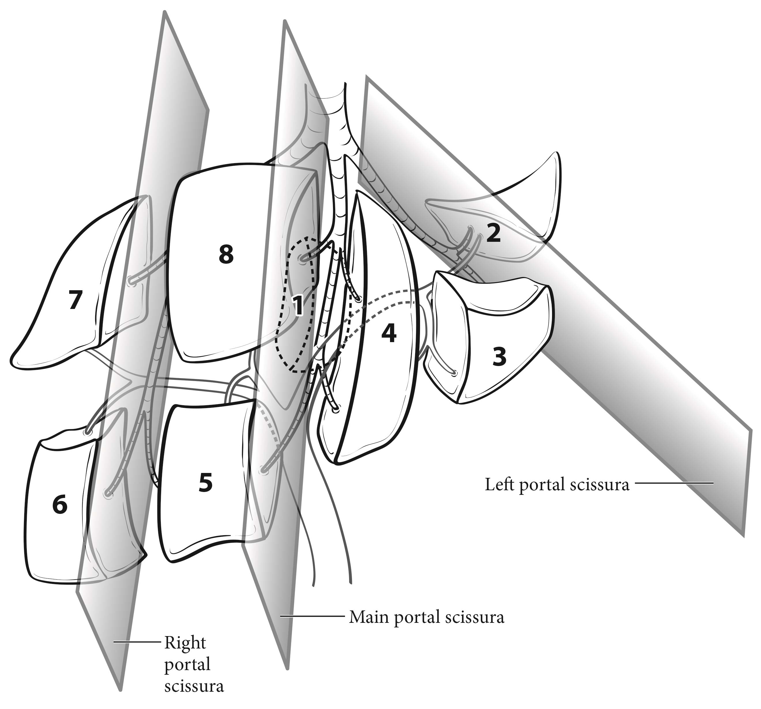
The liver has a dual blood supply from the hepatic artery and portal vein. Tumors are fed by the arterial blood supply. The liver is divided into right and left hemilivers by a plane called the Rex-Cantlie line, which projects between the gallbladder fossa and the vena cava and is defined by the middle hepatic vein. Couinaud refined knowledge about the functional anatomy of the liver and proposed dividing the liver into four sectors and eight segments. In this nomenclature, the liver is divided by vertical and oblique planes, or scissurae, defined by the three main hepatic veins, and a transverse plane or scissura that follows a line drawn through the right and left portal branches, making the four sectors (right paramedian, right lateral, left paramedian, and left lateral), which are further divided into segments by the transverse scissura (Figure 22.1). The eight segments are numbered clockwise in the frontal plane. Recent advances in hepatic surgery have enabled anatomic (also called systematic) resections along these planes.
Histologically, the liver is divided into lobules, each of which is drained by central veins. The portal triads between the lobules contain the intrahepatic bile ducts and the blood supply, which consists of small branches of the hepatic artery and portal vein and intrahepatic lymphatic channels.
HCC may spread through capsular invasion, extracapsular invasion, vascular invasion, and /or intrahepatic metastases. Tumors may extend through the liver capsule to adjacent organs (adrenal gland , diaphragm, and colon) or may rupture, causing acute hemorrhage and peritoneal metastasis.
The regional lymph nodes are the hilar, hepatoduodenal ligament, inferior phrenic, and caval lymph nodes, among which the most prominent are the hepatic artery and portal vein lymph nodes.
Clinical manifestations may include malaise, anorexia, and abdominal pain. A mass effect or cirrhosis-related ascites may cause abdominal fullness. Spontaneous rupture, causing acute abdominal pain and distension, represents a potentially fatal event that warrants prompt diagnosis and management. Hepatitis serologic studieshepatitis B surface antigen, hepatitis B core antibody, and hepatitis C antibodyare warranted. If applicable, a polymerase chain reaction quantitative viral load assay also should be performed. An assessment of liver function and degree of cirrhosis is key; the Child-Pugh scoring system is used most commonly (Table 22.1). In patients treated with systemic therapy, liver biopsy is important for translational research to elucidate key signaling pathways that may be targeted with novel therapies. Liver biopsy is a comparatively safe and well-tolerated procedure.
Points | |||
1 | 2 | 3 | |
Albumin (g/dL) | >3.5 | 2.8-3.5 | <2.8 |
Bilirubin (mg/dL) | <2.0 | 2.0-3.0 | >3.0 |
Prothrombin time | |||
Seconds | <4 | 4-6 | >6 |
INR | <1.7 | 1.7-2.3 | >2.3 |
Ascites | None | Moderate | Severe |
Encephalopathy | None | Grade I-II | Grade III-VI |
Child-Pugh class A | 5-6 points | ||
Child-Pugh class B | 7-9 points | ||
Child-Pugh class C | 10-15 points |
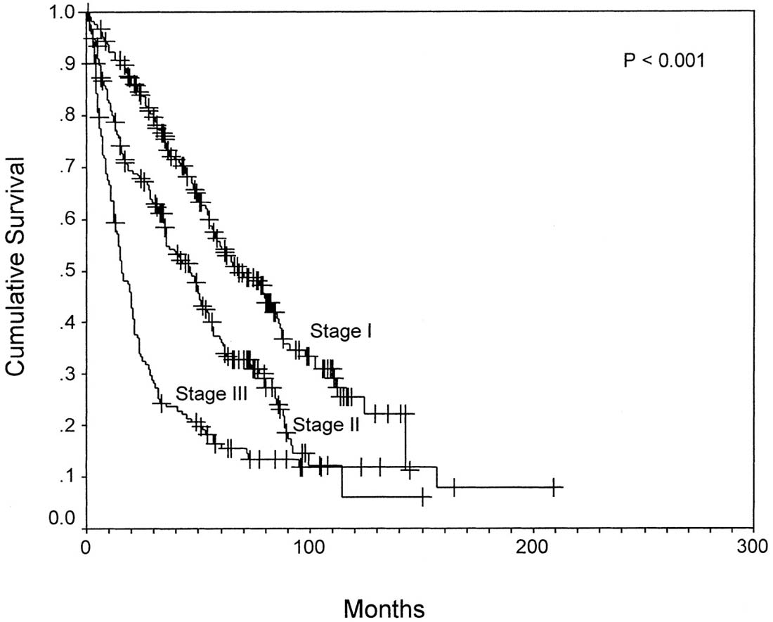
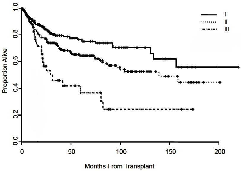
The T classification is based primarily on the results of a multicenter international study of pathological factors affecting prognosis after resection of HCC.3 The classification considers the presence or absence of vascular invasion (as assessed radiographically or microscopically), the number of tumor nodules (single vs. multiple), and the size of the largest tumor. The simplified classification adopted in the AJCC Cancer Staging Manual, 6th Edition and 7th Edition, stratifies patient survival well (Figure 22.2). This staging system subsequently was validated in multiple studies after liver resection4-10 and in a large multicenter series after liver transplantation (Figure 22.3).11
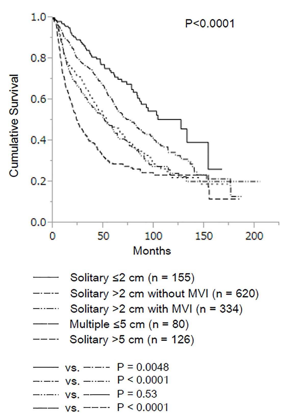
In a recent study of 1,109 patients with solitary HCC measuring up to 2 cm, neither microvascular invasion nor histologic grade had an impact on long-term survival (Figure 22.4).12 Based on these data, the AJCC Cancer Staging Manual, 8th Edition divides T1 disease into two subcategories: T1a, for patients with solitary HCC less than or equal to 2 cm irrespective of microvascular invasion, and T1b for patients with solitary HCC greater than 2 cm without microvascular invasion. The survival curve for solitary HCC greater than 2 cm with microvascular invasion was similar to that for multiple HCCs less than or equal to 5 cm. Therefore, these two groups were classified together in a revised T2 category.
In another long-term survival study of 754 patients, there was no survival difference between patients with T3a and those with T3b tumors (p = 0.073), or between patients with T3b and those with T4 tumors (p = 0.227).13 Thus, the revised 8th Edition reclassifies T3a as T3 and adds T3b to the T4 category.
Major vascular invasion is defined as invasion of the branches of the main portal vein (right or left portal vein, excluding the sectoral and segmental branches),3 one or more of the three hepatic veins (right, middle, or left),3 or the main branches of the proper hepatic artery (right or left hepatic artery).
Multiple tumors include satellitosis, multifocal tumors, and intrahepatic metastases. Assessment of lymph node involvement by clinical or radiographic means is a challenge, as reactive lymph nodes may be present. Invasion of adjacent organs other than the gallbladder or perforation of the visceral peritoneum is considered T4.
Several imaging modalities have relatively high sensitivity and specificity for diagnosis or staging of HCC, although test performance is suboptimal for small or well-differentiated HCC. Computed tomography (CT) and magnetic resonance (MR) imaging with intravenous contrast are the preferred examinations to detect HCC, and constitute key elements in defining the TNM stage.14-16 CT scanning should be performed with hepatic arterial, portal venous, and delayed venous phases. Similarly, if MR imaging is used, precontrast, arterial, venous, and delayed phases are essential. CT scanning frequently is the first examination, particularly if MR imaging is not available or is contraindicated. Ultrasound has lower sensitivity for detection of HCC, although it may be used to evaluate for vascular invasion of the portal and hepatic veins through color Doppler imaging.
Suggested Report FormatComplete pathological staging consists of evaluation of the primary tumor, including histologic grade, regional lymph node status, and underlying liver disease. Tumor size, number, and margin add to the critical prognostic data. Portal venous tumor thrombus should be clearly documented, as it carries a poor prognosis. Tumor grade is based on the degree of nuclear pleomorphism, as described by Edmonson and Steiner. Because of the prognostic significance of underlying liver disease in HCC, it is recommended that the results of the histopathologic analysis of the adjacent (nontumorous) liver be reported. Advanced fibrosis/cirrhosis (modified Ishak score of 5-6) is associated with a worse prognosis than absence of or moderate fibrosis (modified Ishak score of 0-4). Although grade and underlying liver disease have prognostic significance, they are not included in the current staging system.
Regional lymph node involvement is rare (5%). Positive lymph nodes are classified as Stage IV because they carry the same prognosis as cases with distant metastases. For pathological classification, vascular invasion includes gross as well as microscopic involvement of vessels.
Prognostic Factors Required for Stage Grouping
Beyond the factors used to assign T, N, or M categories, no additional prognostic factors are required for stage grouping.
Additional Factors Recommended for Clinical Care
Although there is clear agreement on the prognostic value of the extent of hepatic fibrosis, how to incorporate it into clinically relevant prognostic systems remains a controversy.2 Child-Pugh remains the most commonly used scoring system for assessing prognosis of cirrhosis and has been used in most clinical trials. The Okuda staging system17 was the first clinical system to join tumor extent parameters with cirrhosis-related ones. Other systems include the Cancer of the Liver Italian Program (CLIP),18 the Chinese University Prognostic Index (CUPI) scoring system, the Groupe d'Etude et de Traitement du Carcinoma Hepatocellulaire (GETCH) staging system, the Japan Integrated Staging (JIS) system, and the Barcelona Clinic Liver Cancer (BCLC) classification system.19 The BCLC couples prognosis with treatment assignment.
Multiple fibrosis scoring systems have been described for use in pathological evaluation of liver disease. The system most commonly used by US pathologists is the Batts-Ludwig system20; other systems include the modified Ishak scoring system21 and the METAVIR score.22 The latter is used more widely in Europe than in the United States.
The Ishak scoring system uses a 0-6 scale.
F0:Fibrosis score 0-4 (no to moderate fibrosis)
F1: Fibrosis score 5-6 (severe fibrosis or cirrhosis)
The Batts-Ludwig system uses a 0-4 scale, with a score of 3 defined as fibrous septa with architectural distortion but no obvious cirrhosis, and a score of 4 defined as cirrhosis.
α-Fetoprotein (AFP) is a nonspecific serum protein that generally is elevated in the setting of HCC, especially hepatitis B-related HCC.23 It has been an integral part of different scoring and staging systems, including the CLIP and CUPI. However, because of its nonspecificity, levels should be interpreted in the context of other findings, such as results of imaging studies. AFP is reported to be useful as a predictive marker for response to therapy; however, this application requires prospective study evaluation.24
Model for End-stage Liver Disease (MELD) scoring is useful in determining prognosis and prioritizing for receipt of a liver transplant.25 MELD uses serum bilirubin, serum creatinine, and international normalized ratio (INR) to predict survival. MELD is used by the United Network for Organ Sharing (UNOS) to help allocate livers for transplant.
c-Met receptor tyrosine kinase is the focus of several clinical trials of tyrosine kinase inhibitors and monoclonal antibodies as a therapeutic target. A prognostic impact has been suggested,26 but this has yet to be assessed in larger, prospective studies.
Insulin-like growth factor 1 (IGF-1) has been shown to correlate with hepatic reserve in patients with HCC.27
Higher plasma levels of vascular endothelial growth factor (VEGF) have been shown to correlate significantly with advanced clinicopathologic parameters and poor overall survival.28
The AJCC recently established guidelines that will be used to evaluate published statistical prediction models for the purpose of granting endorsement for clinical use.29 Although this is a monumental step toward the goal of precision medicine, this work was published only very recently. Therefore, the existing models that have been published or may be in clinical use have not yet been evaluated for this cancer site by the Precision Medicine Core of the AJCC. In the future, the statistical prediction models for this cancer site will be evaluated, and those that meet all AJCC criteria will be endorsed.
The following stratification criteria stem from the prognostic factor analyses that are suggested for use in clinical trials focusing on HCC, depending on the specific objectives of the study, the cancer stage(s), and the population under study, including sample size.
Definition of Primary Tumor (T)
T Category | T Criteria |
---|---|
TX | Primary tumor cannot be assessed |
T0 | No evidence of primary tumor |
T1 | Solitary tumor less than or equal to 2 cm, or greater than 2 cm without vascular invasion |
T1a | Solitary tumor less than or equal to 2 cm |
T1b | Solitary tumor greater than 2 cm without vascular invasion |
T2 | Solitary tumor greater than 2 cm with vascular invasion, or multiple tumors, none greater than 5 cm |
T3 | Multiple tumors, at least one of which is greater than 5 cm |
T4 | Single tumor or multiple tumors of any size involving a major branch of the portal vein or hepatic vein, or tumor(s) with direct invasion of adjacent organs other than the gallbladder or with perforation of visceral peritoneum |
Definition of Regional Lymph Node (N)
N Category | N Criteria |
---|---|
NX | Regional lymph nodes cannot be assessed |
N0 | No regional lymph node metastasis |
N1 | Regional lymph node metastasis |
Definition of Distant Metastasis (M)
M Category | M Criteria |
---|---|
M0 | No distant metastasis |
M1 | Distant metastasis |
Registry Data Collection Variables
Fibrolamellar carcinoma, previously known as fibrolamellar variant of HCC, lacks a specific staging system; thus, the current HCC staging system should be used. Lymph node involvement is much more common in fibrolamellar carcinoma than in HCC. In view of the common involvement of lymph nodes in fibrolamellar carcinoma, lymphadenectomy commonly is considered part of its surgical treatment.
The staging classification does not apply to biliary tumors, specifically intrahepatic cholangiocarcinomas, including combined hepatocellular-cholangiocarcinoma, which are considered in a separate staging system (see Chapter 23). It also does not apply to primary sarcoma or metastatic tumors.
G | G Definition |
---|---|
GX | Grade cannot be assessed |
G1 | Well differentiated |
G2 | Moderately differentiated |
G3 | Poorly differentiated |
G4 | Undifferentiated |