section name header

Disease Prologue

Summary

Cancers Staged Using This Staging System

This staging system applies to well-differentiated neuroendocrine tumors arising in the pancreas.

Cancers Not Staged Using This Staging System

These histopathologic types of cancer…Are staged according to the classification for…and can be found in chapter…
Carcinomas of the pancreas, including high-grade (grade 3), poorly differentiated neuroendocrine carcinomaExocrine pancreas28
Well-differentiated neuroendocrine tumors arising in the duodenum (C17.0) or ampulla of Vater (C24.1)Neuroendocrine tumors of the duodenum and ampulla of Vater30

Summary of Changes

ChangeDetails of ChangeLevel of Evidence
New chapterThis staging system was included in the exocrine and endocrine pancreas chapters in previous editions.N/A
AJCC Prognostic Stage GroupsPancreatic neuroendocrine tumors are now staged using a TNM staging system predominantly based on size; the criterion of peripancreatic soft tissue invasion was eliminated.II
Definition of Primary Tumor (T)The Tis distinction was eliminated.II
Definition of Distant Metastasis (M)M1 is subdivided as follows:

M1a: metastasis confined to the liver

M1b: metastases in at least one extrahepatic site (e.g., lung, ovary, nonregional lymph node, peritoneum, bone)

M1c: both hepatic and extrahepatic metastases

IV

ICD-O-3 Topography Codes

CodeDescription
C25.0Head of pancreas
C25.1Body of pancreas
C25.2Tail of pancreas
C25.4Islets of Langerhans (endocrine pancreas)
C25.7Other specified parts of pancreas
C25.8Overlapping lesion of pancreas
C25.9Pancreas, NOS

WHO Classification of Tumors

This list includes histology codes and preferred terms from the WHO Classification of Tumors and the International Classification of Diseases for Oncology (ICD-O). Most of the terms in this list represent malignant behavior. For cancer reporting purposes, behavior codes /3 (denoting malignant neoplasms), /2 (denoting in situ neoplasms), and in some cases /1 (denoting neoplasms with uncertain and unknown behavior) may be appended to the 4-digit histology codes to create a complete morphology code.

CodeDescription
8150Non-functioning pancreatic neuroendocrine tumor
8151Insulinoma
8152Glucagonoma
8153Gastrinoma
8155VIPoma
8156Somatostatinoma
8158ACTH-producing tumor
8240Neuroendocrine tumor (NET) G1 (carcinoid)
8249Neuroendocrine tumor (NET) G2

Lloyd RV, Osamura RY, Klöppel G, Rosai J, eds. World Health Organization Classification of Tumours of Endocrine Organs. Lyon: IARC; 2017. Used with permission.

Bosman FT, Carneiro F, Hruban RH, Theise ND, eds. World Health Organization Classification of Tumours of the Digestive System. Lyon: IARC; 2010. Used with permission.

International Agency for Research on Cancer, World Health Organization. International Classification of Diseases for Oncology. ICD-O-3-Online.http://codes.iarc.fr/home. Accessed September 29, 2017. Used with permission.

Introduction

Pancreatic neuroendocrine tumors (NETs) exhibit neuroendocrine differentiation and comprise less than 2% of all pancreatic malignancies. Although these tumors are rare, their relatively indolent nature translates into a relatively high prevalence: approximately 10% of all pancreatic tumors.1 An analysis of the Surveillance, Epidemiology, and End Results (SEER) database, 1973 to 2004, suggests that the incidence of pancreatic NETs is increasing.2 In 1973, the age-adjusted incidence of pancreatic NETs in the US population was 0.18 per 100,000; in 2003, it was 0.30 per 100,000.2 A recent analysis of the national population-based Cancer Registry of Norway revealed a similar trend.3 The age-stand ardized incidence rate of pancreatic NETs in the population overall (1993-2010) was 0.47 per 100,000 (95% CI, 0.43-0.52); in 2006 to 2010, it was 0.71 per 100,000 (95% CI, 0.61-0.82). The estimated annual percentage change was +6.9%. The reason for the increasing incidence likely is multifactorial; it probably is a result, at least in part, of more accurate classification by pathologists and improved diagnostic tools (cross-sectional and functional imaging), the latter of which has led to an increase in incidentally discovered tumors.4 Pancreatic NETs appear to be slightly more common in men (53%).1 They may occur at any age but are most commonly detected in the fifth to eighth decades; the median age at diagnosis is 60.2 With the exception of patients with insulinoma, patients with pancreatic NETs often present with advanced disease.2,5 Increased detection of incidental tumors has led to a reduction in the proportion of patients diagnosed with metastatic disease at presentation.6 Autopsy studies assessing the presence of small (less than 1 cm) NETs reported frequencies ranging from 0.8-10%.7

The grading classification scheme for pancreatic NETs has evolved over the years to encompass all NETs arising in the pancreas and gastrointestinal tract. Developed by the European Neuroendocrine Tumor Society (ENETS) and adopted by the World Health Organization (WHO) in 2010, the most common classification system consists of three grades (G1, G2, and G3), which correspond to well-differentiated (G1 and G2) and poorly differentiated neoplasms (G3).8-11

Grade is a significant and independent predictor of outcome.12,13 Most NETs arising in the pancreas are well differentiated (G1 and G2) tumors, which are termed pancreatic neuroendocrine tumors, pancreatic NETs, or panNETs. Poorly differentiated neoplasms are termed pancreatic neuroendocrine carcinomas or pancreatic NECs. Although this classification implies that all high-grade tumors are poorly differentiated, recent data suggest that a significant fraction of patients with well-differentiated tumors have a ki-67 index greater than 20%, and usually less than 50%. These “well-differentiated, high-grade” tumors represent a favorable prognostic category compared with poorly differentiated NECs.14

Approximately 20% of pancreatic NETs are associated with a clinical syndrome due to hormone excess. These “functional” tumors (F-pancreatic NETs) thus are defined based on the clinical syndrome, as asymptomatic production of hormones also may be detected in nonfunctional tumors (NF-pancreatic NETs).15 Among functional tumors, the most common hormones produced are insulin and gastrin (Table 34.1).16 Overproduction of glucagon, vasoactive intestinal peptide, or proinsulin is less common.17 Other, rarer hormone-mediated syndromes also have been reported, including pancreatic NETs secreting adrenocorticotropic hormone (ACTH), leading to Cushing's syndrome (ACTHomas); pancreatic NETs causing the carcinoid syndrome; and pancreatic NETs causing hypercalcemia (PTHrp-omas).18,19 Pancreatic NETs associated with calcitonin or ACTH production appear to be relatively aggressive, as do those that switch from one functional syndrome to another.

More than 50% of functional tumors are located in the tail of the pancreas, the exception being gastrinomas, which are more likely (63%) to be located in the head of the pancreas (Table 34.1).1 Interestingly, most gastrinomas (60-80%) actually arise in the duodenum; 75-85% are located in the “gastrinoma triangle” involving the duodenum and pancreatic head.17 Insulinomas typically are small, well-circumscribed tumors that are diagnosed at an early stage as the result of symptoms associated with hypoglycemia. The vast majority do not recur after resection. Importantly, F-pancreatic NETs may produce more than one hormone, and hormone production may change over the course of tumor progression.20

34.1 Clinical features of functional pancreatic neuroendocrine tumors

NameBiologically active peptide(s)Incidence(new cases/106 population/year)Tumor locationMost common symptoms/signs
Most common syndromes
InsulinomaInsulin1-3Pancreas (greater than 99%)Hypoglycemic symptoms (Whipple's triad)
Zollinger-Ellison syndromeGastrin0.5-2Duodenum (70%); pancreas (25%); other sites (5%)Abdominal pain, gastroesophageal reflux, diarrhea, duodenal ulcers, PUD/GERD
Less common syndromes (additional, rarer syndromes also exist)
VIPoma (Verner-Morrison syndrome, pancreatic cholera, WDHA syndrome)Vasoactive intestinal peptide0.05-0.2Pancreas (90%, adult); other (10%, neural, adrenal, periganglionic)Diarrhea, hypokalemia, dehydration
GlucagonomaGlucagon0.01-0.1Pancreas (100%)Rash, glucose intolerance, weight loss
SomatostatinomaSomatostatinRarePancreas (55%); duodenum/jejunum (44%)Diabetes mellitus, cholelithiasis, diarrhea
ACTHomaACTHRarePancreas (4-16% all ectopic Cushing's)Cushing's syndrome
Pancreatic NET causing carcinoid syndromeSerotoninRarePancreas (less than 1% all carcinoid syndrome)Flushing, diarrhea
PTHrp-oma (hypercalcemia)PTHrp, others unknownRarePancreasSymptoms due to hypercalcemia
Abbreviations: GERD, gastroesophageal reflux disease; PTHrp, parathyroid hormone-related protein; PUD, peptic ulcer disease; WDHA, watery diarrhea, hypokalemia, and achlorhydria. (Adapted from Jensen et al.17)
Pancreatic NETs frequently secrete several substances into the serum, including chromogranin A (CgA), pancreatic polypeptide (PP), pancreastatin, and neuron-specific enolase, without obvious clinical consequence. As such (assuming they are not secreting any additional hormones, as listed in Table 34.1), these tumors are considered NF-pancreatic NETs.18,21-23 NF-pancreatic NETs occur at least twice as frequently as F-pancreatic NETs in most series.

The etiology of pancreatic NETs largely is unknown. Most pancreatic NETs are thought to be sporadic. Of these, approximately 43% harbor DAXX/ATRX mutations, 44% harbor somatic inactivating mutations of MEN1, and 15% contain mutations in genes encoding mTOR pathway components.24 The prognostic significance of these mutations remains to be determined definitively.24,25 A small fraction of pancreatic NETs (less than 10%) arise in the context of a hereditary cancer syndrome, the most common of which is multiple endocrine neoplasia type 1 (MEN1).26 MEN1 is caused by mutations in the MEN1 gene located at chromosome 11q13 region, thus altering transcriptional regulation, genomic stability, cell division, and cell cycle control.27 Affected patients develop hyperplasia or neoplasia of multiple endocrine and nonendocrine tissues, including parathyroid adenomas (95-100%) resulting in hyperparathyroidism, pituitary adenomas (54-65%), adrenal adenomas (27-36%), various NETs (gastric, lung, thymic; 0-10%), thyroid adenomas (up to 10%), various skin tumors (80-95%), central nervous system tumors (up to 8%), and smooth muscle tumors (up to 10%).17,28 Pancreatic NETs develop in 80-100% of MEN1 patients and are nearly always multifocal. Pancreatic NETs in this setting often are small and nonfunctional. Gastrinomas (greater than 80% duodenal) develop in 54% of MEN1 patients, insulinomas in 18%, and glucagonomas, vasoactive intestinal peptide-secreting tumors (VIPomas), growth hormone-releasing factor-secreting tumors (GRFomas), and somatostatinomas in less than 5%.17 Pancreatic NETs also occur in up to 10% of patients with von Recklinghausen's disease (also known as neurofibromatosis type 1 [NF1]), 10-17% of patients with von Hippel-Lindau (VHL) syndrome, and rarely in patients with tuberous sclerosis. The likelihood of an underlying genetic syndrome depends on the type of tumor as well as the patient's personal and family history, which should be recorded for every patient presenting with a pancreatic NET. Multifocal disease is a risk factor, as is the type of hormone produced. MEN1 is present in 20-30% of patients with Zollinger-Ellison syndrome (ZES; usually associated with a duodenal gastrinoma) and 5% of patients with an insulinoma.17 Given the high incidence of parathyroid adenomas in MEN1 patients, assessment of ionized calcium and serum parathyroid hormone (PTH) may be used as a screening tool in appropriate patients, with the caveat that secondary elevation of PTH may occur in the setting of vitamin D deficiency.29,30

The staging of pancreatic NETs depends on the size and extent of the primary tumor (including whether there is lymph node involvement and /or distant metastasis). Importantly, the American Joint Committee on Cancer (AJCC) TNM classification system for pancreatic NETs was first introduced in the AJCC Cancer Staging Manual, 7th Edition and was based on the staging algorithm for exocrine pancreatic carcinomas. Recognizing that exocrine and neuroendocrine tumors of the pancreas are distinct entities in terms of underlying tumor biology and prognosis, other staging classification systems have been proposed.12,13,31-33 The 7th Edition AJCC staging system is significantly associated with survival, but its value is limited by the inability to discriminate between the intermediate stages (i.e., Stages II and III are prognostically indistinguishable).12 Furthermore, some of the parameters necessary for staging (e.g., presence of extrapancreatic extension) are difficult to assess pathologically because of the expansile growth pattern common in pancreatic NETs. The system developed by ENETS in 2006 incorporates a narrower T definition. It has proven prognostic value and appears to provide superior distinction among stages (I, II, III, and IV) compared with the AJCC/Union for International Cancer Control (UICC)/WHO 2010 system.12 Therefore, the AJCC Cancer Staging Manual, 8th Edition staging system has been modified to be consistent with the ENETS system.

Importantly, even the current ENETS staging system is imperfect; patients with Stage IIIB (any T N1 M0) disease fare better than those with Stage IIIA (T4 N0 M0) disease, and clear discrimination between stages has not been evident in all validation studies.12,13 There are several potential reasons for this, including the generally favorable survival outcomes of patients with nonmetastatic disease, which limits the ability to distinguish prognostic groups, especially in the absence of very long-term follow-up. In addition, inconsistent lymph node sampling may underlie the conflicting findings of the prognostic importance of lymph node involvement.34 Further refinement of the ENETS staging system is likely, although inadequate data exist for modifications at present.12,33

Surgical resection remains the only potentially curative treatment for well-differentiated (G1/G2) pancreatic NETs. The natural history of these tumors is poorly understood because of their relative rarity, but accepted prognostic factors include patient age, distant metastases, tumor grade, and tumor differentiation.12 Recent studies suggest that lymph node involvement also may be an important prognostic factor.35,36 For accurate staging, routine lymph node sampling is critical for most patients with pancreatic NETs undergoing surgery.

Well-differentiated insulinomas rarely metastasize and have a particularly good prognosis (greater than 90% have a benign clinical course); the prognosis of other functional tumors appears to match that of nonfunctional tumors in most series, although this is not consistent in all studies.12 The type of surgery performed depends on the tumor stage, location, and functional status, and ranges from enucleation to pancreaticoduodenectomy.37 Because insulinomas typically are small and pursue a benign clinical course, enucleation of tumors located away from the main pancreatic duct usually is curative. For insulinomas close to the main pancreatic duct, a pancreatectomy, such as distal pancreatectomy for left-sided lesions or pancreaticoduodenectomy for right-sided lesions, may be required. Insulinomas located in the pancreatic neck region may be treated with a central pancreatectomy. Because most insulinomas are very indolent, a lymphadenectomy typically is not necessary, and spleen preservation may be considered.

In contrast, most NF-pancreatic NETs and other F-pancreatic NETs (i.e., not insulinomas) are capable of malignant behavior. The optimal treatment of incidentally identified small NF-pancreatic NETs less than 1.5 cm is unclear. Recent analyses suggest that surveillance, rather than surgery, is appropriate in many cases. A careful analysis of the potential risks and benefits of pancreatic resection is required, particularly in asymptomatic elderly patients with significant comorbidities.4,38

Most larger NF-NETs or localized pancreatic NETs with an elevated proliferative index have a higher risk of invasion and metastases; thus, resection with a lymphadenectomy should be strongly considered for these neoplasms. Even small pancreatic NETs may be associated with significant lymph node and /or liver metastases.39 Several factors influence the choice of surgical procedure, including primary tumor size, Ki-67 labeling index, mitotic index, location, and medical comorbidities. For left-sided lesions, a distal pancreatectomy and , if necessary, an en bloc splenectomy should be done to ensure adequate lymphadenectomy. A pancreaticoduodenectomy (Whipple procedure) should be considered for right-sided lesions. Rarely, a total pancreatectomy with en bloc splenectomy is required for large lesions that occupy much of the pancreas, but given the high morbidity of such a procedure, a thorough evaluation should be performed first to rule out metastatic disease. A central pancreatectomy with regional lymphadenectomy might be considered for lesions located in the pancreatic neck. If enucleation is considered for these neoplasms, a regional lymphadenectomy may be done to ensure adequate lymph node sampling for staging. Removal of a pancreatic primary tumor in the setting of resectable liver metastases may be considered, particularly if a pancreaticoduodenectomy is not required. Although complete resection and /or palliative debulking surgeries (i.e., primary tumor and liver metastases) are not necessarily curative, data from nonrand omized studies suggest they may be associated with improved hormone-mediated symptoms and improved survival in carefully selected patients.40,41

Several treatment options exist for patients with advanced, unresectable pancreatic NETs. Somatostatin analogs have cytostatic activity and may be used to treat hormone-mediated symptoms.42-44 Chemotherapy has been used with some success, although the optimal regimen remains unclear.45-47 Two targeted therapies are approved for this indication: everolimus (an inhibitor of mTOR signaling) and sunitinib (an oral inhibitor of vascular endothelial growth factor signaling) both delay progression of progressive panNETs.48,49 The use of liver-directed therapy or other treatments depends on several factors, including the tumor's growth rate, extent of disease, and whether the tumor is functional. See published guidelines for additional information regarding the workup and treatment of panNETs.17,37,50
Anatomy

Primary Site(s)

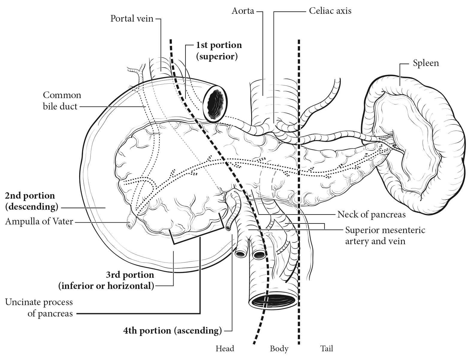
The pancreas is a long, coarsely lobulated gland that lies transversely across the posterior abdomen and extends from the duodenum to the splenic hilum. The organ is divided into a head with a small uncinate process, a neck, a body, and a tail. These are contiguous regions without sharp anatomic distinctions. The pancreas neck lies anterior to the superior mesenteric vessels. The anterior aspect of the body of the pancreas is covered by peritoneum and is in direct contact with the posterior wall of the stomach; posteriorly, the pancreas extends within the retroperitoneal soft tissue to the inferior vena cava, superior mesenteric vein, splenic vein, and left adrenal and kidney.

Pancreatic NETs are distributed throughout the pancreas.1,11,12 Tumors of the head of the pancreas are those arising to the right of the superior mesenteric-portal vein confluence (Figure 34.1). The uncinate process is the part of the pancreatic head that extends behind the superior mesenteric vessels. The neck overlies the superior mesenteric vessels. Tumors of the body of the pancreas are defined as those arising to the left of the neck. Laterally to the left side, the body becomes the tail of the pancreas without any clear junction point.

34.1 Anatomy of the pancreas.

Regional Lymph Nodes

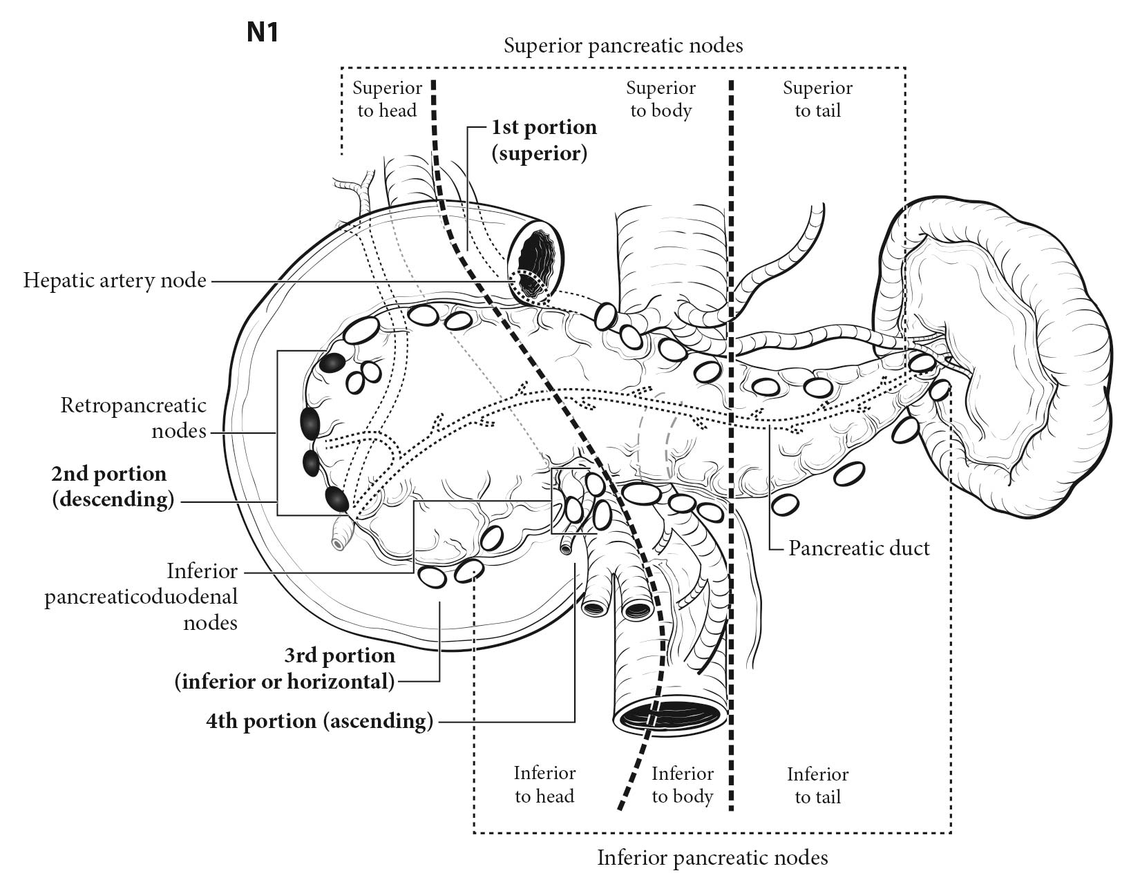
The stand ard regional lymph node basins and soft tissues resected for tumors located in the head and neck of the pancreas include lymph nodes along the common bile duct, common hepatic artery, portal vein, posterior and anterior pancreatoduodenal arcades, and the superior mesenteric vein and right lateral wall of the superior mesenteric artery (Figure 34.2). For cancers located in the body and tail, regional lymph node basins include lymph nodes along the common hepatic artery, celiac axis, splenic artery, and splenic hilum (Figure 34.2). Involvement of peripancreatic lymph nodes is considered regional disease and classified as N1.

34.2 Regional lymph nodes of the pancreas (anterior view).

Metastatic Sites

Distant spread is common on presentation and most commonly involves the liver. Metastases to other sites, such as lung, bones, and peritoneum, also may occur. Involvement of the para-aortic or other distant lymph nodes (i.e., retroperitoneal, retrocrural, and mesenteric lymph nodes) is considered M1 disease (see Figure 34.2 for regional lymph nodes). Seeding of the peritoneum (even if limited to the lesser sac region) is considered M1.

Classification Rules

Clinical Classification

Guidelines have been established for the workup of patients with pancreatic NETs.37,50,51 In general, patients should be evaluated by multiphasic computed tomography (CT) or magnetic resonance (MR) imaging to assess 1) the proximity of the primary pancreatic NET to major vessels and 2) the clinical T, N, and M staging of the lesion before any surgical or medical therapy is considered. In addition, biochemical assessment, somatostatin receptor scintigraphy (SRS), and endoscopic ultrasonography (EUS) should be performed as appropriate. For localized tumors, a biopsy is not necessarily required before surgical resection. However, if a biopsy (e.g., endoscopic biopsy, percutaneous core needle biopsy, fine-needle aspiration) is performed, the results should be incorporated when assessing clinical stage.

By definition, patients with F-pancreatic NETs present with hormone-mediated symptoms consistent with a characteristic syndrome.17,37 As such, the diagnosis of F-pancreatic NETs requires demonstration of a significantly elevated hormone combined with clinical signs or symptoms of oversecretion (e.g., ulcers in the setting of gastrinoma, hypoglycemia in the setting of insulinoma).37 Specifically, for insulinomas, assessment of plasma insulin, proinsulin, and C-peptide is needed at the time of glucose determinations, usually during a 72-hour supervised fast.17 For ZES, fasting serum gastrin (FSG) should be assessed, unstimulated or during a secretin provocation test.18 The diagnosis of ZES requires demonstration of an inappropriate elevation of gastrin in the presence of hyperchlorhydria or an acidic pH (less than 2). FSG alone is not diagnostic, because hypergastrinemia may be caused by hypochlorhydria/achlorhydria (chronic atrophic fundic [autoimmune] gastritis, often associated with pernicious anemia) and is a common consequence of proton pump inhibitor (PPI) use. Ideally, PPIs should be stopped in order to make the diagnosis of gastrinoma, but this may be difficult in patients with severe gastroesophageal reflux disease/gastrinoma and necessitates switching to an H2 blocker (ideally in the context of a specialty unit with experience in diagnosing ZES). Furthermore, other disorders cause hypergastrinemia with hyperchlorhydria (e.g., Helicobacter pylori infection, gastric outlet obstruction, renal failure, antral G-cell syndromes, G-cell hyperplasia, short bowel syndrome, retained antrum). FSG level alone cannot distinguish ZES from achlorhydric states including PPI use.

For VIPomas, the plasma vasoactive intestinal peptide level must be determined. For glucagonoma, measurement of plasma glucagon levels is appropriate. For GRFomas, plasma growth hormone and growth hormone-releasing factor levels should be measured. For Cushing's syndrome, urinary cortisol, plasma ACTH, and appropriate ACTH suppression studies should be performed. For pancreatic NET-associated hypercalcemia, measurement of both serum PTH and PTHrP levels is indicated, and for a pancreatic NET associated with carcinoid syndrome, urinary or plasma 5-hydroxyindoleacetic acid (5-HIAA) should be measured.18,52 Assessment for hormones associated with rarer syndromes should be performed as clinically indicated.19

In contrast, unless incidentally discovered during a workup for an unrelated problem, NF-pancreatic NETs typically present with symptoms due to the tumor itself, including abdominal pain (40-60%), weight loss, or jaundice.18,23 53 Although NF-pancreatic NETs do not secrete peptides causing a clinical syndrome, they characteristically secrete several other peptides, such as CgA, PP, neuron-specific enolase, and /or pancreastatin (a subunit of chromogranin), which may be helpful for the diagnosis and monitoring of affected patients.18,23 At this time, there is insufficient evidence regarding the impact of any individual tumor marker on clinical decision making to recommend a particular assay.

Imaging

Information necessary for the clinical staging of pancreatic NETs may be obtained from physical examination; cross-sectional radiographic imaging studies, including triphasic (noncontrast, arterial, and venous) contrast-enhanced CT or MR imaging; and SRS.5 (Refer to established guidelines for details.)37 The detection rate of pancreatic primary tumors is in the range of 75-79% with cross-sectional imaging, and the sensitivity for detection of liver metastases is up to 80% with contrast-enhanced, multiphasic CT or MR imaging.5,54 SRS with indium-111 pentetreotide imaging (Octreoscan; Mallinckrodt Pharmaceuticals, Dublin, Ireland ) has a sensitivity of up to 90% for pancreatic NETs, depending on tumor size and type (e.g., 20-60% sensitivity for detecting insulinomas).54 Stand ard fluorodeoxyglucose positron emission tomography (PET) with fluorine-18 glucose has limited value in the evaluation of well-differentiated pancreatic NETs. EUS also provides useful information for detection of small and /or multifocal pancreatic NETs and is the procedure of choice for performing fine-needle aspiration biopsy of the pancreas. Studies suggest detection rates of 90-100% for pancreatic lesions and 45-60% for tumors arising in the duodenum.55

Unlike its exocrine counterpart (pancreatic ductal adenocarcinoma), tumor involvement of the celiac axis or superior mesenteric artery is rare in pancreatic NETs. The stand ard radiographic assessment of resectability includes evaluation for distant metastases (e.g., peritoneal, liver, bone); the patency of the superior mesenteric vein and portal vein, as well as the relationship of these vessels and their tributaries to the tumor; and the relationship of the tumor to the superior mesenteric artery, celiac axis, and hepatic artery.

NET imaging using PET with gallium-68 (68Ga)-labeled somatostatin analogs appears promising (same-day results, potential for increased sensitivity, broader affinity profile, better spatial resolution, easier quantification of tracer uptake).56, 57,58 Studies to assess the value of 68Ga-labeled somatostatin analog-based PET/CT and PET/MR imaging relative to stand ard somatostatin scintigraphy are ongoing. This technology represents an emerging imaging tool for NETs in the United States; a kit for the preparation of 68Ga-dotatate injection for PET imaging recently received U.S. Food and Drug Administration approval.59

Pathological Classification

Pathological staging is based on surgical resection specimens. The most sensitive pathological staging is obtained by examining surgically resected primary tumor(s), lymph nodes, and distant metastases according to an established minimum pathology dataset.10,60-62

Partial resection (pancreaticoduodenectomy or distal pancreatectomy) or complete resection of the pancreas, including the tumor and associated regional lymph nodes, provides the optimal information for pathological staging. In pancreaticoduodenectomy specimens, the bile duct, pancreatic duct, and superior mesenteric artery margins should be evaluated grossly and microscopically. The superior mesenteric artery margin also has been termed the retroperitoneal, vascular, or uncinate margin. In total pancreatectomy specimens, the bile duct and retroperitoneal margins should be assessed. Duodenal (with pylorus-preserving pancreaticoduodenectomy) and gastric (with stand ard pancreaticoduodenectomy) margins rarely are involved, but their status should be included in the surgical pathology report. Reporting of margins may be facilitated by ensuring documentation of the pertinent margins: 1) common bile (hepatic) duct, 2) pancreatic neck, 3) superior mesenteric artery, 4) other soft tissue margins (i.e., posterior pancreatic, duodenum, and stomach).

A rich lymphatic network surrounds the pancreas, and accurate tumor staging requires analysis of all lymph nodes removed. Optimal histologic examination of a pancreaticoduodenectomy specimen should include analysis of a minimum of 12 lymph nodes. However, the number of lymph nodes removed depends on the type of surgery performed and may not be feasible in the setting of a distal pancreatectomy without en bloc splenectomy. Therefore, spleen preservation should not be done when there is a high chance of malignancy (i.e., for any pancreatic NET other than a small insulinoma). Furthermore, lymph nodes typically are not sampled in the setting of an enucleation procedure. The number of lymph nodes examined should be specified in the pathology report. Anatomic division of regional lymph nodes is not necessary; however, separately submitted lymph nodes should be reported as labeled by the surgeon. Finally, an N category (N1 or N0) should be assigned as long as at least one lymph node has been assessed, even if the optimal number of lymph nodes have not been examined. Nx should be applied only if no lymph nodes were assessed (e.g., if enucleation was performed). Positive peritoneal cytology is considered M1.

The pathological diagnosis of pancreatic NETs may be established by histologic evaluation alone if classic morphologic features are present. However, the morphology of pancreatic NETs is highly variable, and alternative diagnoses, such as acinar cell carcinoma (or mixed acinar NEC), solid pseudopapillary neoplasm, or ductal adenocarcinoma, may be considered in many cases. Immunolabeling for the general neuroendocrine markers chromogranin and synaptophysin is helpful to support the diagnosis of pancreatic NETs, provided other markers are performed to exclude the alternative diagnoses, some of which share expression of chromogranin or synaptophysin with pancreatic NETs.

Immunohistochemistry for hormones is optional and does not have prognostic significance. Positive immunostaining for a hormone does not necessarily indicate the presence of a hormonal syndrome.

For pancreatic NETs, the r prefix is used for recurrent tumor status (rTNM) following a disease-free interval after treatment.

Prognostic Factors

Prognostic Factors Required for Stage Grouping

Beyond the factors used to assign T, N, or M categories, no additional prognostic factors are required for stage grouping.

Additional Factors Recommended for Clinical Care

Mitotic Count

Tumor grade is determined by mitotic count and Ki-67 labeling index and correlates with progression-free survival, overall survival, and lymph node status in pancreatic NETs.12,63,64 Mitotic count should be assessed as the number of mitoses per 10 high-power fields (HPF): HPF = 2 mm2, at least 50 fields (at 40× magnification) evaluated.

Mitotic count, # of mitoses per 10 HPF (specify:)

  • less than 2
  • 2 to 20
  • greater than 20
  • Not performed
AJCC Level of Evidence: I
Ki-67 Labeling Index

Tumor grade is determined by mitotic count and Ki-67 labeling index and correlates with progression-free survival, overall survival, and lymph node status in pancreatic NETs. The proliferation index as measured by Ki-67 also correlates with these outcome measures in pancreatic NETs.12,38,63,64 The Ki-67 index typically is measured using the MIB1 antibody, by counting the number of immunolabeled tumor cells per 500 to 2,000 cells in areas of highest nuclear labeling; the Ki-67 index is expressed as a percentage.

  • Ki-67 labeling index (specify:)
    • less than 3%
    • 3% to 20%
    • greater than 20%
  • Other (specify)
  • Not performed
AJCC Level of Evidence: I
Associated Genetic Syndrome

Gastroenteropancreatic NETs sometimes arise in the setting of an inherited cancer syndrome characterized by a germline mutation. Tumors arising in the setting of an inherited cancer syndrome may be multiple and appear to be associated with a better prognosis than sporadic tumors, at least in the setting of MEN1.12

This factor should be recorded as follows:

  • Familial syndrome
    • Multiple endocrine neoplasia type 1 (alteration in MEN1)
    • Von Hippel-Lindau disease (mutation in VHL gene)
    • Neurofibromatosis type 1 (mutation inNf1)
    • Tuberous sclerosis complex (mutation in TSC1or TSC2)
    • Mahvash disease (pancreatic NET caused by inactivating glucagon receptor mutation)65
    • Other syndrome
  • Sporadic tumor
  • Unknown/unable to assess
AJCC Level of Evidence: II
Chromogranin A (CgA)

CgA is a 49-kDa acidic polypeptide present in the secretory granules of all neuroendocrine cells. CgA is a general NET marker, and plasma or serum CgA may be used as a marker in patients with either F- or NF-pancreatic NETs.18,66-68 CgA has prognostic significance, with higher levels indicating a worse prognosis.69 Furthermore, changes over time may be useful in assessing for recurrence after surgery or response to therapy in patients with metastatic disease.66,67,70,71

Despite the potential merits of monitoring CgA levels, the clinical utility of CgA is limited by the fact that it is falsely elevated in the setting of PPI use, chronic atrophic gastritis, renal failure, severe hypertension, and other conditions.8 Moreover, levels may fluctuate based on time of collection and fasting versus nonfasting states. Also, the upper limit of normal (ULN) varies widely depending on the assay used and whether plasma or serum is assessed; thus, both the assay and type of sample should be considered when comparing CgA values over time.72 As a result, routine measurement of CgA is not a consensus National Comprehensive Cancer Network (NCCN) recommendation. Multiple Clinical Laboratory Improvement Amendments (CLIA)-licensed and College of American Pathology (CAP)-accredited reference laboratories in the United States can measure CgA levels.

AJCC Level of Evidence: II
Functionality

Tumors with hormone expression noted on immunohistochemistry but not associated with a clinically relevant syndrome or signs should be recorded as nonfunctional. Similarly, tumors associated with elevated blood levels of hormones that are not associated with clinical symptoms also should be recorded as nonfunctional. Insulinomas typically have a low risk of metastasis and thus carry a good prognosis; the outcome of other F-pancreatic NETs appears to be similar to that of nonfunctional tumors in most studies.12,13 Importantly, the clinical manifestations and morbidity of F-pancreatic NETs may differ, and in some cases, mortality may relate to the hormonal syndrome rather than to the extent of the neoplasm.

Functionality should be characterized as follows:

  • Functional
    • Insulinoma
    • Gasrinoma (ZES)
    • Glucagonoma
    • VIPoma (Verner-Morrison syndrome)
    • Somatostatinoma
    • ACTHoma
    • PanNET causing carcinoid syndrom (5-HIAA, serotonin excess)
    • PanNET causing hypercalcemia (PTHrp or other)
    • Other
  • Nonfunctional
  • Unknown/unable to assess
AJCC Level of Evidence: III

Pancreastatin

Pancreastatin is a posttranslational product of CgA whose blood level inversely correlates with prognosis.67,73 Levels also correlate with the number of liver metastases and may be useful in monitoring for recurrence after surgery or for response to therapy.67,73 Perioperative pancreastatin level is an independent predictor of outcome in resected small bowel NETs and pancreatic NETs.74 Additional studies are needed to validate these findings. Compared with CgA, pancreastatin may have better specificity in diagnosing NETs because it is not affected by PPI use or atrophic gastritis.8,9,75 There are at least three large CLIA-certified and CAP-accredited reference laboratories that routinely measure pancreastatin.

AJCC Level of Evidence: III

Risk Assessment

Risk Assesment Models

The AJCC recently established guidelines that will be used to evaluate published statistical prediction models for the purpose of granting endorsement for clinical use.76 Although this is a monumental step toward the goal of precision medicine, this work was published only very recently. Therefore, the existing models that have been published or may be in clinical use have not yet been evaluated for this cancer site by the Precision Medicine Core of the AJCC. In the future, the statistical prediction models for this cancer site will be evaluated, and those that meet all AJCC criteria will be endorsed.

Recommendations

Histologic grade (G1, G2 vs. G3)

TNM Definitions

Definition of Primary Tumor (T)

T CategoryT Criteria
TXTumor cannot be assessed
T1Tumor limited to the pancreas, less than 2 cm
T2Tumor limited to the pancreas, 2-4 cm
T3Tumor limited to the pancreas, greater than 4 cm; or tumor invading the duodenum or common bile duct
T4Tumor invading adjacent organs (stomach, spleen, colon, adrenal gland ) or the wall of large vessels (celiac axis or the superior mesenteric artery)

Limited to the pancreas means there is no invasion of adjacent organs (stomach, spleen, colon, adrenal gland ) or the wall of large vessels (celiac axis or the superior mesenteric artery). Extension of tumor into peripancreatic adipose tissue is NOT a basis for staging.

Multiple tumors should be designated as such (the largest tumor should be used to assign T category): If the number of tumors is known, use T(#); e.g., pT3(4) N0 M0. If the number of tumors is unavailable or too numerous, use the m suffix, T(m); e.g., pT3(m) N0 M0.

Definition of Regional Lymph Node (N)

N CategoryN Criteria
NXRegional lymph nodes cannot be assessed
N0No regional lymph node involvement
N1Regional lymph node involvement

Definition of Distant Metastasis (M)

M CategoryM Criteria
M0No distant metastasis
M1Distant metastases
M1aMetastasis confined to liver
M1bMetastases in at least one extrahepatic site (e.g., lung, ovary, nonregional lymph node, peritoneum, bone)
M1cBoth hepatic and extrahepatic metastases

Stage Prognostic

!!Calculator!!

AJCC PROGNOSTIC STAGE GROUPS

When T is…and N is…and M is…Then the stage group is…
T1N0M0I
T2N0M0II
T3N0M0II
T4N0M0III
Any TN1M0III
Any TAny NM1IV

Registry Data

Registry Data Collection Variables

  1. Size of tumor (value, unknown)
  2. Presence of invasion into adjacent organs/structures (Y/N)
    1. If yes, which ones (pick all that apply):
      1. Stomach (Y/N)
      2. Duodenum (Y/N)
      3. Spleen (Y/N)
      4. Colon (Y/N)
      5. Other:
    2. If yes, were multiple adjacent organs involved (Y/N)
  3. Presence of necrosis
  4. Number of tumors (multicentric disease at primary site)
  5. Lymph node status (including number of lymph nodes assessed and number of positive nodes)
  6. Grade (based on Ki-67 and /or mitotic count; G1, G2, G3, unknown)
  7. Mitotic count (value, unknown)
  8. Ki-67 Labeling Index (value, unknown)
  9. Perineural invasion (Y/N)
  10. Lymphovascular invasion (Y/N)
  11. Margin status (+/-)
  12. Functional status (Y/N, type of syndrome)
  13. Genetic syndrome (Y/N, type of syndrome)
  14. Location in pancreas (head, tail, body, junction body/tail, junction body/head, unknown)
  15. Type of surgery (enucleation; distal pancreatectomy with splenectomy; distal pancratectomy without splenectomy; central pancreatectomy;; pancreaticoduodenectomy (Whipple procedure); unknown; other)
  16. Preoperative CgA level (absolute value with ULN; unknown)
  17. Preoperative pancreastatin level (absolute value with ULN; unknown)
  18. Age of patient

Histopathologic type

This staging system applies to well-differentiated NETs arising in the pancreas.

Histologic grade

HISTOLOGIC GRADE (G)

Grading of pancreatic NETs is required for prognostic stratification and should be performed on all resection specimens and on biopsy specimens containing sufficient tumor tissue to allow accurate measurement of proliferation (50 HPF for mitotic counting and 500 cells to determine the Ki-67 index). If multiple disease sites are sampled (e.g., a primary tumor as well as a metastasis), the grade of each site should be recorded separately. If multiple foci are sampled within a single anatomic site (e.g., multiple liver metastases), the highest grade may be recorded. The grading scheme described here is currently endorsed by ENETS/WHO for gastrointestinal and pancreatic neuroendocrine neoplasms.

GG Definition
GXGrade cannot be assessed
G1Mitotic count (per 10 HPF) less than 2 and Ki-67 index (%) less than 3
G2Mitotic count (per 10 HPF) equal to 2-20 or Ki-67 index (%) equal to 3-20
G3Mitotic count (per 10 HPF) greater than 20 or Ki-67 index (%) greater than 20

10 HPF = 2 mm2; at least 50 HPF (at 40× magnification) must be evaluated in areas of highest mitotic density in order to match WHO 2010 criteria.

MIB1 antibody; % of 500-2,000 tumor cells in areas of highest nuclear labeling.

The Ki-67 index is based on the region with the highest labeling rate (“hot spot”), determined by examining the Ki-67 stain at low magnification. In the event of discordance between the grade indicated by the mitotic count and that suggested by the Ki-67 index, the higher grade should be assigned. Nuclear pleomorphism is not a useful feature for grading neuroendocrine neoplasms. Although necrosis has been regarded as a prognostic factor in some studies, its presence is not incorporated into the grading scheme.

Well-differentiated NETs are subdivided into G1 and G2 tumors based on proliferative and mitotic index. G1 and G2 refer to well-differentiated NETs displaying diffuse and intense expression of two general immunohistochemical neuroendocrine markers (i.e., CgA and synaptophysin). G3 usually indicates a poorly differentiated NEC, which should be staged using the system for pancreatic carcinomas (Chapter 28). High-grade (G3) tumors typically are characterized by a high mitotic count/Ki-67 index, nuclear pleomorphism, and extensive necrosis. Immunohistochemical expression of CgA and /or synaptophysin may be weak.

In some cases, pancreatic neuroendocrine neoplasms with well-differentiated histologic features have a Ki-67 index (or, more rarely, a mitotic count) within the G3 range.14,77-80 Although these neoplasms currently are considered high grade in the WHO grading scheme, emerging data suggest that they are not as aggressive as poorly differentiated NECs with undifferentiated, small cell, or large cell morphology, and their response to therapy is more in line with that of other well-differentiated pancreatic NETs.77 Progression of a well-differentiated pancreatic NET from G1 or G2 to G3 also has been documented. It has been proposed that these well-differentiated neuroendocrine neoplasms be classified as well-differentiated NETs, G3. As such, they should be staged using the parameters of pancreatic NETs, rather than those of pancreatic carcinomas.

Survival

Four years of data (diagnosis years 2010-2013) from the National Cancer Data Base were used to assess survival. Maximum follow-up time for these patients was 3 years. Selection criteria included primary site codes C25.0, C25.1, C25.2, C25.4, C25.7, C25.8, and C25.9; grade 1 and grade 2; histology codes 8150, 8151, 8152, 8153, 8155, 8156, 8158, 8240, 8249, and 8246; ages 18 and older; and only primary or first of multiple primaries.

In this study, 1,174 pancreatic NETs were identified and recategorized according to the 8th Edition (ENETS) staging system as indicated: Stage 1, n =262; Stage IIA, n = 221; Stage IIB, n = 191; Stage IIIA, n = 32; Stage IIIB, n = 346; and Stage IV, n = 122). Product limit survival curves (Kaplan-Meier) and 95% confidence intervals adjusted for age were produced; however, there were not enough cases or follow-up to assess survival accurately. Therefore, survival tables and charts are not included.

Bibliography

  1. Yao JC, Eisner MP, Leary C, et al. Population-based study of islet cell carcinoma. Annals of surgical oncology. Dec 2007;14(12):3492-3500.
  2. Yao JC, Hassan M, Phan A, et al. One hundred years after "carcinoid": epidemiology of and prognostic factors for neuroendocrine tumors in 35,825 cases in the United States. J Clin Oncol. Jun 20 2008;26(18):3063-3072.
  3. Boyar Cetinkaya R, Aagnes B, Thiis-Evensen E, Tretli S, Bergestuen DS, Hansen S. Trends in Incidence of Neuroendocrine Neoplasms in Norway: A Report of 16,075 Cases from 1993 through 2010. Neuroendocrinology. 2015.
  4. Crippa S, Partelli S, Zamboni G, et al. Incidental diagnosis as prognostic factor in different tumor stages of nonfunctioning pancreatic endocrine tumors. Surgery. 2014;155(1):145-153.
  5. Falconi M, Bartsch DK, Eriksson B, et al. ENETS Consensus Guidelines for the management of patients with digestive neuroendocrine neoplasms of the digestive system: well-differentiated pancreatic non-functioning tumors. Neuroendocrinology. 2012;95(2):120-134.
  6. Zerbi A, Falconi M, Rindi G, et al. Clinicopathological features of pancreatic endocrine tumors: a prospective multicenter study in Italy of 297 sporadic cases. Am J Gastroenterol. Jun 2010;105(6):1421-1429.
  7. Kimura W, Kuroda A, Morioka Y. Clinical pathology of endocrine tumors of the pancreas. Analysis of autopsy cases. Digestive diseases and sciences. Jul 1991;36(7):933-942.
  8. Rindi G, Klersy C, Inzani F, et al. Grading the neuroendocrine tumors of the lung: an evidence-based proposal. Endocrine-related cancer. Feb 2014;21(1):1-16.
  9. Rindi G, Arnold R, Capella C, et al. Nomenclature and classification of digestive neuroendocrine tumours. World Health Organization classification of tumours, pathology and genetics of tumours of the digestive system. IARC Press, Lyon. 2010:10-12.
  10. Bosman FT, Carneiro F, Hruban RH, Theise ND. WHO classification of tumours of the digestive system. World Health Organization; 2010.
  11. Qadan M, Ma Y, Visser BC, et al. Reassessment of the current American Joint Committee on Cancer staging system for pancreatic neuroendocrine tumors. Journal of the American College of Surgeons. Feb 2014;218(2):188-195.
  12. Rindi G, Falconi M, Klersy C, et al. TNM staging of neoplasms of the endocrine pancreas: results from a large international cohort study. Journal of the National Cancer Institute. May 16 2012;104(10):764-777.
  13. Strosberg JR, Cheema A, Weber J, Han G, Coppola D, Kvols LK. Prognostic validity of a novel American Joint Committee on Cancer Staging Classification for pancreatic neuroendocrine tumors. J Clin Oncol. Aug 1 2011;29(22):3044-3049.
  14. Basturk O, Yang Z, Tang LH, et al. The high-grade (WHO G3) pancreatic neuroendocrine tumor category is morphologically and biologically heterogenous and includes both well differentiated and poorly differentiated neoplasms. The American journal of surgical pathology. May 2015;39(5):683-690.
  15. Choti MA, Bobiak S, Strosberg JR, et al. Prevalence of functional tumors in neuroendocrine carcinoma: An analysis from the NCCN NET database. Paper presented at: ASCO Annual Meeting Proceedings2012.
  16. Vinik AI, Woltering EA, Warner RR, et al. NANETS consensus guidelines for the diagnosis of neuroendocrine tumor. Pancreas. Aug 2010;39(6):713-734.
  17. Jensen RT, Cadiot G, Brand i ML, et al. ENETS Consensus Guidelines for the management of patients with digestive neuroendocrine neoplasms: functional pancreatic endocrine tumor syndromes. Neuroendocrinology. 2012;95(2):98-119.
  18. Metz DC, Jensen RT. Gastrointestinal neuroendocrine tumors: pancreatic endocrine tumors. Gastroenterology. Nov 2008;135(5):1469-1492.
  19. Vinik AI, Chaya C. Clinical Presentation and Diagnosis of Neuroendocrine Tumors. Hematol Oncol Clin North Am. Feb 2016;30(1):21-48.
  20. Nahmias A, Grozinsky-Glasberg S, Salmon A, Gross DJ. Pancreatic neuroendocrine tumors with transformation to insulinoma: an unusual presentation of a rare disease. Endocrinology, diabetes & metabolism case reports. 2015;2015:150032.
  21. Panzuto F, Severi C, Cannizzaro R, et al. Utility of combined use of plasma levels of chromogranin A and pancreatic polypeptide in the diagnosis of gastrointestinal and pancreatic endocrine tumors. Journal of endocrinological investigation. 2004;27(1):6-11.
  22. Kloppel G, Anlauf M. Epidemiology, tumour biology and histopathological classification of neuroendocrine tumours of the gastrointestinal tract. Best practice & research. Clinical gastroenterology. Aug 2005;19(4):507-517.
  23. Oberg K, Eriksson B. Nuclear medicine in the detection, staging and treatment of gastrointestinal carcinoid tumours. Best practice & research. Clinical endocrinology & metabolism. Jun 2005;19(2):265-276.
  24. Jiao Y, Shi C, Edil BH, et al. DAXX/ATRX, MEN1, and mTOR pathway genes are frequently altered in pancreatic neuroendocrine tumors. Science. Mar 4 2011;331(6021):1199-1203.
  25. Marinoni I, Kurrer AS, Vassella E, et al. Loss of DAXX and ATRX are associated with chromosome instability and reduced survival of patients with pancreatic neuroendocrine tumors. Gastroenterology. Feb 2014;146(2):453-460 e455.
  26. Oberg K. The genetics of neuroendocrine tumors. Semin Oncol. Feb 2013;40(1):37-44.
  27. Eriksson B, Renstrup J, Imam H, Oberg K. High-dose treatment with lanreotide of patients with advanced neuroendocrine gastrointestinal tumors: clinical and biological effects. Ann Oncol. Oct 1997;8(10):1041-1044.
  28. Thakker RV, Newey PJ, Walls GV, et al. Clinical practice guidelines for multiple endocrine neoplasia type 1 (MEN1). The Journal of clinical endocrinology and metabolism. Sep 2012;97(9):2990-3011.
  29. Souberbielle J-C, Cavalier E, Cormier C. How to manage an isolated elevated PTH? Paper presented at: Annales d'endocrinologie2015.
  30. Vinik AI, Silva MP, Woltering EA, Go VL, Warner R, Caplin M. Biochemical testing for neuroendocrine tumors. Pancreas. Nov 2009;38(8):876-889.
  31. Rindi G. The ENETS guidelines: the new TNM classification system. Tumori. Sep-Oct 2010;96(5):806-809.
  32. Martin RC, Kooby DA, Weber SM, et al. Analysis of 6,747 pancreatic neuroendocrine tumors for a proposed staging system. Journal of gastrointestinal surgery : official journal of the Society for Surgery of the Alimentary Tract. Jan 2011;15(1):175-183.
  33. Scarpa A, Mantovani W, Capelli P, et al. Pancreatic endocrine tumors: improved TNM staging and histopathological grading permit a clinically efficient prognostic stratification of patients. Modern pathology : an official journal of the United States and Canadian Academy of Pathology, Inc. Jun 2010;23(6):824-833.
  34. Parekh JR, Wang SC, Bergsland EK, et al. Lymph node sampling rates and predictors of nodal metastasis in pancreatic neuroendocrine tumor resections: the UCSF experience with 149 patients. Pancreas. Aug 2012;41(6):840-844.
  35. Hashim YM, Trinkaus KM, Linehan DC, et al. Regional lymphadenectomy is indicated in the surgical treatment of pancreatic neuroendocrine tumors (PNETs). Annals of surgery. 2014;259(2):197.
  36. Krampitz GW, Norton JA, Poultsides GA, Visser BC, Sun L, Jensen RT. Lymph nodes and survival in pancreatic neuroendocrine tumors. Archives of surgery. Sep 2012;147(9):820-827.
  37. Kulke MH, Shah MH, Benson AB, 3rd, et al. Neuroendocrine tumors, version 1.2015. Journal of the National Comprehensive Cancer Network : JNCCN. Jan 2015;13(1):78-108.
  38. Strosberg JR, Cheema A, Weber JM, et al. Relapse-free survival in patients with nonmetastatic, surgically resected pancreatic neuroendocrine tumors: an analysis of the AJCC and ENETS staging classifications. Annals of surgery. Aug 2012;256(2):321-325.
  39. Massimino KP, Han E, Pommier SJ, Pommier RF. Laparoscopic surgical exploration is an effective strategy for locating occult primary neuroendocrine tumors. The American Journal of Surgery. 2012;203(5):628-631.
  40. Chamberlain RS, Canes D, Brown KT, et al. Hepatic neuroendocrine metastases: does intervention alter outcomes? Journal of the American College of Surgeons. Apr 2000;190(4):432-445.
  41. Mayo SC, de Jong MC, Pulitano C, et al. Surgical management of hepatic neuroendocrine tumor metastasis: results from an international multi-institutional analysis. Annals of surgical oncology. Dec 2010;17(12):3129-3136.
  42. Caplin ME, Pavel M, Cwikla JB, et al. Lanreotide in metastatic enteropancreatic neuroendocrine tumors. N Engl J Med. Jul 17 2014;371(3):224-233.
  43. Rinke A, Muller HH, Schade-Brittinger C, et al. Placebo-controlled, double-blind, prospective, rand omized study on the effect of octreotide LAR in the control of tumor growth in patients with metastatic neuroendocrine midgut tumors: a report from the PROMID Study Group. J Clin Oncol. Oct 1 2009;27(28):4656-4663.
  44. Oberg K. Somatostatin analog octreotide LAR in gastro-entero-pancreatic tumors. Expert review of anticancer therapy. May 2009;9(5):557-566.
  45. Strosberg JR, Fine RL, Choi J, et al. First-line chemotherapy with capecitabine and temozolomide in patients with metastatic pancreatic endocrine carcinomas. Cancer. Jan 15 2011;117(2):268-275.
  46. Moertel CG, Lefkopoulo M, Lipsitz S, Hahn RG, Klaassen D. Streptozocin-doxorubicin, streptozocin-fluorouracil or chlorozotocin in the treatment of advanced islet-cell carcinoma. N Engl J Med. Feb 20 1992;326(8):519-523.
  47. Cheng PN, Saltz LB. Failure to confirm major objective antitumor activity for streptozocin and doxorubicin in the treatment of patients with advanced islet cell carcinoma. Cancer. Sep 15 1999;86(6):944-948.
  48. Raymond E, Dahan L, Raoul JL, et al. Sunitinib malate for the treatment of pancreatic neuroendocrine tumors. N Engl J Med. Feb 10 2011;364(6):501-513.
  49. Yao JC, Shah MH, Ito T, et al. Everolimus for advanced pancreatic neuroendocrine tumors. N Engl J Med. Feb 10 2011;364(6):514-523.
  50. Kulke MH, Anthony LB, Bushnell DL, et al. NANETS treatment guidelines: well-differentiated neuroendocrine tumors of the stomach and pancreas. Pancreas. Aug 2010;39(6):735-752.
  51. Ramage JK, Ahmed A, Ardill J, et al. Guidelines for the management of gastroenteropancreatic neuroendocrine (including carcinoid) tumours (NETs). Gut. Jan 2012;61(1):6-32.
  52. O'Toole D, Saveanu A, Couvelard A, et al. The analysis of quantitative expression of somatostatin and dopamine receptors in gastro-entero-pancreatic tumours opens new therapeutic strategies. European journal of endocrinology / European Federation of Endocrine Societies. Dec 2006;155(6):849-857.
  53. Plockinger U, Rindi G, Arnold R, et al. Guidelines for the diagnosis and treatment of neuroendocrine gastrointestinal tumours. A consensus statement on behalf of the European Neuroendocrine Tumour Society (ENETS). Neuroendocrinology. 2004;80(6):394-424.
  54. Teunissen JJ, Kwekkeboom DJ, Valkema R, Krenning EP. Nuclear medicine techniques for the imaging and treatment of neuroendocrine tumours. Endocrine-related cancer. Oct 2011;18 Suppl 1:S27-51.
  55. and erson MA, Carpenter S, Thompson NW, Nostrant TT, Elta GH, Scheiman JM. Endoscopic ultrasound is highly accurate and directs management in patients with neuroendocrine tumors of the pancreas. The American journal of gastroenterology. 2000;95(9):2271-2277.
  56. Lebtahi R, Cadiot G, Sarda L, et al. Clinical impact of somatostatin receptor scintigraphy in the management of patients with neuroendocrine gastroenteropancreatic tumors. Journal of nuclear medicine : official publication, Society of Nuclear Medicine. Jun 1997;38(6):853-858.
  57. Hofman MS, Lau WF, Hicks RJ. Somatostatin receptor imaging with 68Ga DOTATATE PET/CT: clinical utility, normal patterns, pearls, and pitfalls in interpretation. Radiographics : a review publication of the Radiological Society of North America, Inc. Mar-Apr 2015;35(2):500-516.
  58. Toumpanakis C, Kim MK, Rinke A, et al. Combination of cross-sectional and molecular imaging studies in the localization of gastroenteropancreatic neuroendocrine tumors. Neuroendocrinology. 2014;99(2):63-74.
  59. Chatalic KL, Kwekkeboom DJ, de Jong M. Radiopeptides for Imaging and Therapy: A Radiant Future. Journal of nuclear medicine : official publication, Society of Nuclear Medicine. Dec 2015;56(12):1809-1812.
  60. Travis WD, Brambilla E, Muller-Hermelink HK, Harris CC. Pathology and genetics of tumours of the lung, pleura, thymus and heart. 2004.
  61. Klimstra DS, Modlin IR, Adsay NV, et al. Pathology reporting of neuroendocrine tumors: application of the Delphic consensus process to the development of a minimum pathology data set. The American journal of surgical pathology. Mar 2010;34(3):300-313.
  62. Klimstra DS. Pathology reporting of neuroendocrine tumors: essential elements for accurate diagnosis, classification, and staging. Semin Oncol. Feb 2013;40(1):23-36.
  63. Panzuto F, Boninsegna L, Fazio N, et al. Metastatic and locally advanced pancreatic endocrine carcinomas: analysis of factors associated with disease progression. J Clin Oncol. Jun 10 2011;29(17):2372-2377.
  64. Panzuto F, Merola E, Rinzivillo M, et al. Advanced digestive neuroendocrine tumors: metastatic pattern is an independent factor affecting clinical outcome. Pancreas. Mar 2014;43(2):212-218.
  65. Lucas MB, Yu VEYR. Mahvash disease: pancreatic neuroendocrine tumor syndrome caused by inactivating glucagon receptor mutation. Journal of Molecular and Genetic Medicine. 2013.
  66. de Herder WW. Biochemistry of neuroendocrine tumours. Best practice & research. Clinical endocrinology & metabolism. Mar 2007;21(1):33-41.
  67. Stronge RL, Turner GB, Johnston BT, et al. A rapid rise in circulating pancreastatin in response to somatostatin analogue therapy is associated with poor survival in patients with neuroendocrine tumours. Annals of clinical biochemistry. Nov 2008;45(Pt 6):560-566.
  68. Yang X, Yang Y, Li Z, et al. Diagnostic value of circulating chromogranin a for neuroendocrine tumors: a systematic review and meta-analysis. PloS one. 2015;10(4):e0124884.
  69. Han X, Zhang C, Tang M, et al. The value of serum chromogranin A as a predictor of tumor burden, therapeutic response, and nomogram-based survival in well-moderate nonfunctional pancreatic neuroendocrine tumors with liver metastases. European journal of gastroenterology & hepatology. May 2015;27(5):527-535.
  70. Massironi S, Rossi RE, Casazza G, et al. Chromogranin A in diagnosing and monitoring patients with gastroenteropancreatic neuroendocrine neoplasms: a large series from a single institution. Neuroendocrinology. 2014;100(2-3):240-249.
  71. Yao JC, Pavel M, Phan AT, et al. Chromogranin A and neuron-specific enolase as prognostic markers in patients with advanced pNET treated with everolimus. The Journal of clinical endocrinology and metabolism. Dec 2011;96(12):3741-3749.
  72. Glinicki P, Jeske W, Kapuscinska R, Zgliczynski W. Comparison of chromogranin A (CgA) levels in serum and plasma (EDTA2K) and the respective reference ranges in healthy males. Endokrynologia Polska. 2015;66(1):53-56.
  73. Bloomston M, Al-Saif O, Klemanski D, et al. Hepatic artery chemoembolization in 122 patients with metastatic carcinoid tumor: lessons learned. Journal of gastrointestinal surgery : official journal of the Society for Surgery of the Alimentary Tract. Mar 2007;11(3):264-271.
  74. Sherman SK, Maxwell JE, O'Dorisio MS, O'Dorisio TM, Howe JR. Pancreastatin predicts survival in neuroendocrine tumors. Annals of surgical oncology. Sep 2014;21(9):2971-2980.
  75. Xourafas D, Tavakkoli A, Clancy TE, Ashley SW. Distal pancreatic resection for neuroendocrine tumors: is laparoscopic really better than open? Journal of gastrointestinal surgery : official journal of the Society for Surgery of the Alimentary Tract. May 2015;19(5):831-840.
  76. Kattan MW, Hess KR, Amin MB, et al. American Joint Committee on Cancer acceptance criteria for inclusion of risk models for individualized prognosis in the practice of precision medicine. CA: a cancer journal for clinicians. Jan 19 2016.
  77. Sorbye H, Strosberg J, Baudin E, Klimstra DS, Yao JC. Gastroenteropancreatic high-grade neuroendocrine carcinoma. Cancer. Sep 15 2014;120(18):2814-2823.
  78. Velayoudom-Cephise FL, Duvillard P, Foucan L, et al. Are G3 ENETS neuroendocrine neoplasms heterogeneous? Endocrine-related cancer. Oct 2013;20(5):649-657.
  79. Hijioka S, Hosoda W, Mizuno N, et al. Does the WHO 2010 classification of pancreatic neuroendocrine neoplasms accurately characterize pancreatic neuroendocrine carcinomas? Journal of gastroenterology. 2014;50(5):564-572.
  80. Heetfeld M, Chougnet CN, Olsen IH, et al. Characteristics and treatment of patients with G3 gastroenteropancreatic neuroendocrine neoplasms. Endocrine-related cancer. Aug 2015;22(4):657-664.