Authors: Karyn A. Goodman, Marc Gollub, Cathy Eng, James Brierley, Joel Palefsky, Richard Goldberg, Mary Kay Washington, for the American Joint Committee on Cancer Expert Panel on Cancers of the Lower GI, Anus Disease Site.
Other Contributors: Lauren Kosinski and Michael Kattan.
Acknowledgments and disclaimers: With guidance from Mary Kay Washington, Heidi Nelson, Donna M. Gress, Aleisha J. Williams, Elaine Alexander, AJCC Staff, AJCC Expert Panel for the Lower GI Anus Disease Site, AJCC Editorial Committee, and AJCC Executive Committee.
Author contributions: KAG wrote the manuscript. RG, MKW performed critical revision of the manuscript for important intellectual content. The AJCC Editorial Committee approved the manuscript.
Alice Chen Kitterman revised the illustrations.
This Cancer Staging Protocol was prepared and published through the generous support of the American Cancer Society.
Cancers Staged Using This Staging System
All carcinomas arising in the anal canal, including high-grade neuroendocrine carcinomas (small cell neuroendocrine carcinoma and large cell neuroendocrine carcinoma), carcinomas that arise within anorectal fistulas and those arising in the perianal area are staged using this system.
Cancers Not Staged Using This Staging System
These histopathologic types of cancer | Are staged according to the classification for |
---|---|
Sarcomas | Soft tissue sarcoma of the abdomen and thoracic visceral organs |
Mucosal melanoma of the anus | No AJCC staging system |
Well-differentiated neuroendocrine tumors | No AJCC staging system |
Premalignant lesions [i.e., LSIL, HSIL, anal intraepithelial neoplasia (AIN) Pagets disease, Bowens disease] | Not part of AJCC staging system |
Introductory Comments:
The following protocol is intended to standardize communication of critical components of cancer staging. It includes corresponding explanatory notes that provide the level of evidence for each critical element. While the focus of this protocol with synoptic report format is on cancer staging for clinical care and registry support, information on additional and emerging prognostic factors is included. Additional information on staging may be found in the AJCC 8th Edition Chapter 1: Principles of Staging on the AJCC website cancerstaging.org.
Staging Report Format:
Explanatory Nodes
Supplemental Information
Staging Report Format
Instructions
Instructions for the use of this synoptic staging report:
This synoptic staging report format was designed to demonstrate documentation of critical elements for AJCC stage classifications of primary carcinomas of the anus.
Explanatory notes are provided for further descriptions and specifications for each data element.
Summary of Changes
Change | Details of Change | Level of Evidence |
---|---|---|
AJCC Prognostic Stage Groups | Revised Stage IIB from T3N0M0 to T1-2N1M0, Stage IIIA from T1-2N1M0 to T3N0-1M0, and Stage IIIC from T3-4N1M0 to T4N1M0 | II |
Definition of Primary Tumor (T) and AJCC Prognostic Stage Groups | Removal of Tis from T Category and Stage Group 0 from Prognostic Stage Groups | II |
Emerging Factors for Collection | Recommended p16 status for collection in registries | II |
Note N: Regional Lymph Nodes | Obturator nodal regions were added to the list of regional nodes and included in illustrations | II |
Illustrations | New and revised illustrations including diagrams of cross-sectional imaging to aid in identifying nodal regions | N/A |
Modalities Used for Diagnosis and Staging
Clinical Examination (Note CE)
Imaging (Note 1)
Diagnostic Procedures/Surgical Treatment
Note CE: Clinical Examination
Carcinomas of the anal canal typically are staged clinically according to the size and extent of the untreated primary tumor. Patients with cancer of the anal canal typically are staged at the time of presentation with inspection, palpation, and biopsy of the mass; palpation (and biopsy as needed) of regional lymph nodes; and radiologic imaging of the chest, abdomen, and pelvis.
Note I: Imaging
Anal squamous cell carcinomas are staged clinically with physical exam, endoscopy and contrast-enhanced CT scan of the chest, abdomen and pelvis to determine the T (tumor), N (node) and M (metastasis) categories of the AJCC staging system. Imaging is the primary modality for diagnosing nonpalpable nodal disease and distant metastatic disease. Nonregional nodes or involvement of extrapelvic sites on imaging studies are considered M1 disease. 18F-Fluorodeoxyglucose (FDG)-PET scanning may detect metastases not noted on CT or MRI in approximately 2-5% cases with 5-37% upstaging and 8-27% downstaging leading to 13-60% changes in management.13 The relative uses of PET/CT and pelvic MRI for local staging are evolving. Given a dearth of high-level evidence and differing guidelines, practice patterns vary. For T-categorization - determined by size and adjacent organ involvement - CT with poor soft-tissue contrast is inadequate. Pelvic MRI is likely more accurate, perhaps explaining its entry into various guidelines beginning in 2010.14 But nodal assessment is limited by MRI and is more accurate with PET/CT (sensitivity 0.93 and specificity 0.76 vs. MRI 0.89 and 0.62).15,16 PET may also assess metabolic tumor activity allowing prognostication.17-22 It also more accurately delineates target volumes for radiation.16 Of note, HIV-positive patients may have falsely positive nodes on PET imaging.
Post-treatment imaging is not standardized. Response (complete in most individuals), has been traditionally determined by non-radiologic clinical assessment.23 However, for incomplete responders, MRI for local surgical planning and PET/ CT to exclude distant metastases form the basis for disease assessment.13
Clinical Staging and Workup
This table is a simplified algorithm of the investigations and procedures required to generate anal cancer clinical TNM staging information.
Its purpose is to provide clarity regarding appropriate modalities to use in determining the individual categories of the anal cancer clinical TNM staging.
Disclaimer:The table represents common approaches to staging and work up for this cancer. Some or all of these tests are used in staging the cancer and are provided as a reference. The table is not a guideline for treatment and should not be used in this manner but instead utilized to identify how each of these tests contribute to the determination of T, N, M categories and Stage.
DIAGNOSTIC WORKUP | DESCRIPTION | SPECIFIC CONTRIBUTION TO TNM CATEGORY |
---|---|---|
Clinical exam | ||
Inspection of perianal skin, Digital anorectal exam | Size, local spread | T1-T4 |
Lymph node exam | Regional spread | N1 |
Anoscopy | Size, local spread | T1-T3 |
Biopsy | Microscopic confirmation | T1-T4, N1, M1 |
Exam under anesthesia (EUA) | Size, involvement of sphincter, and/or spread to vagina, pelvic wall, bladder involvement | T1-T4 |
Colonoscopy | Size, local spread, synchronous rectal cancer | T1-T4 |
Pelvic exam and PAP smear | Local spread, involvement of vagina/cervix, synchronous cervical dysplasia/cancer | T4 |
Imaging | ||
CT | Chest/abdomen/pelvis with IV contrast for all stages | Small tumors may not be well seen on CT imaging |
PET/CT (whole body) | Base of neck to mid-thigh | T1-T4, N0-N1a-c, M0-M1 |
MRI | Pelvis define extent of local disease including inguinal lymph nodes | Role evolving and unclear. Best used in large tumors with possible organ invasion and in cases of incomplete response requiring surgery. T1-T4, N0-N1a-c |
Laboratory studies | ||
HPV p16 | Immunohistochemistry (IHC), microscopy | Histopathological classification |
HIV status, CD4 count | Viral load, flow cytometry |
Pathological Staging and Workup
This table is a simplified algorithm of the investigations and procedures required to generate anal cancer pathological TNM staging information.
Its purpose is to provide clarity regarding appropriate modalities for the pathologist and managing physician to use in determining the individual categories of the anal cancer pathological TNM staging.
Disclaimer:The table represents common approaches to staging and work up for this cancer. Some or all of these tests are used in staging the cancer and are provided as a reference. The table is not a guideline for treatment and should not be used in this manner but instead utilized to identify how each of these tests contribute to the determination of T, N, M categories and Stage.
CATEGORY | SPECIMEN | PATHOLOGIST | MANAGING PHYSICIAN (Stage Documented by Cancer Registry) |
General Information |
|
| |
pTX | Not for use by pathologist; assigned only by managing physician | May assign if unable to determine pT category after surgical resection | |
pT0 | No tumor found in specimen and never identified on diagnostic biopsies | No tumor found in specimen and never identified on diagnostic biopsies | |
pT1 | Local excision or surgical resection | Information from surgical specimens | Local excision of small lesions with negative margins may be curative |
pT2 | Surgical resection, usually abdominoperineal resection | Pathology reports plus intraoperative findings and imaging studies | |
pT3 | |||
pT4 | May require biopsy-proven documentation of spread to vagina, urethra, bladder to assign pT4 | ||
pNX | Not for use by pathologist; assigned only by managing physician |
| |
pN0 | Fine Needle Aspiration (FNA), core needle biopsy, sentinel node biopsy, lymph node dissection (including procedures performed prior to definitive surgical resection) Note: These procedures in the absence of a surgical resection are cN | Requires:
| Requires:
|
pN1 | |||
pN1a | |||
pN1b | |||
pN1c | |||
cM0 | Not assigned by pathologist | When no clinical or pathologic evidence of metastatic disease, assign cM0 | |
cM1 | Not assigned by pathologist | Signs/symptoms of distant metastasis, and/or imaging findings, assign cM1 | |
pM1 | Pathologic confirmation of metastatic disease by any method |
|
|
Introduction
This classification applies to carcinomas of the anal canal and perianus (formerly anal margin). The landmarks that define the anal canal and perianus are discussed and illustrated in the Note S: Identification of Primary Site section. This protocol also covers the classification and staging of carcinomas of the perianal region.
Anal cancer is rare, representing only 0.5% of all new cancer cases in the United States.1 The incidence is higher in women than men, 2.3 versus 1.6 per 100,000, respectively, with an overall rate of 2.0 per 100,000 persons. The incidence of anal cancer in both men and women has been rising steadily in the United States over the past decade, increasing roughly 2.1% per year. In 2021 alone, 9,090 new anal cancer cases were estimated in the United States, with 1,430 deaths.1,2
The primary management of squamous cell carcinomas (SCCs) of the anal canal, the most common histologic type, is nonoperative, involving combined chemotherapy and radiation therapy. Fortunately, localized SCC of the anal canal has been associated with excellent outcomes with combined-modality therapy, with a 5-year overall survival rate of 78% in the positive arm of a US phase III randomized trial, Radiation Therapy Oncology Group (RTOG) 98-11.3
In contrast, the management of perianal carcinomas remains mixed, with both operative and nonoperative treatments used selectively, based on involvement of adjacent structures, tumor size, and histology of the primary lesion. Complete pathological staging is possible for a perianal primary tumor treated surgically. The remainder of the staging of regional lymph nodes and distant disease in perianal tumors is as described for anal cancers.
AJCC Levels of Evidence
Level I | The available evidence includes consistent results from multiple large, well-designed, and well-conducted national and international studies in appropriate patient populations, with appropriate endpoints and appropriate treatments. Both prospective studies and retrospective population-based registry studies are acceptable; studies should be evaluated on the basis of methodology rather than chronology. |
Level II | The available evidence is obtained from at least 1 large, well-designed, and well-conducted study in appropriate patient populations with appropriate endpoints and with external validation. |
Level III | The available evidence is somewhat problematic because of a factor such as the number, size, or quality of individual studies; inconsistency of results across individual studies; appropriateness of patient population used in 1 or more studies; or the appropriateness of outcomes used in 1 or more studies. |
Level IV | The available evidence is insufficient because appropriate studies have not yet been performed. |
Table adapted from Amin et al.52
General Staging Rules
These general rules apply to the application of T, N, and M categories for all anatomic sites and classifications.
Topic | Rules |
---|---|
Microscopic confirmation |
Example: Lung cancer diagnosed by CT scan only, that is, without a confirmatory biopsy |
Time frame/staging window for determining clinical stage | Information gathered about the extent of the cancer is part of clinical classification:
|
Time frame/staging window for determining pathological stage | Information including clinical staging data and information from surgical resection and examination of the resected specimensif surgery is performed before the initiation of radiation and/or systemic therapyfrom the date of diagnosis:
Note: Patients who receive radiation and/or systemic therapy (neoadjuvant therapy) before surgical resection are not assigned a pathological category or stage, and instead are staged according to post neoadjuvant therapy criteria. |
Time frame/staging window for staging post neoadjuvant therapy or posttherapy | After completion of neoadjuvant therapy, patients should be staged as:
After completion of neoadjuvant therapy followed by surgery, patients should be staged as:
The time frame should be such that the post neoadjuvant surgery and staging occur within a time frame that accommodates disease-specific circumstances, as outlined in the specific disease sites and in relevant guidelines. Note: Clinical stage should be assigned before the start of neoadjuvant therapy. |
Progression of disease | If there is documented progression of cancer before therapy or surgery, only information obtained before the documented progression is used for clinical and pathological staging. Progression does not include growth during the time needed for the diagnostic workup, but rather a major change in clinical status. Determination of progression is based on managing physician judgment, and may result in a major change in the treatment plan. |
Uncertainty among T, N, or M categories, and/or stage groups: rules for clinical decision making | If uncertainty exists regarding how to assign a category, subcategory, or stage group, the lower of the two possible categories, subcategories, or groups is assigned for
Stage groups are for patient care and prognosis based on data. Physicians may need to make treatment decisions if staging information is uncertain or unclear. Note: Unknown or missing information for T, N, M or stage group is never assigned the lower category, subcategory, or group. |
Uncertainty rules do not apply to cancer registry data | If information is not available to the cancer registrar for documentation of a subcategory, the main (umbrella) category should be assigned (e.g., T1 for a breast cancer described as <2 cm in place of T1a, T1b, or T1c). If the specific information to assign the stage group is not available to the cancer registrar (including subcategories or missing prognostic factor categories), the stage group should not be assigned but should be documented as unknown. |
Prognostic factor category information is unavailable | If a required prognostic factor category is unavailable, the category used to assign the stage group is:
|
Grade | The recommended histologic grading system for each disease site and/or cancer type, if applicable, is specified in each disease site and should be used by the pathologist to assign grade. The cancer registrar will document grade for a specific site according to the coding structure in the relevant disease site. |
Synchronous primary tumors in a single organ: (m) suffix | If multiple tumors of the same histology are present in one organ:
Note: The (m) suffix applies to multiple invasive cancers. It is not applicable for multiple foci of in situ cancer or for a mixed invasive and in situ cancer. |
Synchronous primary tumors in paired organs | Cancers occurring at the same time in each of paired organs are staged as separate cancers. Examples include breast, lung, and kidney. Exception: For tumors of the thyroid, liver, and ovary, multiplicity is a T-category criterion, thus multiple synchronous tumors are not staged independently. |
Metachronous primary tumors | Second or subsequent primary cancers occurring in the same organ or in different organs outside the staging window are staged independently and are known as metachronous primary tumors. Such cancers are not staged using the y prefix. |
Unknown primary or no evidence of primary tumor | If there is no evidence of a primary tumor, or the site of the primary tumor is unknown, staging may be based on the clinical suspicion of the organ site of the primary tumor, with the tumor categorized as T0. The rules for staging cancers categorized as T0 are specified in the relevant disease sites. Example: An axillary lymph node with an adenocarcinoma in a woman, suspected clinically to be from the breast, may be categorized as T0 N1 (or N2 or N3) M0 and assigned Stage II (or Stage III). Examples of exception: The T0 category is not used for head and neck squamous cancer sites, as such patients with an involved lymph node are staged as unknown primary cancers using the Cervical Nodes and Unknown Primary Tumors of the Head and Neck system (T0 remains a valid category for human papillomavirus [HPV]- and EpsteinBarr virus [EBV]-associated oropharyngeal and nasopharyngeal cancers). |
Date of diagnosis | It is important to document the date of diagnosis, because this information is used for survival calculations and time periods for staging. The date of diagnosis is the date a physician determines the patient has cancer. It may be the date of a diagnostic biopsy or other microscopic confirmation or of clear evidence on imaging. This rule varies by disease site and shares similarities with the earlier discussion on microscopic confirmation. |
Stage Classifications
Stage classifications are determined according to the point in time of the patients care in relation to diagnosis and treatment. The five stage classifications are clinical, pathological, posttherapy/post neoadjuvant therapy, recurrence/ retreatment, and autopsy.
Classification | Designation | Details |
---|---|---|
Clinical | cTNM or TNM | Criteria: used for all patients with cancer identified before treatment It is composed of diagnostic workup information, until first treatment, including:
Note: Exceptions exist by site, such as complete excision of primary tumor for melanoma. |
Pathological | pTNM | Criteria: used for patients if surgery is the first definitive therapy It is composed of information from:
|
Posttherapy or post neoadjuvant therapy | ycTNM and ypTNM | For purposes of posttherapy or post neoadjuvant therapy, neoadjuvant therapy is defined as systemic and/or radiation therapy given before surgery; primary radiation and/or systemic therapy is treatment given as definitive therapy without surgery. yc The yc classification is used for staging after primary systemic and/or radiation therapy, or after neoadjuvant therapy and before planned surgery Criteria: First therapy is systemic and/or radiation therapy yp The yp classification is used for staging after neoadjuvant therapy and planned post neoadjuvant therapy surgery. Criteria: First therapy is systemic and/or radiation therapy and is followed by surgery. |
Recurrence or retreatment | rTNM | This classification is used for assigning stage at time of recurrence or progression until treatment is initiated. Criteria: Disease recurrence after disease-free interval or upon disease progression if further treatment is planned for a cancer that:
rc Clinical recurrence staging is assigned as rc. rp Pathological staging information is assigned as rp for the rTNM staging classification. This classification is recorded in addition to and does not replace the original previously assigned clinical (c), pathological (p), and/or posttherapy (yc, yp) stage classifications, and these previously documented classifications are not changed. |
Autopsy | aTNM | This classification is used for cancers not previously recognized that are found as an incidental finding at autopsy, and not suspected before death (i.e., this classification does not apply if an autopsy is performed in a patient with a previously diagnosed cancer). Criteria: No cancer suspected prior to death Both clinical and pathological staging information is used to assign aTNM. |
Note N: Regional Lymph Nodes
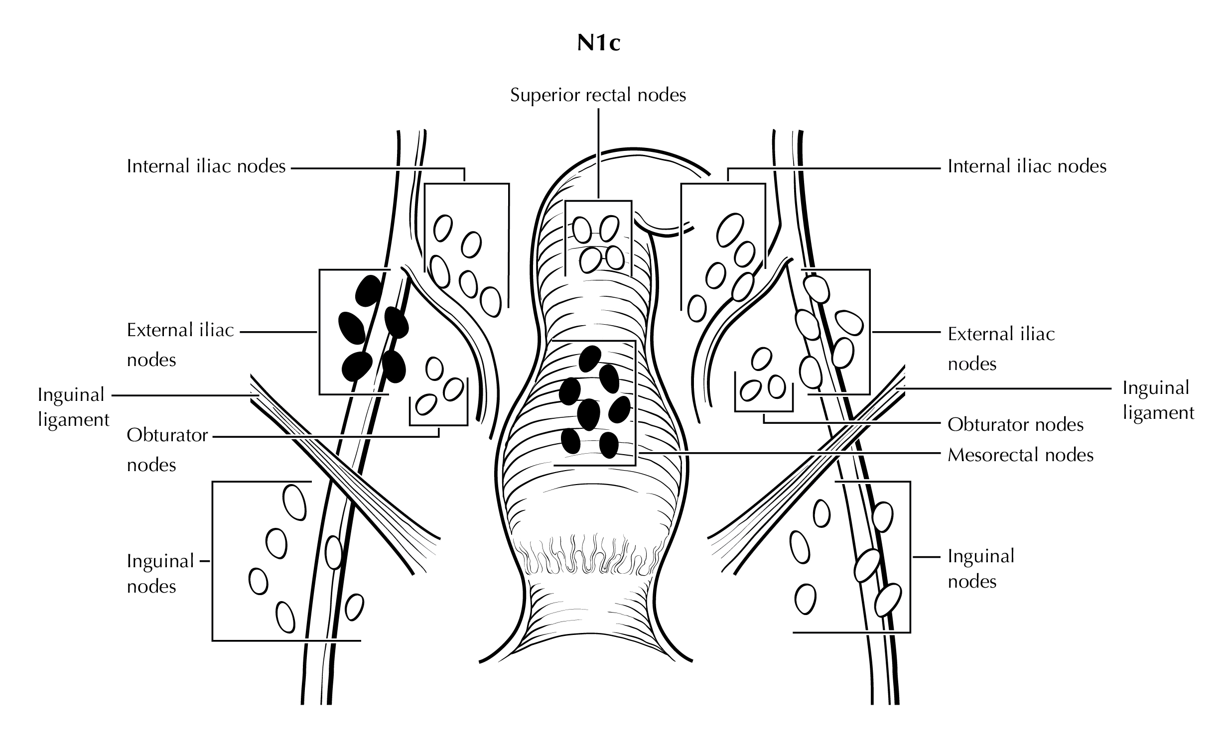
Lymphatic drainage and nodal involvement of anal cancers often depend on the location of the primary tumor. Tumors above the dentate line spread primarily to the mesorectal and internal iliac nodes, whereas tumors below the dentate line also may spread to the inguinal and external iliac nodes.
Assessment of lymph node involvement in anal cancer is even more challenging than in other cancers, where it has similarly been difficult to agree on radiographic criteria, because of the non-surgical nature of anal cancer preventing a surgical gold standard. Level I evidence is lacking for that reason. Many anal cancer articles included or are dominated by clinical follow up of nodes over time as a surrogate of involvement or not. Some include biopsies, such as from sentinel lymph node imaging or without this additional imaging. Furthermore, due to the demographics of this disease and its HPV association, men and women with HIV form an intersection of the affected population and may have non-specific HIV related lymphadenopathy further complicating interpretation.
Sizes and imaging appearances of nodes in CT and MR have been used for anal cancer as in other tumors and metabolic activity too is proposed through the use of PET scanning. In rectal cancer, a hybrid combination of these (The Dutch Consensus Criteria) have already shown more accurate staging and possibly safer patient outcomes.25 Possibly this will be the correct direction for anal cancer as well. Such a hybrid set of criteria was created for the non-HIV participants of the currently ongoing DECREASE trial (https://www.clinicaltrials.gov/ct2/show/NCT00056407) which incorporates PET and MRI (Table Anus-Suggested Definitions of Involved Lymph Nodes), but of course, the number of patients undergoing surgery will not be any greater than historically and so the lack of a pathologic gold standard still exists. However, this system can be considered as one created by a group of experts based on an exhaustive literature review and mainly extrapolation from higher level data available on other malignancies and might be generally recommended in the absence of a good alternative.
TABLE ANUS-SUGGESTED DEFINITION OF INVOLVED LYMPH NODES
Anatomic Location | CT/MRI- based Size OR | CT/MRI-based Morphology OR | PET-based FDG Uptake |
---|---|---|---|
Mesorectal, Presacral | Short axis > 5mm | Irregular Border OR Central necrosis (only for LN > 3mm on MRI) | > Blood pool (Deauville 3-5) |
Internal Iliac, Obturator | Short axis > 7mm | Irregular Border OR Central necrosis | > Blood pool (Deauville 3-5) |
Common Iliac and External Iliac | Short axis > 10mm | Irregular Border OR Central necrosis | > Blood pool (Deauville 3-5) |
Inguinal | No size criteria | Irregular Border OR Central necrosis | > Liver (Deauville 4-5) |
Note that the Deauville Criteria were created for 18F-FDG PET evaluation of lymphoma and have been borrowed here.26
Data using PET/CT for surgical nodal staging in other malignancies is extrapolated to anal cancer and revealed NPVs of 90-100% and PPVs of 43-100%.27-29 For mesorectal nodes, extrapolating from rectal cancer node-for-node validation studies, a combination of signal and border characteristics allowed a sensitivity of 85% and a specificity of 98%.30 PET adds specificity to MRI for involved mesorectal nodes in anal cancer when there is greater FDG uptake than normal tissues (PPV = 84%).31 For inguinal nodes, combining PET/CT and sentinel lymph node biopsy it was shown that size is an unreliable criterion in 123 nodes that were removed with negative nodes size average 1.16 cm and positive nodes
size average of 1.19 cm.28 Regarding inguinal nodes in HIV+ patients; symmetric enlargement has been found more often (100%) than when compared with non-HIV patients (<50%).32
The regional lymph nodes are as follows (Figure Anus-Nodal Map):
Mesorectal
Inguinal
Superior rectal (hemorrhoidal)
External iliac
Internal iliac
Obturator
All other nodal groups represent sites of distant metastasis.
If the vessel wall or its remnant is identifiable on H&E, elastin, or any other stain, the lesion should be classified as lymphovascular invasion (LVI) present (a CAP-required data element).
FIGURE ANUS-NODAL MAP. Schematic of regional draining lymph nodes for tumors of the anus, coronal view (A). Depiction of cross-sectional imaging of regional lymph nodes in the pelvis (B).
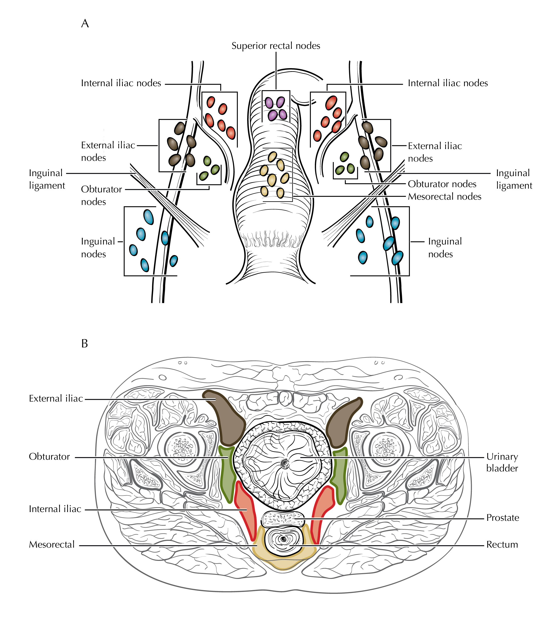
FIGURE ANUS-N1a. N1a is defined as tumor involvement of inguinal, mesorectal (as illustrated), superior rectal, internal iliac, or obturator lymph nodes.
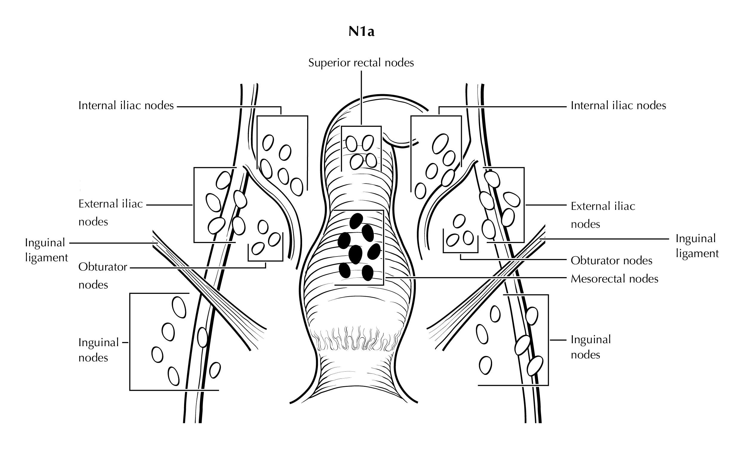
FIGURE ANUS-N1b. N1b is defined as tumor involvement of external iliac lymph nodes.
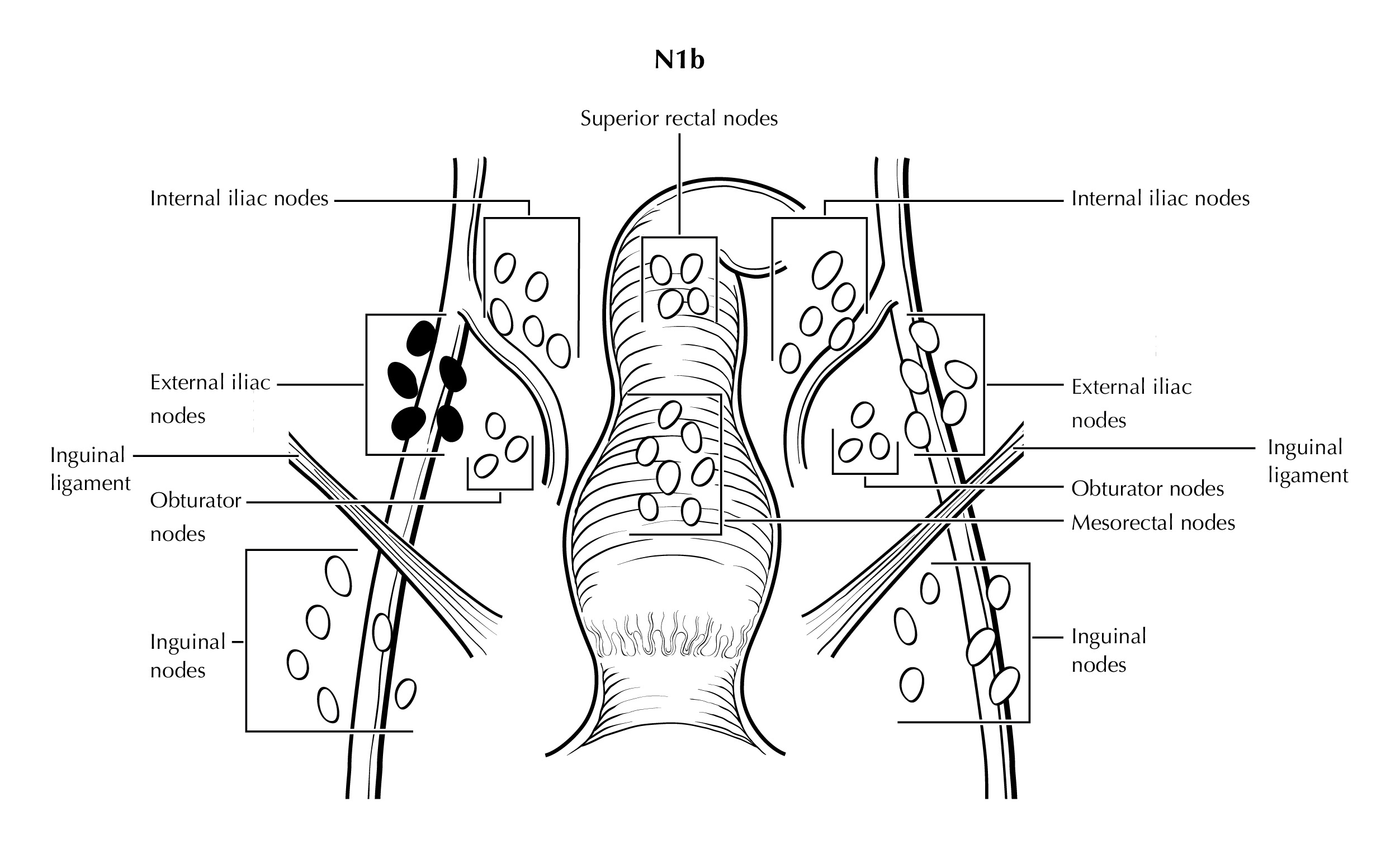
FIGURE ANUS-N1c. N1c is defined as tumor involvement of external iliac (N1b) with any N1a nodes (mesorectal nodes are illustrated).
Identification of Primary Site (Note S)
NOTE: This list includes topography codes and terms from the International Classification of Diseases for Oncology (ICD-O).2
Note S: Identification of Primary Site
The anus is the distal opening to the lower gastrointestinal tract (Figure Anus-Anatomy Coronal and Figure Anus-Anatomy Sagittal Female). The anal canal begins where the rectum enters the puborectalis sling at the apex of the anal sphincter complex (palpable as the anorectal ring on digital rectal examination and approximately 1 to 2 cm proximal to the dentate line). The anus ends with the squamous mucosa blending with the perianal skin, which coincides roughly with the palpable intersphincteric groove or the outermost boundary of the internal sphincter muscle, easily visualized on endoanal ultrasound. The anus encompasses true mucosa of three different histologic types: colorectal-type glandular, transitional, and squamous (proximal to distal, respectively). The most proximal aspect of the anal canal is lined by a continuation of the colorectal mucosa. The anal transitional zone (ATZ) is interposed between this colorectal zone and the distal squamous zone. The ATZ mucosa variably consists of discontinuous islands of squamous and rectal glandular epithelium, immature squamous metaplasia and a multi-layered transitional epithelium. The anal glands arise in the ATZ and may extend into the internal sphincter smooth muscle. The squamous (distal) zone of the anal canal extends from the dentate line to the mucocutaneous junction with the perianal skin and is lined by a nonkeratinizing squamous epithelium devoid of epidermal appendages (hair follicles, apocrine glands, and sweat glands).
Tumors that develop from mucosa (of any of the three types) that cannot be visualized in their entirety while gentle traction is placed on the buttocks are termed anal cancers, whereas those that arise within the skin at or distal to the squamous mucocutaneous junction, can be seen in their entirety with gentle traction placed on the buttocks, and are within 5 cm of the anus are termed perianal cancers.
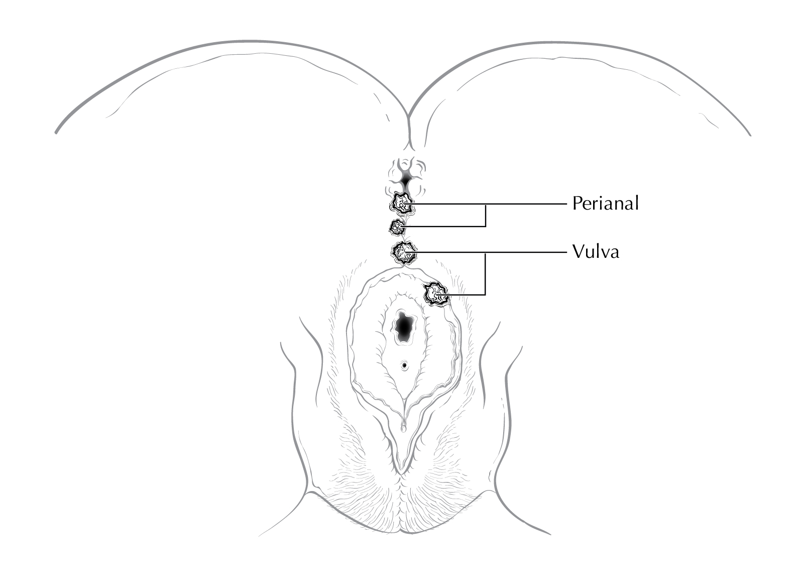
Squamous cell carcinomas (SCCs) overlying the perineal body may be classified as perianal or vulvar, and the treatment plans may be quite dissimilar. For this reason, we recommend the following: lesions that clearly arise from the vulva and extend onto the perineum and potentially involve the anus should be classified as vulvar. Similarly, lesions that clearly arise from the distal anal squamous mucosa and extend onto the perineum should be classified as perianal. Lesions localized to the perineum that are not clearly arising from either the vulva or the anus should be categorized based on the clinicians clinical impression. Thus, we recommend the following terminology: perineum favor vulva and perineum favor perianus. We also recommend consulting with colleagues in gynecologic oncology, colorectal or general surgery, or surgical oncology, because classification has a significant impact on treatment.
FIGURE ANUS-ANATOMY CORONAL. Anus and surrounding structures. Anal cancers (A-C) and peri-anal cancer (D).
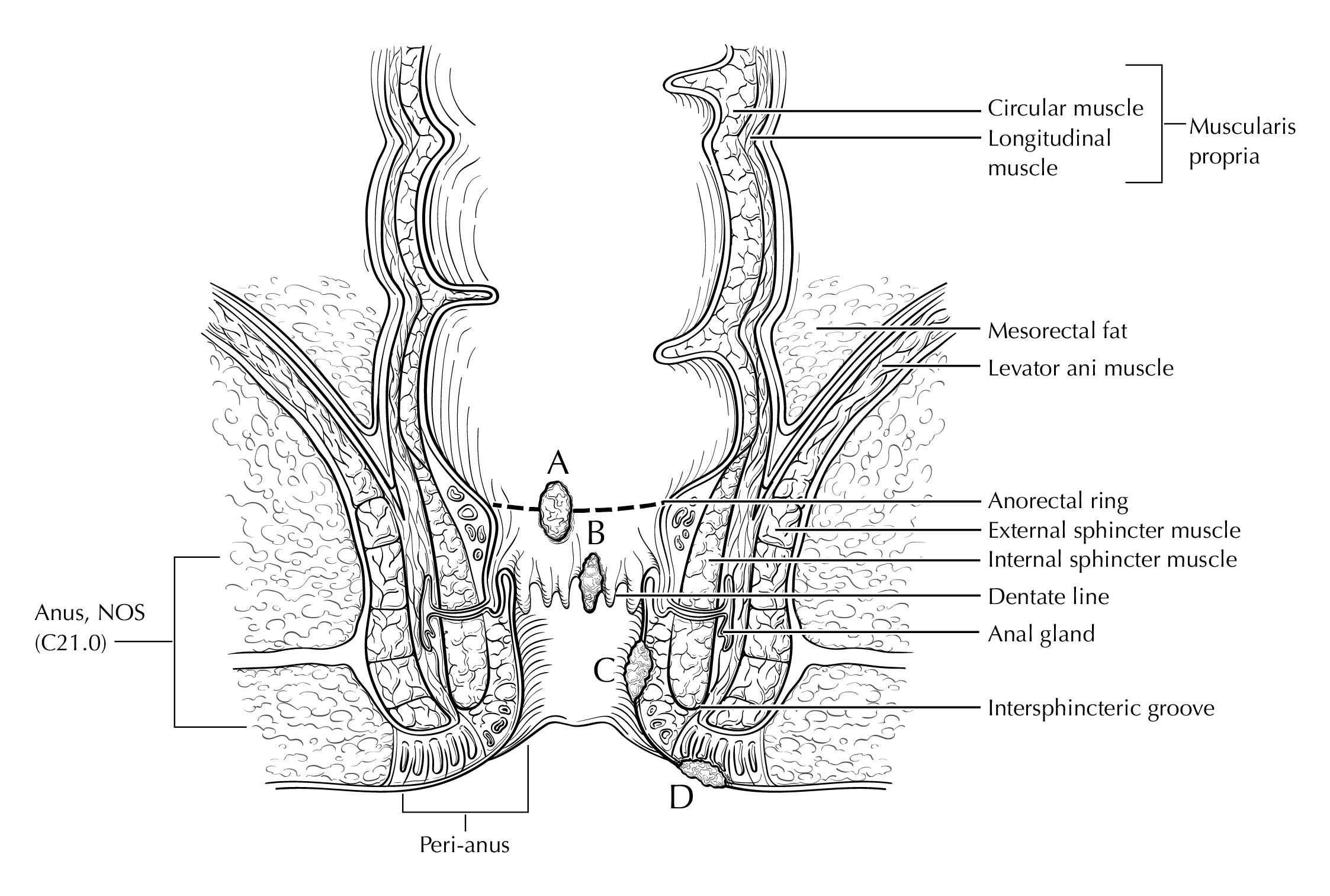
FIGURE ANUS-ANATOMY SAGITTAL FEMALE. Anus with surrounding structures.
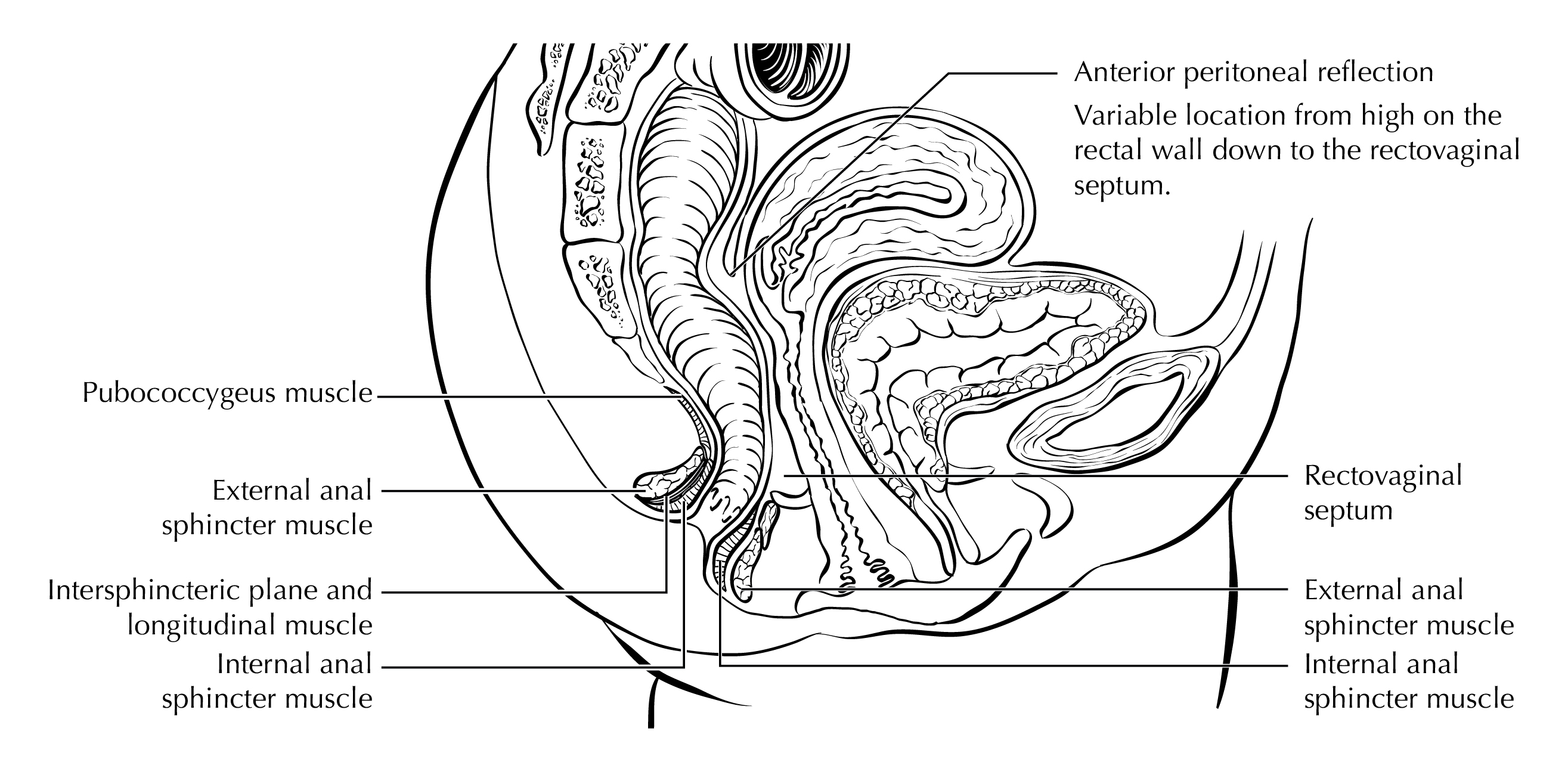
FIGURE ANUS-SURFACE LESIONS MALE. Anal and perianal lesions staged with this protocol. Skin cancers are not staged with this protocol. The diagram demonstrates a tumor straddling anal verge (anal), a tumor within 5 cm of anal verge (perianal), and a tumor >5 cm from anal verge (skin).
FIGURE ANUS-SURFACE LESIONS FEMALE. Anatomic site distinctions between perianal and vulva.
Note T: Primary Tumor (T)
The anatomy of the anus is described in Note S: Identification of Primary Site with a figure of the anatomy.
In the AJCC 8th edition staging system of anal cancer, the disease was staged as Stage 0 when the lesion is in situ (Tis), meaning that it is entirely intra-epithelial and has not crossed the basement membrane, i.e., is not invasive cancer. In addition, Tis lesions have not spread to nearby lymph nodes (N0) or distant sites (M0). Tis lesions are considered to be a form of high-grade squamous intraepithelial lesions (HSIL) which, like other HSIL lesions may progress to cancer if left untreated but are themselves not malignant.5 Use of the term carcinoma in situ may lead to confusion through the use of the word carcinoma, which has led to some patients being erroneously treated with protocols for bona fide cancers, including chemo-and/or radiation therapy. Inclusion of the Tis in the TNM staging system may also lead clinicians to incorrectly believe that these pre-malignant lesions are malignant and need to be treated as such. In contrast, patients with Tis lesions may benefit from local removal to prevent progression to cancer. The Anal Cancer/HSIL Outcomes Research (ANCHOR) Study recently reported that treatment of anal HSIL, consisting primarily of local ablation, reduces the risk of progressing to anal cancer compared with no treatment. There are no reliable data on survival after treatment of Tis lesions. There is no routine, standard of care screening system for Tis lesions and reporting of these lesions is not reliable. Thus, in the AJCC Version 9 of the Anus Protocol, Stage 0 has been removed.
Of note, direct invasion of the rectal wall, perianal skin, subcutaneous tissue or the anal sphincter muscles is not classified as T4.
FIGURE ANUS-T1. T1 is defined as tumor that is less than or equal to 2 cm in greatest dimension.
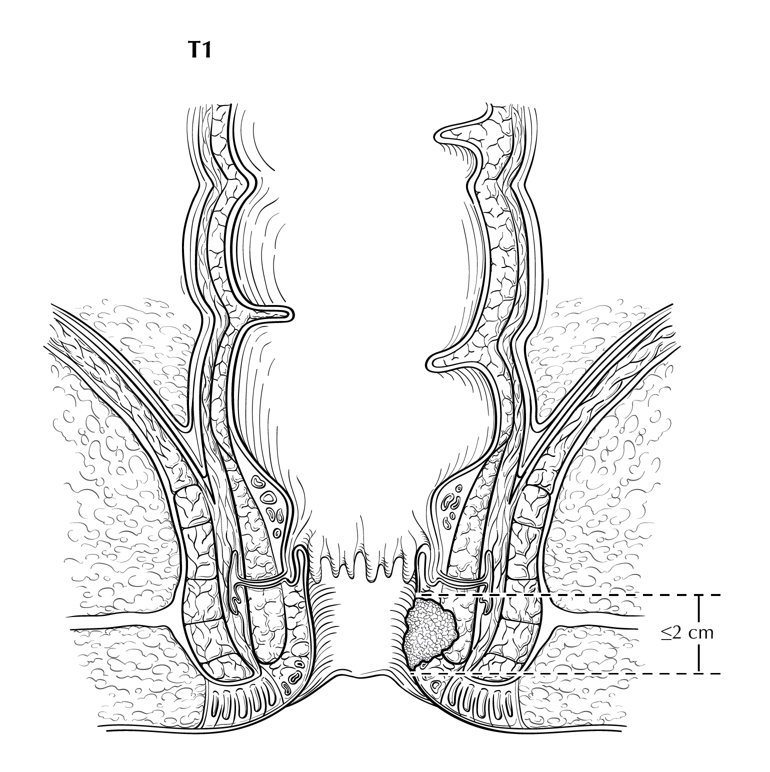
FIGURE ANUS-T2. Two views of T2 showing tumor that is greater than 2 cm but less than or equal to 5cm in greatest dimension. On the right side of the diagram, the tumor extends above the dentate line.
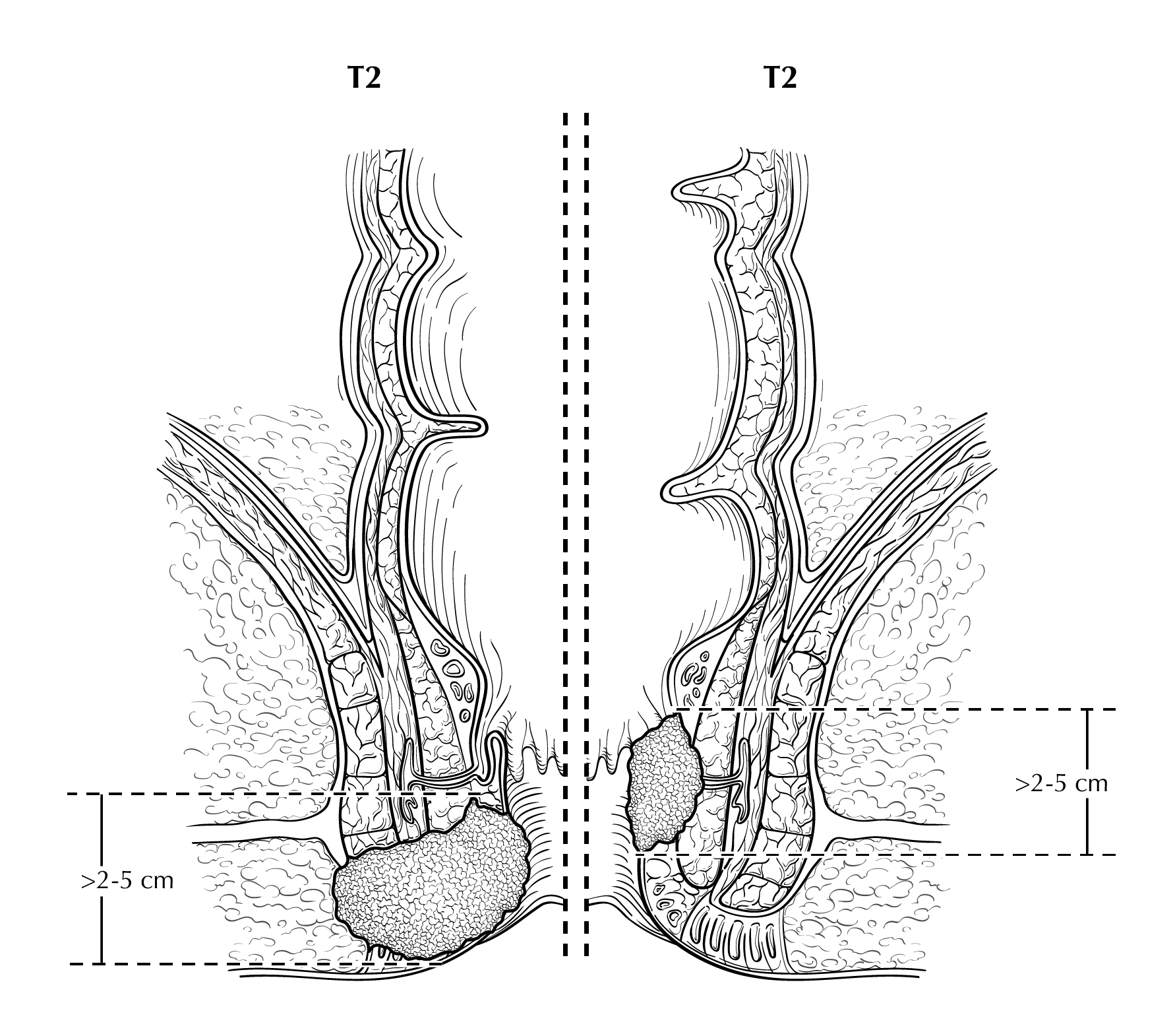
FIGURE ANUS-T3. T3 is defined as tumor that is greater than 5 cm in greatest dimension.
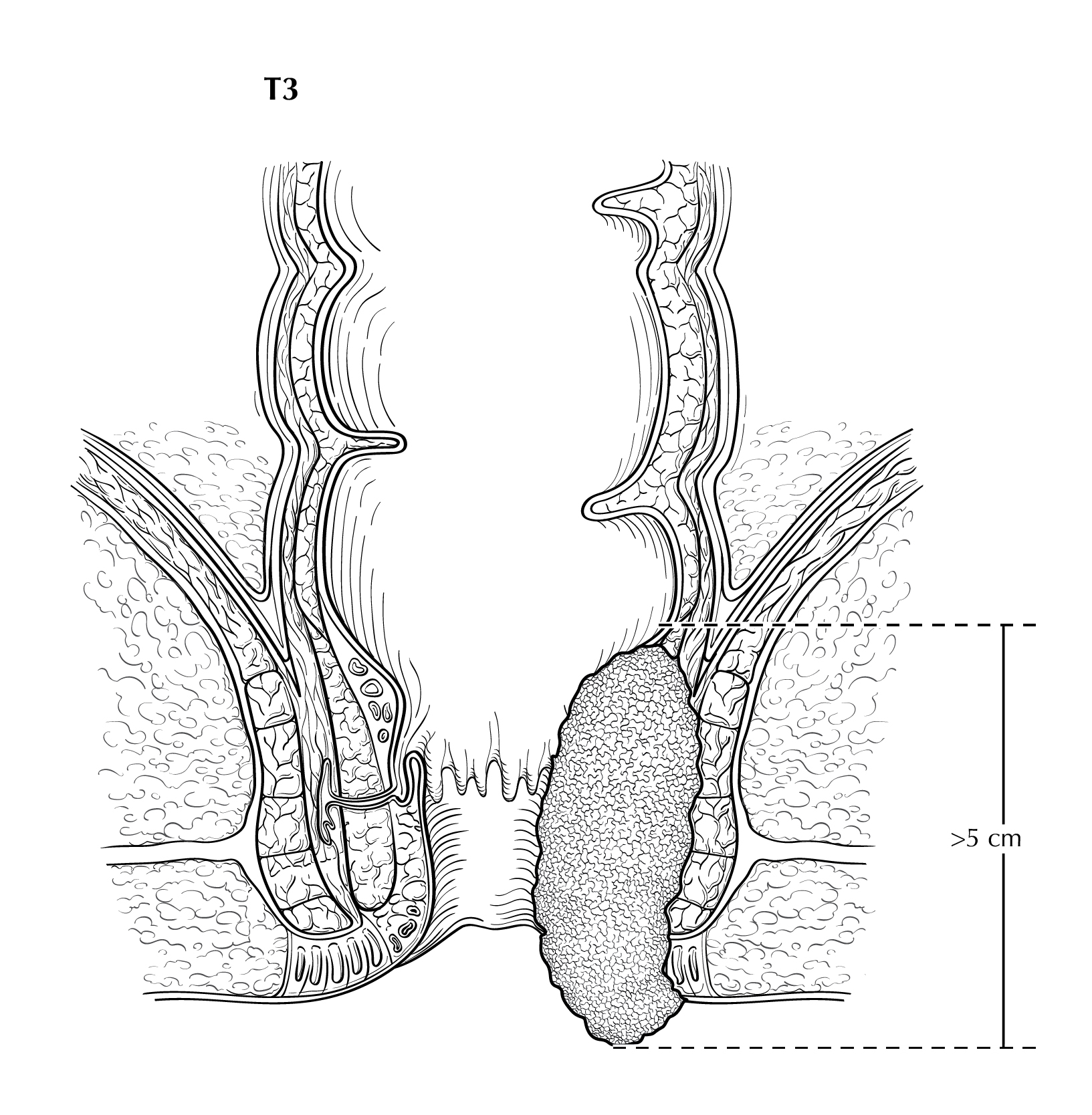
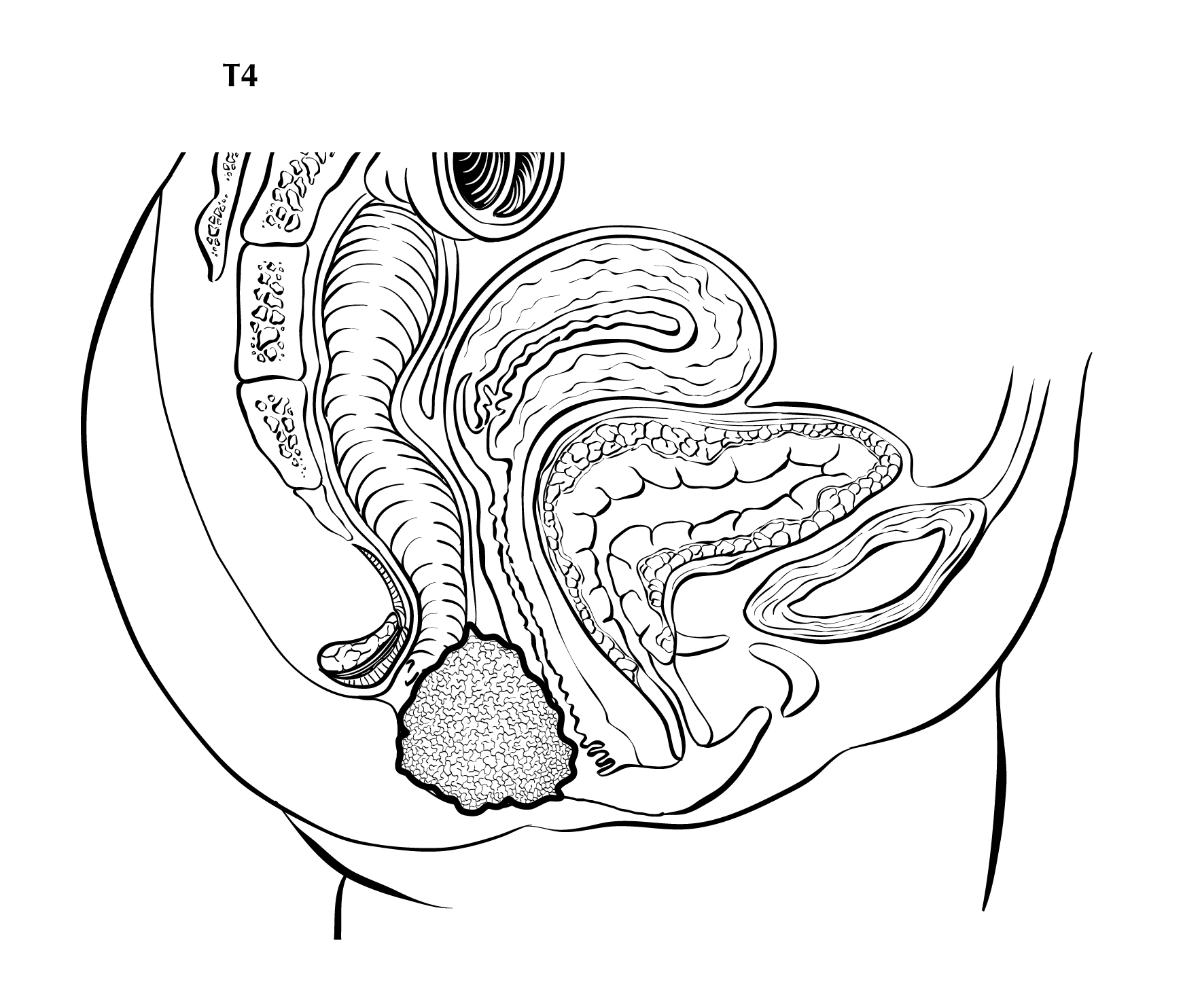
FIGURE ANUS-T4. T4 is defined as tumor of any size invading adjacent organ(s), such as the vagina (as illustrated), urethra, and bladder.
Staging Rules for Anus
Since the primary treatment for anal canal squamous cell carcinomas is usually upfront definitive chemoradiation, the clinical staging (cTNM) is more commonly used, since a surgical resection specimen is not available for complete evaluation by the pathologist providing information for the managing physician to assign pathological stage. If there is persistent or recurrent local tumor after chemoradiation, the clinical stage (cTNM) prior to initiating chemoradiation is the primary stage, but a posttherapy pathological stage is generated after surgical resection (usually combined abdominoperineal resection) annotated by the y prefix (ypTNM). For those early stage tumors treated primarily with surgery without chemoradiation, pathological stage (pTNM) is used. Additional details to help guide staging are shown in the figure below and described in the treatment scenarios.
Common staging scenarios:
Anal lesion treated with definitive chemoradiation
The most common scenario is the patient has an anal lesion found on exam. Based on clinical exams and imaging, the physician assigns the clinical staging (A in figure above) of cT for the primary anal lesion, cN for any nodal involvement, and cM for distant metastasis found on exam or imaging and pM for microscopic proof of distant metastasis. The treatment plan based on the clinical stage is primary chemotherapy and radiation therapy. Posttherapy clinical staging (C in figure above) may be assigned after completion of the primary treatment to assess the response based on physical exam, imaging, and biopsies assigning ycT for residual tumor, ycN for nodal assessment, and the M category as assigned in the clinical stage.
In rare cases of persistent disease, the patient may undergo a surgical resection. The pathologist will assign posttherapy pathological ypT based on assessment of the primary tumor, and ypN for regional nodes. The managing physician will then use the posttherapy yc stage combined with the operative findings and the pathology report to assign the posttherapy pathological staging (D in figure above) ypT, ypN, and the cM or pM.
Anal lesion treated surgically
Less commonly, after clinical staging it may be determined that a small anal lesion may be resected. Clinical staging (A in figure above), cT, cN, and cM/pM are assigned based on physical exam, imaging findings, and any biopsies. The patient then has a surgical resection. The pathologist assigns pT, pN, and pM (when lymph nodes and any distant metastases are sampled) based on the resected specimen. The managing physician then assigns the pathological staging (B in figure above) based on the clinical stage information, the operative findings, and the resected specimen pathology report information. Since many of these early stage tumors are resected with a wide local excision, the pT may be the only pathological staging available and the cN and cM may still need to be assigned based on the imaging findings for patient care.
Rules for Classification
Classifications specify the timeframe in the patients care and the criteria used to assign TNM. The same classification should be used throughout the assignment of TNM and stage group.
Clinical Classification (c) (Note C)
Pathological Classification (p) (Note P)
Posttherapy Clinical Classification (yc) (Note YC)
Posttherapy Pathological Classification (yp) (Note YP)
Additional classifications for recurrence/retreatment and autopsy:
Recurrence/Retreatment TNM Classification (r) (Note R)
Autopsy TNM Classification (a) (Note A)
Introduction to TNM Staging Classification
Stage may be defined at several time points in the care of the cancer patient. To properly stage a patient's cancer, it is essential to first determine the time point in a patients care. These points in time are termed classifications and are based on time during the continuum of evaluation and management of the disease. Then, T, N, and M categories are assigned for a particular classification (clinical, pathological, posttherapy, recurrence, and/or autopsy) by using information obtained during the relevant time frame, sometimes also referred to as a staging window. These staging windows are unique to each particular classification and are set forth explicitly in the Supplemental Information. The prognostic stage groups then are assigned using the T, N, and M categories, and sometimes also site-specific prognostic and predictive factors.
Among these classifications, the two predominant are clinical classification (i.e., pretreatment) and pathological classification (i.e., after surgical treatment as initial therapy).
Note C: Rules for Clinical TNM Classification
Clinical stage classification is based on patient history, physical examination, and any imaging done before initiation of treatment. Imaging study information may be used for clinical staging, but clinical stage may be assigned based on whatever information is available. No specific imaging is required to assign a clinical stage for any cancer site. When performed within this framework, biopsy information on regional lymph nodes and/or other sites of metastatic disease may be included in the clinical classification. The TNM is denoted by use of a lowercase c prefix: cT, cN, and cM0, cM1, or pM1. The M category use of cM or pM is based on method of assessment.
See General Staging Rules Table and Stage Classifications Table in Supplemental Information for additional guidance, including the time frame/staging window for determining clinical stage.
Clinical stage is important to record for all patients because:
clinical stage is essential for selecting initial therapy, and
clinical stage is critical for comparison across patient cohorts when some have surgery as a component of initial treatment and others do not.
Clinical stage may be the only stage classification by which comparisons can be made across all patients, because not all patients will undergo surgical treatment before other therapy, and response to treatment varies. Differences in primary therapy make comparing groups of patients difficult if that comparison is based on pathological assessment. For example, it is difficult to compare patients treated with primary surgery with those treated with chemotherapy or radiotherapy without surgery or neoadjuvant therapy.
Clinical classification is based on evidence acquired from the date of diagnosis until initiation of primary treatment. Examples of primary treatment include definitive surgery, radiation therapy, systemic therapy, and neoadjuvant radiation and systemic therapy.
Clinical Classification
Clinical assessment is based on medical history, physical examination, radiology, and endoscopy, with biopsy for histologic confirmation of malignancy. HIV testing should be performed in patients with established risk factors. Given patterns of spread, a thorough examination of lymph node areas, including inguinofemoral regions, should be performed. If possible, suspicious inguinal lymphadenopathy should be biopsied, generally through fine-needle aspiration, to further facilitate diagnosis and tumor staging, particularly in the HIV-positive population, in whom reactive nodes are common. In female patients, vaginal examination should be performed to rule out posterior vaginal invasion and fistula, and the cervix should be evaluated to rule out gynecologic malignancy. Whole body imaging methods such as CT, PET/CT and PET/MR are recommended to assign clinical M category.
Note P: Rules for Pathological TNM Classification
Classification of T, N, and M after surgical treatment is denoted by use of a lowercase p prefix: pT, pN, and cM0, cM1, or pM1. The purpose of pathological classification is to provide additional precise and objective data for prognosis and outcomes, and to guide subsequent therapy.
Pathological stage classification is based on clinical stage information supplemented/modified by operative findings and pathological evaluation of the resected specimens. This classification is applicable when surgery is performed before initiation of adjuvant radiation or systemic therapy.
See General Staging Rules Table and Stage Classifications Table in Supplemental Information for additional guidance.
Pathological Classification
Because the primary treatment for anal canal SCC usually is combined chemoradiation, a surgical resection specimen is not commonly available for complete evaluation by the pathologist (pTNM), unless there is persistent or recurrent local tumor after chemoradiation. For the latter group of patients who were assigned a clinical stage (cTNM) before beginning primary chemoradiation, a posttherapy pathological stage is determined after surgical resection (usually combined abdominoperineal resection) and annotated by the y prefix (ypTNM) (See Note YP). For tumors treated initially by surgery without neoadjuvant chemoradiation, pTN is assigned on pathological examination of the resection specimen, in consultation with the surgeon, who may comment on intraoperative findings and residual disease status and assign the c/pM.
Note YC: Rules for Posttherapy Clinical TNM Classification
Stage determined after treatment for patients receiving systemic and/or radiation therapy alone or as a component of their initial treatment, or as neoadjuvant therapy before planned surgery, is referred to as posttherapy classification. It also may be referred to as post neoadjuvant therapy classification.
See General Staging Rules Table and Stage Classifications Table in Supplemental Information for additional guidance.
Observed changes between the clinical classification and the posttherapy classification may provide clinicians with information regarding the response to therapy. The clinical extent of response to therapy may guide the scope of planned surgery, and the clinical and pathological extent of response to therapy may provide prognostic information and guide the use of further adjuvant radiation and/or systemic therapy.
Classification of T, N, and M after systemic and/or radiation treatment intended as definitive therapy is denoted by use of a lowercase yc prefix: ycT, ycN, c/pM. The c/pM category may include cM0, cM1, or pM1. The posttherapy clinical assessment of the T and N (ycTNM) categories uses specific criteria. In contrast, the M category for posttherapy classification remains the same as that assigned in the clinical stage before initiation of systemic/radiation therapy (e.g., if there is a complete clinical response to therapy in a patient previously categorized as cM1, the M1 category is used for final yc and yp staging).
See Stage Classifications Table in Supplemental Information for additional guidance.
Posttherapy Clinical Classification
After primary chemoradiation therapy has been completed, assessment for response to treatment is conducted. The posttherapy assessment and evaluation is typically performed starting at 6 weeks post-therapy. As long as the tumor is decreasing in size, the patient should be followed closely without any interventions. By six months post-chemoradiation, the tumor can be fully evaluated for response. In most cases, there is a clinical complete response based on physical exam, imaging, and potentially, biopsies and a posttherapy clinical stage (ycTNM) is assigned as ycT0N0M0 for no residual tumor. The ycT can be assigned for any residual primary tumor. The ycN is assigned for the posttherapy nodal status. The M category is the same as that assigned (cM0, cM1, or pM1) in the clinical stage.
If there is residual tumor persistent beyond 6 months following completion of chemoradiation therapy, a surgical resection is indicated, usually abdominoperineal resection (APR). Information from that surgical resection is assigned posttherapy pathological stage (Note YP).
Note YP: Rules for Posttherapy Pathological TNM Classification
Classification of T, N, and M after primary systemic and/or radiation treatment followed by surgery is denoted by use of a lowercase yp prefix: ypT, ypN, c/pM. The c/pM category may include cM0, cM1, or pM1. The posttherapy pathological assessment of the T and N (ypTNM) categories uses specific criteria. In contrast, the M category for posttherapy classification remains the same as that assigned in the clinical stage before initiation of therapy (e.g., if there is a complete clinical response to therapy in a patient previously categorized as cM1, the M1 category is used for final yc and yp staging).
The time frame for assignment of ypT and ypN should be such that the posttherapy surgery and staging occur within a period that accommodates disease-specific circumstances.
Criteria: First therapy is systemic and/or radiation therapy followed by surgery.
y-pathological (yp) classification is based on the:
y-clinical (yc) stage information, and supplemented/modified by
operative findings, and
pathological evaluation of the resected specimen.
Examples of treatments that satisfy the definition of posttherapy for anal carcinomas may be found in sources such as the NCCN Guidelines, ASCO guidelines, or other treatment guidelines. Systemic therapy includes chemotherapy and immunotherapy. Not all medications given to a patient meet the criteria for therapy or neoadjuvant therapy (e.g., a short course of therapy that is provided for variable and often unconventional reasons, should not be categorized as neoadjuvant therapy).
See Stage Classifications Table in Supplemental Information for additional guidance.
Posttherapy Pathological Classification
Residual tumor after primary chemoradiation therapy may require surgical resection, usually abdominoperineal resection (APR). The stage classification assigned is posttherapy pathological (ypTNM) and consists of the ycTNM information obtained from physical exam, imaging, and biopsies, combined with the surgeons operative findings, and the information supplied from the pathologist on the resected specimen pathology report. The ypT and ypN utilizes all this information. The M category is the same as that assigned (cM0, cM1, or pM1) in the clinical stage.
Tumor Regression after Preoperative Radiotherapy and/or Chemotherapy for Anal Carcinoma
The pathological response to preoperative radiotherapy and/or chemotherapy is an important prognostic factor for most carcinomas. This response is assessed by the pathologist and is reported with the prefix y: ypT and ypN. Regression may be evaluated in excision or resection specimens (see the College of American Pathologists Protocol for the Examination of Resection Specimens from Patients with Carcinomas of Anus). Specimens from patients receiving preoperative chemoradiation should be examined thoroughly at the primary tumor site, in regional nodes, and for peritumoral satellite nodules or deposits in the remainder of the specimen.
Although scoring systems for tumor response for anal malignancies have not been established, a three-tier system based upon the modified Ryan scheme is suggested. Response of the primary tumor and lymph node metastases should be assessed.
Modified Ryan Scheme for Tumor Regression Score24
Description | Tumor regression score |
---|---|
No viable cancer cells (complete response) | 0 |
Single cells or rare small groups of cancer cells (near-complete response) | 1 |
Residual cancer with evident tumor regression, but more than single cells or rare small groups of cancer cells (partial response) | 2 |
Extensive residual cancer with no evident tumor regression (poor or no response) | 3 |
Note R: Rules for Recurrence/Retreatment TNM Classification
Staging classifications at the time of retreatment for a recurrence or disease progression is referred to as recurrence classification. It also may be referred to as retreatment classification. Classification of T, N, and M for recurrence or retreatment is denoted by use of the lowercase r prefix: rcT, rcN, rc/rpM, and rpT, rpN, rc/rpM. The rc/rpM may include rcM0, rcM1, or rpM1.
See Stage Classifications Table in Supplemental Information for additional guidance.
Note A: Rules for Autopsy TNM Classification
Staging classification for cancers identified only at autopsy is referred to as autopsy classification. This classification is used when cancer is diagnosed at autopsy and there was no prior suspicion or evidence of cancer before death. All clinical and pathological information obtained at the time of death and through post-mortem examination is included. Classification of T, N, and M at autopsy is denoted by use of the lowercase a prefix: aT, aN, aM.
See Stage Classifications Table in Supplemental Information for additional guidance.
Prognostic Factors Required for Stage Grouping
Beyond the factors used to assign T, N, or M categories, no additional prognostic factors are required for stage grouping.
Note AF: Additional Factors Impacting Treatment Decisions During First Treatment Phase
Prognostic Tumor Characteristics
Beyond the factors used to assign T, N, or M categories, no additional prognostic factors are required for stage grouping. Patient and tumor characteristics affecting prognosis include:
HPV Status
The most important risk factor for squamous cell anal cancer is infection of the anus, cervix, or vulva with HPV, primarily types 16 and 18. Recent evidence indicates that virtually every anal SCC is related to HPV, although uncertainty continues to exist regarding the impact of various subclassifications of the virus.33 Viral proteins in these high-risk HPV types mediate oncogenic transformation of the anal squamous epithelia.34 In contrast to squamous carcinomas of the head and neck, there do not appear to be two major categories with entirely different biologies and prognoses based on the HPV status. A recent meta-analysis suggests that HPV 16 is found more frequently (75%) and HPV 18 less frequently (10%) in anal carcinomas than in cervical carcinomas.35 Although HPV 16 was the primary HPV type found in anal cancers in all groups, in a meta-analysis examining the relationship between disease severity and HPV status, HPV 16 was found to be relatively less common in cancers of men and women living with HIV, compared to those who did not have HIV.36 Overall, the data suggest that the spectrum of HPV types associated with anal cancer may be broader in people living with HIV than among HIV-negative individuals. This does not affect treatment approaches currently but may be of future importance when devising strategies that include HPV typing to screen individuals at increased risk of anal cancer, and if and when HPV type-specific therapeutic approaches are included in cancer treatment regimens.
Other known risk factors for anal SCC include a history of sexually transmitted disease including anal warts, a history of multiple sexual partners, and anal intercourse, all of which may be associated with a higher incidence of HPV infection.37,38 SCCs of the anus are more common in women; however, men have a worse prognosis.38 The poorer prognosis in males also was confirmed in three phase III chemoradiation trials (US GI Intergroup 98-11, European Organisation for Research and Treatment of Cancer [EORTC] 22861, and United Kingdom ACT I).39-41 Chronic immunosuppression, which may be related to HIV-positive status or a history of organ transplantation, and tobacco use also are important risk factors for anal cancer.42-45
Histologic Type
Nonsquamous carcinomas of the anal canal, including adenocarcinoma, high-grade neuroendocrine carcinoma, and undifferentiated carcinoma, are associated with worse 5-year survival (OS) rates than SCCs of the anal canal.46 However, historically recognized histologic SCC variants, such as large cell keratinizing, large cell nonkeratinizing, and basaloid subtypes, have no associated prognostic differences. Basal cell carcinomas of the perianal region tend to have a better prognosis, with lower risk of relapse.12
HIV Status
The impact of HIV status on prognosis remains incompletely defined. The literature remains controversial regarding the perception that people living with HIV tolerate chemoradiation therapy less well and have poorer outcomes compared with HIV-negative patients. Patients whose HIV is well controlled with highly active antiretroviral therapy appear to do as well as HIV-negative patients. Better documentation of HIV status is needed.47-49 HIV status is reported as positive or negative, and it is documented in and abstracted from the medical record but may not always be submitted to central databases. AJCC Level of Evidence: I
Gender
Male sex has a negative impact on prognosis. Although SCCs of the anus are more common in women, men have a worse prognosis.1,50 The poorer prognosis in males also was confirmed in three phase III chemoradiation trials (US GI Intergroup 98-11, EORTC 22861, and United Kingdom ACT I).39-41 Sex is reported as male or female, and it is documented in and abstracted from the medical record. AJCC Level of Evidence: I
Tumor Location
Tumor location defines nodal drainage fields at risk and treatment approach. For anal canal lesions, external beam irradiation fields differ from those for perianal lesions. Perianal cancers would be treated as skin cancers elsewhere in the body with focused irradiation fields without inclusion of regional nodes, unless there was deep invasion of the tumor, necessitating inguinal node coverage. For cancers of the anal canal, irradiation fields commonly include the primary tumor plus all regional node groups felt to be at risk (inguinal, mesorectal/superior rectal, internal iliac, external iliac). Tumor location is reported as anal or perianal, and left/right/anterior/posterior/lateral, and it is documented in and abstracted from the medical record. AJCC Level of Evidence: I
Assignment of AJCC TNM
AJCC data elements required for staging are identified with an asterisk (*).
*Stage classification based on time frame and criteria (see Supplemental Information)
*Definition of Primary Tumor (T)(Note T)
T Category | T Criteria |
---|---|
TX | Primary tumor not assessed |
T0 | No evidence of primary tumor |
T1 | Tumor less than or equal to 2 cm in greatest dimension |
T2 | Tumor greater than 2 cm but less than or equal to 5 cm in greatest dimension |
T3 | Tumor greater than 5 cm in greatest dimension |
T4 | Tumor of any size invading adjacent organ(s), such as the vagina, urethra, or bladder |
Primary Tumor Suffix
*Definition of Regional Lymph Node (N) (Note T)
N Category | N Criteria |
---|---|
NX | Regional lymph nodes cannot be assessed |
N0 | No tumor involvement of regional lymph node(s) |
N1 | Tumor involvement of regional lymph node(s) |
N1a | Tumor involvement of inguinal, mesorectal, superior rectal, internal iliac, or obturator lymph node(s) |
N1b | Tumor involvement of external iliac lymph node(s) |
N1c | Tumor involvement of N1b (external iliac) with any N1a node(s) |
Regional Lymph Nodes Suffix
*Definition of Distant Metastasis (M)
M Category | M Criteria |
---|---|
cM0 | No distant metastasis |
cM1 | Distant metastasis |
pM1 | Microscopic confirmation of distant metastasis |
AJCC Prognostic Stage Groups (Note PSG)
AJCC data elements required for staging are identified with an asterisk (*).
*AJCC prognostic stage group is assigned based on the stage classification and categories chosen
When T is | and N is | and M is | Then the stage group is |
---|---|---|---|
T1 | N0 | M0 | I |
T2 | N0 | M0 | IIA |
T1-T2 | N1 | M0 | IIB |
T3 | N0-N1 | M0 | IIIA |
T4 | N0 | M0 | IIIB |
T4 | N1 | M0 | IIIC |
Any T | Any N | M1 | IV |
Additional Factors Impacting Treatment Decisions During First Treatment Phase (Note AF)
Additional data elements that are clinically significant but not required for staging are identified with a dagger symbol ().
Prognostic Tumor Characteristics
Note PSG: AJCC Prognostic Stage Groups
The managing physician alone is responsible for assigning the patients stage, because only (s)he routinely has access to all the pertinent information from physical examination, imaging studies, biopsies, diagnostic procedures, surgical findings, and pathology reports. Although the pathologist and the radiologist provide important staging information, and may provide important T-, N-, and/or M-related information, stage is defined ultimately from the synthesis of an array of patient history and physical examination findings supplemented by imaging and pathology data.
Data Collection
Registry Data Collection Guidance
Emerging Factors for Collection
Histologic Grade (G) (Note G)
Grade is assigned based on histopathologic assessment.
G | G Definition |
---|---|
GX | Grade cannot be determined |
G1 | Well differentiated (low grade) |
G2 | Moderately differentiated (low grade) |
G3 | Poorly differentiated (high grade) |
G4 | Undifferentiated (high grade) |
Note G: Histologic Grade (G)
Anal squamous cell carcinomas and adenocarcinomas are graded using a three-tier system (well, moderately, and poorly differentiated). For carcinomas showing variation in tumor grade, the highest grade is assigned.
Carcinomas showing solid growth pattern with no squamous or glandular differentiation (undifferentiated carcinomas) are assigned grade 4.
Small cell neuroendocrine carcinoma and large neuroendocrine carcinoma are grade 3.
High-grade (poorly differentiated) squamous carcinomas or adenocarcinomas of the anus have a worse prognosis than low-grade tumors.
Histopathologic Type (Note HT)
Additional data elements that are clinically significant but not required for staging are identified with a dagger symbol ().
Histopathologic Codes NOTE: This list includes histology codes and preferred terms from the WHO Classification of Tumours1 and the International Classification of Diseases for Oncology (ICD-O).2 Most of the terms in this list represent malignant behavior. For cancer reporting purposes, behavior codes /3 (denoting malignant neoplasms), /2 (denoting in situ neoplasms), and in some cases /1 (denoting neoplasms with uncertain and unknown behavior) may be appended to the 4-digit histology codes to create a complete morphology code. The more common types (squamous cell carcinoma and its subtypes) are listed first, followed by less common entities.
Code | Description |
---|---|
8070 | Squamous cell carcinoma |
8071 | Squamous cell carcinoma, keratinizing, NOS |
8072 | Squamous cell carcinoma, large cell, non-keratinizing, NOS |
8085 | Squamous cell carcinoma, HPV-associated |
8086 | Squamous cell carcinoma, HPV-independent |
8051 | Verrucous carcinoma |
8083 | Basaloid squamous cell carcinoma |
8123 | Basaloid carcinoma |
8124 | Cloacogenic carcinoma |
8013 | Large cell neuroendocrine carcinoma (NEC) |
8041 | Small cell neuroendocrine carcinoma (NEC) |
8246 | Neuroendocrine carcinoma (NEC) |
8140 | Adenocarcinoma |
8215 | Adenocarcinoma of anal glands |
8480 | Mucinous adenocarcinoma |
8490 | Signet ring cell carcinoma |
8154 | Mixed neuroendocrine-non-neuroendocrine neoplasm (MiNEN) |
8244 | Mixed adenoneuroendocrine carcinoma |
8020 | Undifferentiated carcinoma |
8032 | Spindle cell carcinoma |
8033 | Carcinoma with sarcomatoid component |
8090 | Basal cell carcinoma, NOS |
8000§ | Neoplasm, malignant |
8010§ | Carcinoma, NOS |
§Histology is not ideal for clinical use in patient care, as it describes an unspecified or outdated diagnosis. Data collectors may use this code only if there is not enough information in the medical record to document a more specific diagnosis.
Sources: WHO Classification of Tumours Editorial Board. Digestive System Tumours. Lyon (France): International Agency for Research on Cancer, 2019. (WHO Classification of Tumours series, 5th ed., vol 1).
World Health Organization. International Classification of Diseases for Oncology. ICD-O-3.2-Online.1Used with permission
Note HT: Histopathologic Type
Most carcinomas of the anal canal and perianal region are squamous cell carcinomas (SCCs). The terms transitional cell and cloacogenic carcinoma are acceptable but not recommended, because these tumors are now recognized as nonkeratinizing types of SCC. While recognition of the various histologic patterns of SCC is important for pathologists to avoid misdiagnosis, subclassification lacks clinical relevance, and may be difficult to do on biopsies. Nonsquamous anal carcinomas include adenocarcinomas, large cell and small cell neuroendocrine carcinomas, and basal cell carcinomas (BCCs).4
Squamous Cell Carcinoma
The predominant histologic type of malignancy arising in the anal canal is SCC. Estimates suggest that 95% of these cancers are caused by oncogenic human papillomavirus (HPV) types, with HPV 16 associated with 89% of cases. Histologic patterns of no clinical import include basaloid, non-keratinizing, and keratinizing forms. The verrucous carcinoma subtype (discussed below) in pure form is locally destructive and does not metastasize, but cases of unequivocal conventional SCC arising in verrucous carcinoma should be classified as conventional SCC.
Non-invasive abnormalities of the anal squamous epithelium associated with HPV infection (dysplasia) are classified using the Lower Anogenital Squamous Terminology (LAST) system,5 in which anal intraepithelial neoplasia I (AIN 1, mild dysplasia), and AIN 2 that is p16-negative are classified as low grade squamous intraepithelial lesion (LSIL) and p16-positive AIN 2 and 3 (moderate and severe dysplasia) are classified as high-grade intraepithelial lesion (HSIL). Direct evidence supports the progression of HSIL to squamous cell carcinoma.6 However, HSIL is a non-invasive precursor lesion, not a malignancy, and should not be coded as such.
Verrucous Carcinoma
Verrucous carcinoma (VC) is a bulky, exophytic well-differentiated squamous cell carcinoma with a pushing, rather than infiltrative, invasive front and minimal cytologic atypia. Historically, verrucous carcinoma has been considered synonymous with giant condyloma of Buschke-Lowenstein. However, these lesions have a different gross morphology (VCs have a coarsely irregular surface compared to the more finely warty surface of giant condyloma), and VC is not associated with HPV infection whereas giant condyloma is associated with HPV6 and HPV 11.7 Any histologic evidence of conventional SCC in a VC or metastasis should lead to the diagnosis of SCC and appropriate therapy.
Other Histologic Types:
Adenocarcinoma
Adenocarcinomas of the anal canal are rare. In an analysis of 57,869 anal and rectal carcinomas identified in the SEER database diagnosed between 1990 and 2011 only 0.8% (462) had anal adenocarcinoma. Prior studies have suggested these patients have a worse overall prognosis relative to their squamous cell carcinoma of the anal canal or rectal cancer counterparts. The outcomes for anal adenocarcinomas, are poorer, stage for stage, than for SCC.8
Adenocarcinomas of the anal canal may be divided into three subtypes: a mucosal subtype phenotypically similar to colorectal adenocarcinoma; those originating in anal glands and primarily located in the submucosa or deeper layers; and those originating in fistulas and generally associated with Crohns disease.4 Anal gland-type and colorectal-type of anal adenocarcinomas may be distinguished by their different patterns of expression of keratins and CDX2, an intestinal marker.9 About half of anal gland adenocarcinomas are associated with high risk HPV infection, whereas the colorectal type is not.9 Anal adenocarcinomas arising in fistulas display variable morphology and may resemble typical colorectal adenocarcinomas, anal gland adenocarcinomas, or have mucinous features.
A small report from the Rare Cancer Network on 82 patients from 11 multinational institutions were staged according to UICC 5th edition, which is identical to AJCC 5th and 7th editions. In a multivariate analysis the 5-year overall survival for T1 category was 72%, for T2 or T3, 37% and for T4 only 9%. For N0 or N1 disease it was 41% and only 13% for N2 or N3.10 This report confirmed that the 7th edition TNM was effective in separating different prognostic groups among anal adenocarcinomas. Thus, although primarily developed based on outcomes for anal squamous cell carcinoma, the Anus TNM staging is appropriate for use in adenocarcinoma of the anus as well. There are no data to confirm or refute the changes between the 7th and 8th editions as they pertain to adenocarcinoma.
Basal Cell Carcinoma
Basal cell carcinoma (BCC) of the perianus is very rare, comprising less than 1% of all anorectal neoplasms.11 It arises in perianal skin and may rarely extend into the anal canal. Differentiating basaloid SCC from BCC may be challenging, as they have similar histologic features. However, SCCs arise from a known precursor lesion (anal squamous intraepithelial neoplasia), whereas BCCs do not have a well-defined precursor. In general, BCCs are associated with a low recurrence rate, and wide local excision with negative margins remains the standard of care.12
Neuroendocrine Carcinoma and Mixed Neuroendocrine-Non-Neuroendocrine Malignancies
Neuroendocrine neoplasms involving the anus are rare; neuroendocrine carcinomas (large cell and small cell neuroendocrine carcinoma) are the most commonly encountered. Mixed neuroendocrine-non-neuroendocrine neoplasms in the anal canal are most commonly neuroendocrine carcinoma and adenocarcinoma. 4
Well-differentiated neuroendocrine neoplasms of the anus are not staged with this staging system.
Survival Data
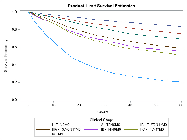
Regarding SCC of the anal canal, based on data from the National Cancer Database (NCDB) for 2010 to 2017, the 5-year observed survival rates for each of the stage groups per Version 9 are as follows: Stage I (n = 6,924), 83.5%; Stage IIA (n = 11,843), 75.5%; Stage IIB (n = 5,750), 69.3%; Stage IIIA (n = 8,188), 59.2%; Stage IIIB (n = 1,380), 55.4%; Stage IIIC (n = 1,782), 51.1%; Stage IV (n = 3,454), 20.5%. Stage-related survival is shown in Table Anus-Survival and Figure Anus-Survival.
TABLE ANUS-SURVIVAL. NCDB Clinical Stage Grouping 2- and 5-year Unadjusted Survival
Stage | Number of Patients (n =39,642) | 2-year Overall Survival (%) | 5-year Overall Survival (%) | Median Survival (months) |
---|---|---|---|---|
I | 6924 | 92.2 | 83.5 | 191.74 |
IIA | 11843 | 87.5 | 75.5 | 165.22 |
IIB | 5750 | 84.1 | 69.3 | 155.70 |
IIIA | 8188 | 75.7 | 59.2 | 96.00 |
IIIB | 1380 | 71.5 | 55.4 | 83.32 |
IIIC | 1782 | 69.5 | 51.1 | 63.44 |
IV | 3454 | 39.3 | 20.5 | 16.53 |
FIGURE ANUS-SURVIVAL. KaplanMeier Overall Survival Curves, 2010-2017