section name header

Authors

Authors: Guido Rindi, Arvind Dasari, Thomas A. Hope, Erik Mittra, Samantha M. Thomas, Alice C. Wei for the American Joint Committee on Cancer Expert Panel for Neuroendocrine Tumors of the Pancreas.

Acknowledgements and disclaimers: With guidance from Mary Kay Washington, Heidi Nelson, Donna M. Gress, Aleisha

J. Williams, Elaine Alexander, AJCC Staff, AJCC Expert Panel for Neuroendocrine Tumors, AJCC Editorial Committee, and AJCC Executive Committee.

Author contributions: GR, AD, TAH, EM, SMT, ACW wrote the manuscript. GR, AD, TAH, EM, SMT, ACW performed critical revision of the manuscript for important intellectual content. The AJCC Editorial Committee approved the manuscript.

Kellie M. Holoski revised the illustrations.

This Cancer Staging Protocol was prepared and published through the generous support of the American Cancer Society.

Chapter Summary

Cancers Staged Using This Staging System

This staging system applies only to well-differentiated neuroendocrine tumors of the pancreas (NET G1, G2, and G3).

Cancers Not Staged Using This Staging System

These histopathologic types of cancer…Are staged according to the classification for…

Carcinomas of the pancreas, including poorly differentiated neuroendocrine carcinoma (NEC), Mixed neuroendocrine non-neuroendocrine (MiNEN) neoplasms

Exocrine pancreas

Well-differentiated neuroendocrine tumors of the duodenum (C17.0) or ampulla of Vater (C24.1)

Neuroendocrine tumors of the duodenum and ampulla of Vater

The following protocol is intended to standardize communication of critical components of cancer staging. It includes corresponding explanatory notes that provide the level of evidence for each critical element. While the focus of this protocol with synoptic report format is on cancer staging for clinical care and registry support, information on additional and emerging prognostic factors is included. Additional information on staging may be found in the AJCC 8th Edition Chapter 1: Principles of Cancer Staging on the AJCC website cancerstaging.org.

Synoptic Staging Report Format

Explanatory Notes Supplemental Information

Staging Report Format

Instructions for the use of this staging report:

This staging report was designed to demonstrate documentation of critical elements for AJCC stage classifications of primary neuroendocrine tumors of the pancreas.

Explanatory notes are provided for further descriptions and specifications for each data field.

Summary of Changes

ChangeDetails of ChangeLevel of Evidence

Histopathologic Type

Updated according to WHO Classification of Tumors, 5th Ed.

II

Prognostic and Non-Tumor Factors

Included DAXX/ATRX, ARX and PDX1 as potential biomarkers and several clinicopathologic factors for Non-Tumor Factors

II

Modalities Used for Diagnosis and Staging,

Clinical Staging and Workup

Included DOTA PET scans in imaging used for diagnosis and the new Clinical Staging and Workup table

II

Introduction

Discussed latest epidemiological data and treatment options including PRRT, capecitabine in combination with temozolomide

I

Classifications specify the timeframe in the patient's care and the criteria used to assign TNM. The same classification should be used throughout the assignment of TNM and stage group.

Diagnostic Phase

Identification of Primary Site (Note S)

NOTE: This list includes topography codes and terms from the International Classification of Diseases for Oncology (ICD-O).

CodeDescription

C25.0

Head of pancreas

C25.1

Body of pancreas

C25.2

Tail of pancreas

C25.4

Islets of Langerhans (endocrine pancreas)

C25.7

Other specified parts of the pancreas

C25.8

Overlapping lesion of the pancreas

C25.9

Pancreas, NOS

Clinical Examination (Note CE)

Imaging (Note I)

Diagnostic Procedures/Surgical Treatment

This table is a simplified algorithm of the investigations and procedures required to generate neuroendocrine tumors of the pancreas clinical TNM staging information.

Its purpose is to provide clarity regarding appropriate modalities to use in determining the individual categories of the neuroendocrine tumors of the pancreas clinical TNM staging.

Disclaimer:The table represents common approaches to staging and workup for this cancer. Some or all of these tests are used in staging the cancer and are provided as a reference. The table is not a guideline for treatment and should not be used in this manner but instead utilized to identify how each of these tests contribute to the determination of T, N, M categories and Stage.

DIAGNOSTIC WORKUPDESCRIPTIONSPECIFIC CONTRIBUTION TO TNM CATEGORY
Clinical Exam

Endoscopic ultrasound (EUS)

Tumor size

T1-T4

Biopsy

Provide histologic diagnosis

None

Imaging

CT (pancreas protocol)

Chest/abdomen/pelvis - define extent of local disease, nodal involvement, metastasis

T3-T4, N1, M1a-c

MRI (multiphasic and multiplanar)

Abdomen/pelvis - define extent of local disease, nodal involvement, metastasis. Hepatobiliary contrast preferred for liver lesions.

T3-T4, N1, M1a-c

Somatostatin receptor (SSTR) PET/ CT or PET/MRI

Skull base to mid-thigh - define extent of local disease, nodal involvement, metastasis. Most useful for low or intermediate grade tumors.

T3-T4, N1, M1a-c

Octreotide scan

Whole body with SPECT/CT-define extent of local disease, nodal involvement, metastasis. Only if SSTR PET not available.

T3-T4, N1, M1a-c

FDG PET/CT or PET/MRI

Skull base to mid-thigh - define extent of local disease, nodal involvement, metastasis. Most useful for high-grade tumors

T3-T4, N1, M1a-c

Laboratory Studies

Biochemical evaluation

Provide information about functional status, to be done as clinically indicated

None

Genetic testing

For germline and somatic alterations

None

This table is a simplified algorithm of the investigations and procedures required to generate neuroendocrine tumors of the pancreas pathological TNM staging information.

Its purpose is to provide clarity regarding appropriate modalities for the pathologist and managing physician to use in determining the individual categories of the neuroendocrine tumors of the pancreas pathological TNM staging.

Disclaimer:The table represents common approaches to staging and workup for this cancer. Some or all of these tests are used in staging the cancer and are provided as a reference. The table is not a guideline for treatment and should not be used in this manner but instead utilized to identify how each of these tests contribute to the determination of T, N, M categories and Stage.

CATEGORYSPECIMENPATHOLOGISTMANAGING PHYSICIAN (Stage Documented by Cancer Registry)

General Information

  • Assignment of pTNM categories are based on surgical resection specimen, as well as intraoperative findings, biopsy procedures and clinical evaluation up to the point of definitive surgical treatment, if available
  • All other surgical procedure specimens use cTNM; for example, biopsy of a positive regional lymph node without surgical resection of the primary carcinoma is classified as cN1
  • Assignment of pTNM categories for the patient requires use of information from all biopsy procedures performed during the clinical evaluation up to and including definitive surgical treatment
  • Requires information from clinical assessment or imaging studies or interoperative findings to assign pTNM categories (may not change pTNM, but must be considered)

pTX

Not for use by pathologist; assigned only by managing physician

May assign if unable to determine pT category after surgical resection

pT0

No tumor found in specimen and never identified on diagnostic biopsies

No tumor found in specimen and never identified on diagnostic biopsies

pT1

Pancreas resection

Pathological information from surgical specimen only

Tumor size

Pathology report +/- appropriate clinical exam, imaging studies, and intraoperative findings

pT2

pT3

Tumor size or duodenum/ ampulla and bile duct invasion

pT4

Tumor invasion of adjacent organs or wall of large vessels

pNX

Not for use by pathologist; assigned only by managing physician

  • May assign if unable to determine pN category
  • No regional node(s) sampled or resected

pN0

Fine Needle Aspiration (FNA), core needle biopsy, sentinel node biopsy, lymph node dissection (including procedures performed prior to definitive surgical resection)

Note: These procedures in the absence of a surgical resection are cN

Requires:

  • At least one lymph node sampled
  • May require information from a previous node biopsy procedure to assign pN category
  • For FNA or core biopsy: use (f) modifier

Primary site surgical resection is required to assign pN

Requires:

  • Same information as the pathologist
  • Supplement with clinically positive nodes from examination or imaging

pN1

cM0

Not assigned by pathologist

When no clinical or pathologic evidence of metastatic disease, assign cM0

cM1

Not assigned by pathologist

Signs/symptoms of distant metastasis, and/or imaging findings, assign cM1

cM1a

Not assigned by pathologist

Signs/symptoms of distant metastasis to liver

cM1b

Not assigned by pathologist

Signs/symptoms of distant metastasis in at least one extrahepatic site

cM1c

Not assigned by pathologist

Signs/symptoms of distant metastasis to both hepatic and extrahepatic sites

pM1

Pathologic confirmation of metastatic disease by any method

  • Do not use pMX or pM0
  • Pathologic confirmation includes procedures performed prior to definitive resection
  • Do not use pMX or pM0
  • pM1 includes all clinically confirmed metastasis if at least one metastatic site is confirmed microscopically

pM1a

Liver sampling

  • Microscopic confirmation of liver metastasis

Requires pathological assessment of liver metastasis

pM1b

Extrahepatic site sampling

  • Microscopic confirmation of extrahepatic site metastasis

Requires pathological assessment of at least one extrahepatic site metastasis

  • pM1 includes all clinically confirmed metastasis if at least one metastatic site is confirmed microscopically

pM1c

Liver and extrahepatic site sampling

  • Microscopic confirmation of extrahepatic site metastasis and liver metastasis

Requires pathological assessment of liver and/or extrahepatic site metastasis

  • pM1 includes all clinically confirmed metastasis if at least one metastatic site is confirmed microscopically

Staging Rules for Neuroendocrine Tumors of the Pancreas

42.fig-AJCCFig52 Diagnostic Workup

Common staging scenarios:

  1. Pancreas tumor treated surgicallyThe most common scenarios are identification of tumor as an incidental finding on cross-sectional imaging or based on patient's symptoms. Based on the imaging and biochemical evaluation the physician assigns the clinical staging (A in figure above) of cT for the primary lesion, cN for any nodal involvement, and cM for distant metastasis found on exam or imaging and pM for microscopic proof of distant metastasis. The treatment plan is based on the clinical stage and grade. For localized tumors, the treatment may include active surveillance for small, non-functional tumors, or resection for functional or larger lesions. When surgical resection is performed, the pathologist assigns pT, pN, and pM (when lymph nodes and any distant metastases are sampled) based on the resection specimen. The managing physician then assigns the pathological staging (B in figure above) of pT and pN based on the clinical stage information, the operative findings, and the resected specimen pathology report information, and the cM category based on physical exam, imaging, and unsampled operative findings or the pM category based on physical exam, imaging, operative findings, and pathology report which provides microscopic evidence.
  2. Unresectable or metastaticLess commonly, after clinical staging it may be determined there is advanced locoregional disease and/or distant metastasis. Clinical staging (A in figure above) of cT, cN, and cM/pM are assigned based on clinical evaluation and imaging findings, along with FNA/biopsy as described in scenario 1.

WHO Classification of Tumors

Additional data elements that are clinically significant but not required for staging are identified with a dagger symbol.(†)

Histopathologic Codes NOTE: This list includes histology codes and preferred terms from the WHO Classification of Tumours and the International Classification of Diseases for Oncology (ICD-O). Most of the terms in this list represent malignant behavior. For cancer reporting purposes, behavior codes /3 (denoting malignant neoplasms), /2 (denoting in situ neoplasms), and in some cases /1 (denoting neoplasms with uncertain and unknown behavior) may be appended to the 4-digit histology codes to create a complete morphology code.

Code

Description

8240

Neuroendocrine tumor, NOS

8240

Neuroendocrine tumor, grade 1

8249

Neuroendocrine tumor, grade 2

8249

Neuroendocrine tumor, grade 3

8150

Pancreatic neuroendocrine tumor, non-functioning

8151

Insulinoma

8153

Gastrinoma

8155

VIPoma

8152

Glucagonoma

8156

Somatostatinoma

8158

ACTH-producing neuroendocrine tumor

8241

Serotonin-producing neuroendocrine tumor

8272

GH-producing neuroendocrine tumor

WHO Classification of Tumours Editorial Board. Endocrine and Neuroendocrine Tumours. Lyon (France): International Agency for Research on Cancer; 2022. (WHO Classification of Tumours series, 5th ed.; vol. 10).

WHO Classification of Tumours Editorial Board. Digestive System Tumours. Lyon (France): International Agency for Research on Cancer; 2019. (WHO Classification of Tumours series, 5th ed.; vol. 1).

World Health Organization. International Classification of Diseases for Oncology. ICD-O-3.2-Online. Used with permission.

Note HT: Histopathologic Type

This staging system applies to well-differentiated NETs arising in the pancreas.

The pathological diagnosis of PanNETs may be established by histologic evaluation alone if classic morphologic features are present. However, the morphology of PanNETs is highly variable, and alternative diagnoses, such as acinar cell carcinoma (or mixed acinar NEC), solid pseudopapillary neoplasm, or ductal adenocarcinoma, may be considered in many cases.

Immunolabeling for the general neuroendocrine markers chromogranin A, synaptophysin and INSM1 is helpful to support the diagnosis of pancreatic NETs, provided other markers are performed to exclude the alternative diagnoses, some of which share expression of chromogranin or synaptophysin with pancreatic NETs. Immunohistochemistry for hormones is optional and does not have prognostic significance. Positive immunostaining for a hormone does not necessarily indicate the presence of a hormonal syndrome.

Introduction

Introduction

Pancreatic neuroendocrine tumors (PanNETs) comprise less than 2% of all pancreatic malignancies by incidence. Although these tumors are rare, their relatively indolent nature translates into a relatively high prevalence: approximately 10% of all pancreatic tumors.1 An analysis of the Surveillance, Epidemiology, and End Results (SEER) database, 1973 to 2012, suggests that the incidence of pancreatic NETs is increasing.2 In 1973, the age-adjusted incidence of pancreatic NETs in the US population was 0.18 per 100,000; in 2003, it was 0.80 per 100,000. Studies from other countries have also shown similar trends.3,4 The reason for the increasing incidence likely is multifactorial; it probably is a result, at least in part, of more accurate classification by pathologists and improved diagnostic tools (cross-sectional and functional imaging), the latter of which has led to an increase in incidentally discovered tumors. Most pancreatic NETs are sporadic although they may be associated with hereditary syndromes such as multiple endocrine neoplasia type I (MEN1), von Hippel-Lindau (VHL) syndrome, neurofibromatosis type I (NF1), and tuberous sclerosis. Risk factors for sporadic pancreatic NETs are poorly understood. Although smoking, diabetes and prior history of chronic pancreatitis have been proposed as potential risk factors, there is weak evidence for a causal relationship.5-8

PanNETs are by definition well-differentiated and graded G1-G3 according to the current WHO 2019 and WHO 2022 classifications (see Grade and Note G).9 Grade is a significant and independent predictor of survival outcomes.9,10

Approximately 20% of pancreatic NETs are associated with a clinical syndrome due to hormone excess and defined as “functional” tumors (F-pancreatic NETs). F-PanNETs are classified based on the clinical syndrome (Table-Syndromes Associated with NET Pancreas). The most common syndromes are due to unregulated production of insulin and gastrin, less common of glucagon, vasoactive intestinal peptide, or proinsulin. 11,12 Other, rarer hormone-mediated syndromes include PanNETs leading to Cushing's syndrome (ACTH-oma), hypercalcemia (PTHrp-omas) and causing the carcinoid syndrome.13,14 The switch from one functional syndrome to another has been observed.15 With the exception of insulinoma, F-PanNETs behave as aggressively as non-functioning (NF) PanNETs.12

NF-PanNETs secrete several substances into the serum, including chromogranin A (CgA), pancreastatin, and neuron-specific enolase, in absence of clinical signs. In most series, NF-PanNETs account for about 70-80% of PanNETs and are thus the most frequent.

TABLE-SYNDROMES ASSOCIATED WITH NET PANCREAS. Clinical features of functional pancreatic neuroendocrine tumors.

Most common syndromes

Name

Biologically active peptide(s)

Incidence (new cases/106 population/year)

Tumor location

Most common symptoms/ signs

Insulinoma

Insulin

1-3

Pancreas (greater than 99%)

Hypoglycemic symptoms (Whipple's triad (1) symptoms of hypoglycemia, (2) hypoglycemia (blood glucose level <50 mg/dL), and (3) relief of symptoms following ingestion of glucose)

Zollinger-Ellison syndrome

Gastrin

0.5-2

Duodenum (70%);

pancreas (25%);

other sites (5%)

Abdominal pain, gastroesophageal reflux, diarrhea, duodenal ulcers, PUD/GERD

Less common syndromes (additional, rarer syndromes also exist)

Name

Biologically active peptide(s)

Incidence (new cases/106 population/year)

Tumor location

Most common symptoms/ signs

VIPoma (Verner- Morrison syndrome, pancreatic cholera, WDHA syndrome)

Vasoactive intestinal peptide

0.05-0.2

Pancreas (90%, adult); other (10%, neural, adrenal, periganglionic)

Diarrhea, hypokalemia, dehydration

Glucagonoma

Glucagon

0.01-0.1

Pancreas (100%)

Rash, glucose intolerance, weight loss

Somatostatinoma

Somatostatin

Rare

Pancreas (55%); duodenum/jejunum (44%)

Diabetes mellitus, cholelithiasis, diarrhea

ACTHoma

ACTH

Rare

Pancreas (4-16% all ectopic Cushing's)

Cushing's syndrome

Pancreatic NET causing carcinoid syndrome

Serotonin

Rare

Pancreas (less than 1% all carcinoid syndrome)

Flushing, diarrhea

PTHrp-oma (hypercalcemia)

PTHrp, others unknown

Rare

Pancreas

Symptoms due to hypercalcemia

Abbreviations: PUD: peptic ulcer disease, GERD: gastroesophageal reflux disease, WDHA: watery diarrhea, hypokalemia, and achlorhydria, ACTH: adrenocorticotrophic hormone, PTHrp: parathyroid hormone-related protein. (Adapted from Jensen et al.16)

Most pancreatic NETs are sporadic. The molecular underpinnings of sporadic PanNETs are complex. Genetic somatic alterations most frequently involve the MEN1(multiple endocrine neoplasia type 1) gene associating with less frequent abnormalities of genes involved in altered telomere length (DAXX, ATRX), MTOR signaling (PTEN, DEPDC5, TSC1, TSC2, PSPN), DNA damage repair (MUTYH, CHECK2, BRCA2) and chromatin modification (SETD2, MLL3).17 Altered telomere length (ALT) status or DAXX and ATRX loss bear prognostic significance.18 Similarly, recent data suggest that alfa vs beta cell lineage specification as defined by ARX and PDX1 genes may also be associated with poor prognosis.19-21 A fraction of pancreatic NETs (17%) arise in the context of a hereditary cancer syndrome, the most common of which is MEN1.17,22,23

The staging of pancreatic NETs depends on the size and extent of the primary tumor, and whether there is lymph node involvement and/or distant metastasis. PanNETs staging was first introduced in the AJCC Cancer Staging Manual, 7th Edition Chapter 24 Exocrine and Endocrine Pancreas. In 8th Edition it was upgraded to Chapter 34 Neuroendocrine Tumors of the Pancreas based on the ENETS proposal.24,10 Its efficacy has been consistently proven, though long-term follow-up extending beyond the canonical five years may likely generate further data to fine tune current parameters. Evidence points to the relevance of lymph node sampling and consequent prognostic impact of lymph node involvement.25-28

Surgical resection is the mainstay for potentially curative treatment for PanNETs and largely depends on grade and stage. The type of surgery performed depends on the stage group, location, and functional status, and ranges from enucleation to pancreaticoduodenectomy.29,30 Surgery is indicated for lesions > 2 cm. The optimal treatment of incidentally identified small NF-pancreatic NETs between 1 and 2 cm is unclear.12,31 In many cases, especially for asymptomatic elderly patients with significant comorbidities, surveillance, rather than surgery, is appropriate. Insulinomas requires surgery independently by size to cure hyper insulinemic-hypoglycemia symptoms. Their particularly good prognosis likely depends on early discovery and cure. The prognosis of other functional tumors largely matches that of nonfunctional tumors although studies on large series are limited.24

Several factors influence the type of surgery, including primary tumor size, Ki-67 index, mitotic count, location, and medical comorbidities. For left-sided lesions, a distal pancreatectomy and, if necessary, an en bloc splenectomy can be done to ensure adequate margins and lymphadenectomy. A pancreaticoduodenectomy (Whipple procedure) should be considered for right-sided lesions. Rarely, a total pancreatectomy may be required for large or multifocal tumors. Parenchymal sparing procedures such as a central pancreatectomy or enucleation may be considered in selected cases.32 Regional lymphadenectomy may be done to ensure adequate lymph node sampling for staging.

Removal of a pancreatic primary tumor in the setting of resectable liver metastases may be considered. Although complete resection and/or palliative debulking surgery are not necessarily curative, data from nonrandomized studies suggest they may be associated with improved survival in selected patients.33,34 Discussion at a multispecialty tumor board is required to reach decision consensus on complex cases.

Several treatment options are available for patients with advanced or unresectable PanNETs or those with recurrent disease. Somatostatin receptor targeted therapies include somatostatin analogs (e.g., octreotide and lanreotide) that have cytostatic activity and may also be used to treat hormone-mediated symptoms and 177-Lutetium-DOTATATE is FDA approved for the treatment of progressive somatostatin receptor positive pancreatic NETs.35-37 Chemotherapy regimens containing an alkylating agent have been used with some success, and the current, preferred regimen is capecitabine in combination with temozolomide.38-41 Two targeted therapies are also approved for this indication: everolimus (an inhibitor of mTOR signaling) and sunitinib (an oral inhibitor of vascular endothelial growth factor signaling); both delay progression of progressive PanNETs.42,43.

The use of locoregional liver-directed therapy or other treatments including liver metastasectomy and trans-arterial therapies such as bland embolization depends on a variety factors, including symptom burden, tumor growth rate, extent of disease, and whether the tumor is functional. Individualized therapies should be discussed in a multispecialty tumor board. See published guidelines for additional information regarding the workup and treatment of panNETs.12,16,29-31,44,45

Survival Data

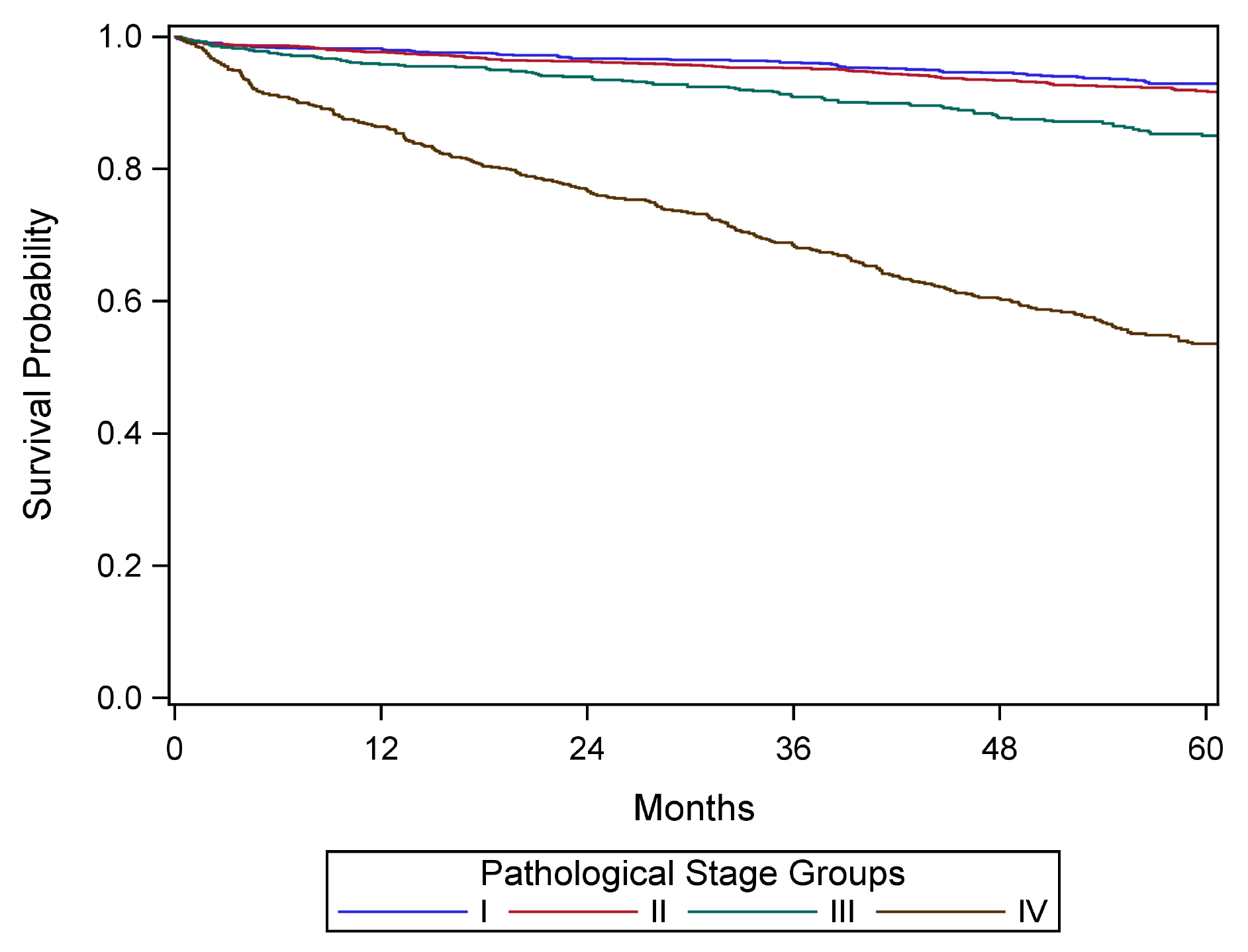
The National Cancer Database was used to select patients with PanNETs diagnosed 2010-2016 (Figure NET Pancreas-Survival, Pathological). Overall survival was defined as the time from diagnosis to death due to any cause. Patients who did not die were censored at date of last follow-up. The Kaplan-Meier method was used to estimate unadjusted overall survival. All survival analyses and figure generation were conducted with SAS version 9.4 (SAS Institute, Cary NC).

42.1 FIGURE NET PANCREAS-SURVIVAL, PATHOLOGICAL. Unadjusted overall survival by pathological stage group. The Kaplan-Meier method was used to estimate unadjusted overall survival. Overall survival time was truncated at 5 years and censored marks were omitted for visualization. Data source: NCDB 2010-2016.

Classification Rules

Introduction to TNM Staging Classification

Stage may be defined at several time points in the care of the cancer patient. To properly stage a patient's cancer, it is essential to first determine the time point in a patient's care. These points in time are termed classifications and are based on time during the continuum of evaluation and management of the disease. Then, T, N, and M categories are assigned for a particular classification (clinical, pathological, posttherapy, recurrence, and/or autopsy) by using information obtained during the relevant time frame, sometimes also referred to as a staging window. These staging windows are unique to each particular classification and are set forth explicitly in the Supplemental Information. The prognostic stage groups then are assigned using the T, N, and M categories, and sometimes also site-specific prognostic and predictive factors.

Among these classifications, the two predominant are clinical classification (i.e., pretreatment) and pathological classification (i.e., after surgical treatment as initial therapy).

Note C: Rules for Clinical TNM Classification

Clinical stage classification is based on patient history, physical examination, and any imaging done before initiation of treatment. Imaging study information may be used for clinical staging, but clinical stage may be assigned based on whatever information is available. No specific imaging is required to assign a clinical stage for any cancer site. When performed within this framework, biopsy information on regional lymph nodes and/or other sites of metastatic disease may be included in the clinical classification.

See General Staging Rules Table and Stage Classifications Table in Supplemental Information for additional guidance, including the time frame/staging window for determining clinical stage.

Clinical stage is important to record for all patients because:

Clinical stage may be the only stage classification by which comparisons can be made across all patients, because not all patients will undergo surgical treatment before other therapy, and response to treatment varies. Differences in primary therapy make comparing groups of patients difficult if that comparison is based on pathological assessment. For example, it is difficult to compare patients treated with primary surgery with those treated with chemotherapy or radiation therapy without surgery or neoadjuvant therapy.

Clinical classification is based on evidence acquired from the date of diagnosis until initiation of primary treatment. Examples of primary treatment include definitive surgery, radiation therapy, systemic therapy, and neoadjuvant radiation and systemic therapy.

Clinical Classification

Clinical assessment is based on medical history, clinical examination, imaging, and biochemical assessment. If a biopsy is performed, the results should be incorporated when assigning clinical stage.

Note P: Rules for Pathologic TNM Classification

Classification of T, N, and M after surgical treatment is denoted by use of a lowercase p prefix: pT, pN, and cM0, cM1, or pM1. The purpose of pathological classification is to provide additional precise and objective data for prognosis and outcomes, and to guide subsequent therapy.

Pathological stage classification is based on clinical stage information supplemented/modified by operative findings and pathological evaluation of the resected specimens. This classification is applicable when surgery is performed before initiation of adjuvant radiation or systemic therapy.

See General Staging Rules Table and Stage Classifications Table in Supplemental Information for additional guidance.

Pathological Classification

Pathological staging is based on surgical resection specimens. The most sensitive pathological staging is obtained by examining surgically resected primary tumor(s), lymph nodes, and distant metastases according to an established minimum pathology dataset whenever possible adopting standard synoptic reporting protocols.9,57-61

Note YC: Rules for Posttherapy Clinical TNM Classification

Stage determined after treatment for patients receiving systemic and/or radiation therapy alone or as a component of their initial treatment, or as neoadjuvant therapy before planned surgery, is referred to as posttherapy classification. It also may be referred to as post neoadjuvant therapy classification.

See General Staging Rules Table and Stage Classifications Table in Supplemental Information for additional guidance.

Observed changes between the clinical classification and the posttherapy classification may provide clinicians with information regarding the response to therapy. The clinical extent of response to therapy may guide the scope of planned surgery, and the clinical and pathological extent of response to therapy may provide prognostic information and guide the use of further adjuvant radiation and/or systemic therapy.

Classification of T, N, and M after systemic or radiation treatment intended as definitive therapy is denoted by use of a lowercase yc prefix: ycT, ycN, c/pM. The c/pM category may include cM0, cM1, or pM1. The post neoadjuvant therapy assessment of the T and N (yTNM) categories uses specific criteria. In contrast, the M category for post neoadjuvant therapy classification remains the same as that assigned in the clinical stage before initiation of neoadjuvant therapy (e.g., if there is a complete clinical response to therapy in a patient previously categorized as cM1, the M1 category is used for final yc and yp staging).

See Stage Classifications Table in Supplemental Information for additional guidance.

Posttherapy Clinical Classification

Neoadjuvant treatment is rarely performed for neuroendocrine tumors.

Note YP: Rules for Posttherapy Pathological TNM Classification

Classification of T, N, and M after systemic or radiation neoadjuvant treatment followed by surgery is denoted by use of a lowercase yp prefix: ypT, ypN, c/pM. The c/pM category may include cM0, cM1, or pM1. The post neoadjuvant therapy assessment of the T and N (yTNM) categories uses specific criteria. In contrast, the M category for post neoadjuvant therapy classification remains the same as that assigned in the clinical stage before initiation of neoadjuvant therapy (e.g., if there is a complete clinical response to therapy in a patient previously categorized as cM1, the M1 category is used for final yc and yp staging).

The time frame for assignment of ypT and ypN should be such that the post neoadjuvant therapy surgery and staging occur within a period that accommodates disease-specific circumstances.

Criteria: First therapy is systemic and/or radiation therapy followed by surgery.

y-pathological (yp) classification is based on the:

  • y-clinical stage information, and supplemented/modified by
  • operative findings, and
  • pathological evaluation of the resected specimen.

Examples of treatments that satisfy the definition of neoadjuvant therapy may be found in sources such as the NCCN Guidelines, ASCO guidelines, or other treatment guidelines. Systemic therapy includes chemotherapy, hormone therapy, and immunotherapy. Not all medications given to a patient meet the criteria for neoadjuvant therapy (e.g., a short course of therapy that is provided for variable and often unconventional reasons, should not be categorized as neoadjuvant therapy).

See Stage Classifications Table in Supplemental Information for additional guidance.

Posttherapy Pathological Classification

Neoadjuvant treatment is rarely performed for neuroendocrine tumors.

Note R: Rules for Recurrence/Retreatment TNM Classification

Staging classifications at the time of retreatment for a recurrence or disease progression is referred to as recurrence classification. It also may be referred to as retreatment classification. Classification of T, N, and M for recurrence or retreatment is denoted by use of the lowercase r prefix: rcT, rcN, rc/rpM, and rpT, rpN, rc/rpM. The rc/rpM may include rcM0, rcM1, or rpM1.

See Stage Classifications Table in Supplemental Information for additional guidance.

Note A: Rules for Autopsy TNM Classification

Staging classification for cancers identified only at autopsy is referred to as autopsy classification. This classification is used when cancer is diagnosed at autopsy and there was no prior suspicion or evidence of cancer before death. All clinical and pathological information obtained at the time of death and through post-mortem examination is included. Classification of T, N, and M at autopsy is denoted by use of the lowercase a prefix: aT, aN, aM.

See Stage Classifications Table in Supplemental Information for additional guidance.

Note CE: Clinical Examination

Guidelines have been established for the workup of patients with PanNETs.29,44,46 In general, patients should be evaluated by multiphasic computed tomography (CT) or magnetic resonance (MR) imaging to assess: 1) the proximity of the primary pancreatic NET to major vessels and 2) the clinical T, N, and M staging of the lesion before any surgical or medical therapy is considered. In addition, biochemical assessment, Ga68-SRS-PET or somatostatin receptor scintigraphy (SRS), and endoscopic ultrasonography (EUS) should be performed as appropriate. EUS provides useful information for detection of small and/or multifocal PanNETs and is the procedure of choice for performing fine-needle aspiration or fine needle biopsy of the pancreas. Studies suggest detection rates of 90-100% for pancreatic lesions.47 For localized tumors, a biopsy is not necessarily required before surgical resection. However, if a biopsy (e.g., endoscopic biopsy, percutaneous core needle biopsy, fine-needle aspiration) is performed, the results should be incorporated when assessing clinical stage.

By definition, patients with F-PanNETs present with hormone-mediated symptoms consistent with a characteristic syndrome.16,29,30 As such, the diagnosis of F-PanNETs requires demonstration of a significantly elevated hormone combined with clinical signs or symptoms of hypersecretion with a specific endocrinological workup.29,30

Assessment for hormones associated with rarer syndromes should be performed as clinically indicated.14

Unless incidentally discovered during a workup for an unrelated problem, NF-PanNETs present with symptoms due to the tumor itself, including abdominal pain, weight loss, or jaundice.13,48,49 NF-PanNETs may secrete peptides, such as CgA and the chromogranin fraction pancreastatin, PP, neuron-specific enolase, which may be helpful for the diagnosis and monitoring of affected patients.13,48 Recently, a nuclei acid-based NETtestR has also been developed with possible prognostic implications.50

Note I: Imaging

Information necessary for the clinical staging of pancreatic NETs may be obtained from physical examination; cross-sectional radiographic imaging studies, including triphasic (non-contrast, arterial, and venous) contrast-enhanced CT or MR imaging; and molecular imaging using SSTR-PET51 (refer to established guidelines for details).29 In particular, CT is best for anatomic staging of the primary tumor and for surgical planning, while MRI, and in particular hepatobiliary phase MRI is best suited for detecting and following disease in the liver.31,52,53 SSTR-PET has the highest detection rate for metastatic disease for neuroendocrine tumors and provides several other advantages including same-day results, potential for increased sensitivity compared to anatomical imaging, broader affinity profile, better spatial resolution, and easier quantification of tracer uptake.54,55,56 There are three US Food and Drug Administration (FDA) approved SSTR-PET imaging agents: 68Ga-DOTATATE (NetSpot, AAA/Novartis), 68Ga-DOTATOC, and 64Cu-DOTATATE (Detectnet, Curium). If avoidable, imaging should no longer be performed with the older radiopharmaceutical indium-111 pentetreotide (Octreoscan, Curium) which has much lower resolution than PET in addition to other disadvantages related to radiation exposure and imaging time. 18F-fluorodeoxyglucose (FDG) PET has limited value in the evaluation of low-grade PanNETs, but has an important role in higher grade tumors where it can outperform SSTR-PET due to a downregulation of SSTRs.

Unlike its exocrine counterpart (pancreatic ductal adenocarcinoma), tumor involvement of the major visceral arteries (i.e. celiac axis or superior mesenteric artery) is rare in PanNETs. The standard radiographic assessment of resectability includes evaluation for distant metastases (e.g., peritoneal, liver, bone); the patency of the superior mesenteric vein and portal vein, as well as the relationship of these vessels and their tributaries to the tumor; and the relationship of the tumor to the superior mesenteric artery, celiac axis, and hepatic artery.

General Staging Rules

These general rules apply to the application of T, N, and M categories for all anatomic sites and classifications.

TopicRules

Microscopic confirmation

  • Microscopic confirmation is necessary for TNM classification, including clinical classification (with rare exception).
  • In rare clinical scenarios, patients who do not have any biopsy or cytology of the tumor may be staged. This is recommended in rare clinical situations, only if the cancer diagnosis is NOT in doubt. In the absence of histologic confirmation, survival analysis may be performed separately from staged cohorts with histologic confirmation. Separate survival analysis is not required if clinical findings support a cancer diagnosis and specific site.

Example: Lung cancer diagnosed by CT scan only, that is, without a confirmatory biopsy

Time frame/staging window for determining clinical stage

Information gathered about the extent of the cancer is part of clinical classification:

  • from date of diagnosis before initiation of primary treatment or decision for watchful waiting or supportive care to one of the following time points, whichever is shortest:
    • 4 months after diagnosis
    • to the date of cancer progression if the cancer progresses before the end of the 4 month window; data on the extent of the cancer is only included before the date of observed progression

Time frame/staging window for determining pathological stage

Information including clinical staging data and information from surgical resection and examination of the resected specimens—if surgery is performed before the initiation of radiation and/or systemic therapy—from the date of diagnosis:

  • within 4 months after diagnosis
  • to the date of cancer progression if the cancer progresses before the end of the 4-month window; data on the extent of the cancer is included only before the date of observed progression
  • and includes any information obtained about the extent of cancer up through completion of definitive surgery as part of primary treatment if that surgery occurs later than

4 months after diagnosis and the cancer has not clearly progressed during the time window

Note: Patients who receive radiation and/or systemic therapy (neoadjuvant therapy) before surgical resection are not assigned a pathological category or stage, and instead are staged according to post neoadjuvant therapy criteria.

Time frame/staging window for staging post neoadjuvant therapy or posttherapy

After completion of neoadjuvant therapy, patients should be staged as:

  • yc: posttherapy clinicalAfter completion of neoadjuvant therapy followed by surgery, patients should be staged as:
  • yp: posttherapy pathological

The time frame should be such that the post neoadjuvant surgery and staging occur within a time frame that accommodates disease-specific circumstances, as outlined in the specific disease sites and in relevant guidelines.

Note: Clinical stage should be assigned before the start of neoadjuvant therapy.

Progression of disease

If there is documented progression of cancer before therapy or surgery, only information obtained before the documented progression is used for clinical and pathological staging.

Progression does not include growth during the time needed for the diagnostic workup, but rather a major change in clinical status.

Determination of progression is based on managing physician judgment, and may result in a major change in the treatment plan.

Uncertainty among T, N, or M categories, and/or stage groups: rules for clinical decision making

If uncertainty exists regarding how to assign a category, subcategory, or stage group, the lower of the two possible categories, subcategories, or groups is assigned for

  • T, N, or M
  • prognostic stage group/stage group

Stage groups are for patient care and prognosis based on data. Physicians may need to make treatment decisions if staging information is uncertain or unclear.

Note: Unknown or missing information for T, N, M or stage group is never assigned the lower category, subcategory, or group.

Uncertainty rules do not apply to cancer registry data

If information is not available to the cancer registrar for documentation of a subcategory, the main (umbrella) category should be assigned (e.g., T1 for a breast cancer described as < 2 cm in place of T1a, T1b, or T1c).

If the specific information to assign the stage group is not available to the cancer registrar (including subcategories or missing prognostic factor categories), the stage group should not be assigned but should be documented as unknown.

Prognostic factor category information is unavailable

If a required prognostic factor category is unavailable, the category used to assign the stage group is:

  • X, or
  • If the prognostic factor is unavailable, default to assigning the anatomic stage using clinical judgment.

Grade

The recommended histologic grading system for each disease site and/or cancer type, if applicable, is specified in each disease site and should be used by the pathologist to assign grade.

The cancer registrar will document grade for a specific site according to the coding structure in the relevant disease site.

Synchronous primary tumors in a single organ: (m) suffix

If multiple tumors of the same histology are present in one organ:

  • the tumor with the highest T category is classified and staged, and
  • the (m) suffix is used
  • An example of a preferred designation is: pT3(m) N0 M0.
  • If the number of synchronous tumors is important, an acceptable alternative designation is to specify the number of tumors. For example, pT3(4) N0 M0 indicates four synchronous primary tumors.

Note: The (m) suffix applies to multiple invasive cancers. It is not applicable for multiple foci of in situ cancer or for a mixed invasive and in situ cancer.

Synchronous primary tumors in paired organs

Cancers occurring at the same time in each of paired organs are staged as separate cancers. Examples include breast, lung, and kidney.

Exception: For tumors of the thyroid, liver, and ovary, multiplicity is a T-category criterion, thus multiple synchronous tumors are not staged independently.

Metachronous primary tumors

Second or subsequent primary cancers occurring in the same organ or in different organs outside the staging window are staged independently and are known as metachronous primary tumors.

Such cancers are not staged using the y prefix.

Unknown primary or no evidence of primary tumor

If there is no evidence of a primary tumor, or the site of the primary tumor is unknown, staging may be based on the clinical suspicion of the organ site of the primary tumor, with the tumor categorized as T0. The rules for staging cancers categorized as T0 are specified in the relevant disease sites.

Example: An axillary lymph node with an adenocarcinoma in a woman, suspected clinically to be from the breast, may be categorized as T0 N1 (or N2 or N3) M0 and assigned Stage II (or Stage III).

Examples of exception: The T0 category is not used for head and neck squamous cancer sites, as such patients with an involved lymph node are staged as unknown primary cancers using the “Cervical Nodes and Unknown Primary Tumors of the Head and Neck” system (T0 remains a valid category for human papillomavirus [HPV]- and Epstein-Barr virus [EBV]-associated oropharyngeal and nasopharyngeal cancers).

Date of diagnosis

It is important to document the date of diagnosis, because this information is used for survival calculations and time periods for staging.

The date of diagnosis is the date a physician determines the patient has cancer. It may be the date of a diagnostic biopsy or other microscopic confirmation or of clear evidence on imaging. This rule varies by disease site and shares similarities with the earlier discussion on microscopic confirmation.

Stage Classifications

Stage classifications are determined according to the point in time of the patient's care in relation to diagnosis and treatment. The five stage classifications are clinical, pathological, posttherapy/post neoadjuvant therapy, recurrence/ retreatment, and autopsy.

ClassificationDesignationDetails

Clinical

cTNM or TNM

Criteria: used for all patients with cancer identified before treatment

It is composed of diagnostic workup information, until first treatment, including:

  • clinical history and symptoms
  • physical examination
  • imaging
  • endoscopy
  • biopsy of the primary site
  • biopsy or excision of a single regional node or sentinel nodes, or sampling of regional nodes, with clinical T
  • biopsy of distant metastatic site
  • surgical exploration without resection
  • other relevant examinations

Note: Exceptions exist by site, such as complete excision of primary tumor for melanoma.

Pathological

pTNM

Criteria: used for patients if surgery is the first definitive therapy

It is composed of information from:

  • diagnostic workup from clinical staging combined with
  • operative findings, and
  • pathology review of resected surgical specimens

Posttherapy or post neoadjuvant therapy

ycTNM and ypTNM

For purposes of posttherapy or post neoadjuvant therapy, neoadjuvant therapy is defined as systemic and/or radiation therapy given before surgery; primary radiation and/or systemic therapy is treatment given as definitive therapy without surgery.

yc

The yc classification is used for staging after primary systemic and/or radiation therapy, or after neoadjuvant therapy and before planned surgery

Criteria: First therapy is systemic and/or radiation therapy

yp

The yp classification is used for staging after neoadjuvant therapy and planned post neoadjuvant therapy surgery.

Criteria: First therapy is systemic and/or radiation therapy and is followed by surgery.

Recurrence or retreatment

rTNM

This classification is used for assigning stage at time of recurrence or progression until treatment is initiated.

Criteria: Disease recurrence after disease-free interval or upon disease progression if further treatment is planned for a cancer that:

  • recurs after a disease-free interval or
  • progresses (without a disease-free interval)

rc

Clinical recurrence staging is assigned as rc.

rp

Pathological staging information is assigned as rp for the rTNM staging classification.

This classification is recorded in addition to and does not replace the original previously assigned clinical (c), pathological (p), and/or posttherapy (yc, yp) stage classifications, and these previously documented classifications are not changed.

Autopsy

aTNM

This classification is used for cancers not previously recognized that are found as an incidental finding at autopsy, and not suspected before death (i.e., this classification does not apply if an autopsy is performed in a patient with a previously diagnosed cancer).

Criteria: No cancer suspected prior to death

Both clinical and pathological staging information is used to assign aTNM.

Note S: Identification of Primary Site(s)

The pancreas is a long, coarsely lobulated gland that lies transversely across the posterior abdomen and extends from the duodenum to the splenic hilum. The organ is divided into a head with an uncinate process, a neck, a body, and a tail. These are contiguous regions without sharp anatomic distinctions. The pancreas neck lies anterior to the superior mesenteric vessels. The anterior aspect of the body of the pancreas is covered by peritoneum and is in contact with the posterior wall of the stomach; posteriorly, the pancreas extends within the retroperitoneal soft tissue to the inferior vena cava, superior mesenteric vein, splenic vein, and left adrenal and kidney. PanNETs are distributed throughout the pancreas. Tumors of the head of the pancreas are those arising to the right of the superior mesenteric-portal vein confluence (Figure NET Pancreas- Anatomy). The uncinate process is the part of the pancreatic head that extends behind the superior mesenteric vessels. The neck overlies the superior mesenteric vessels. Tumors of the body of the pancreas are defined as those arising to the left of the neck. Laterally to the left side, the body becomes the tail of the pancreas without any clear junction point.

42.fig-AJCCFig52 FIGURE NET PANCREAS-ANATOMY. Anatomy of the pancreas. This protocol stages neuroendocrine tumors of the pancreas.
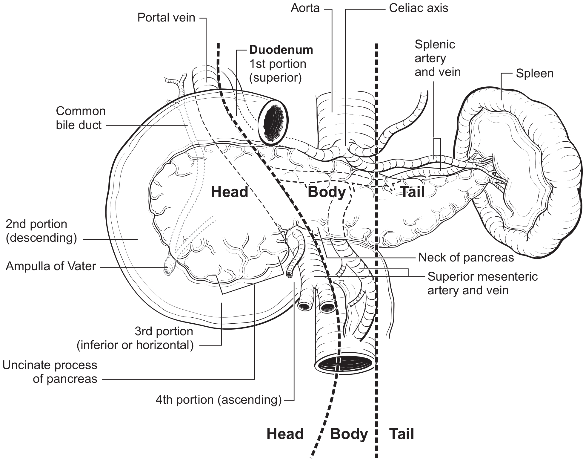
The anatomy of the pancreas is described in Note S: Identification of Primary Site(s) with a figure of the anatomy.

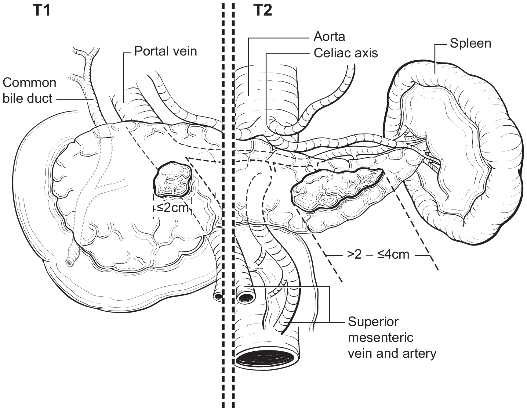
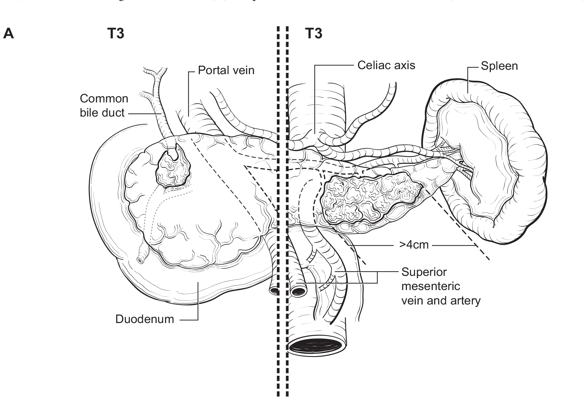
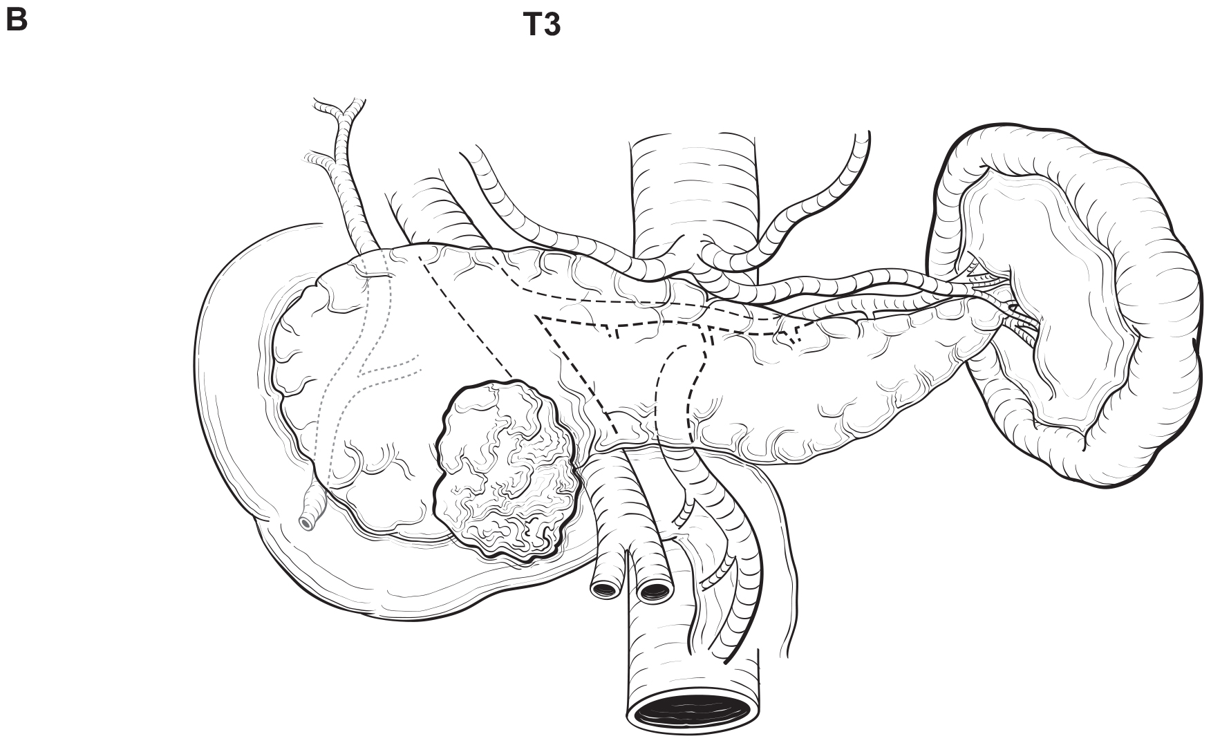
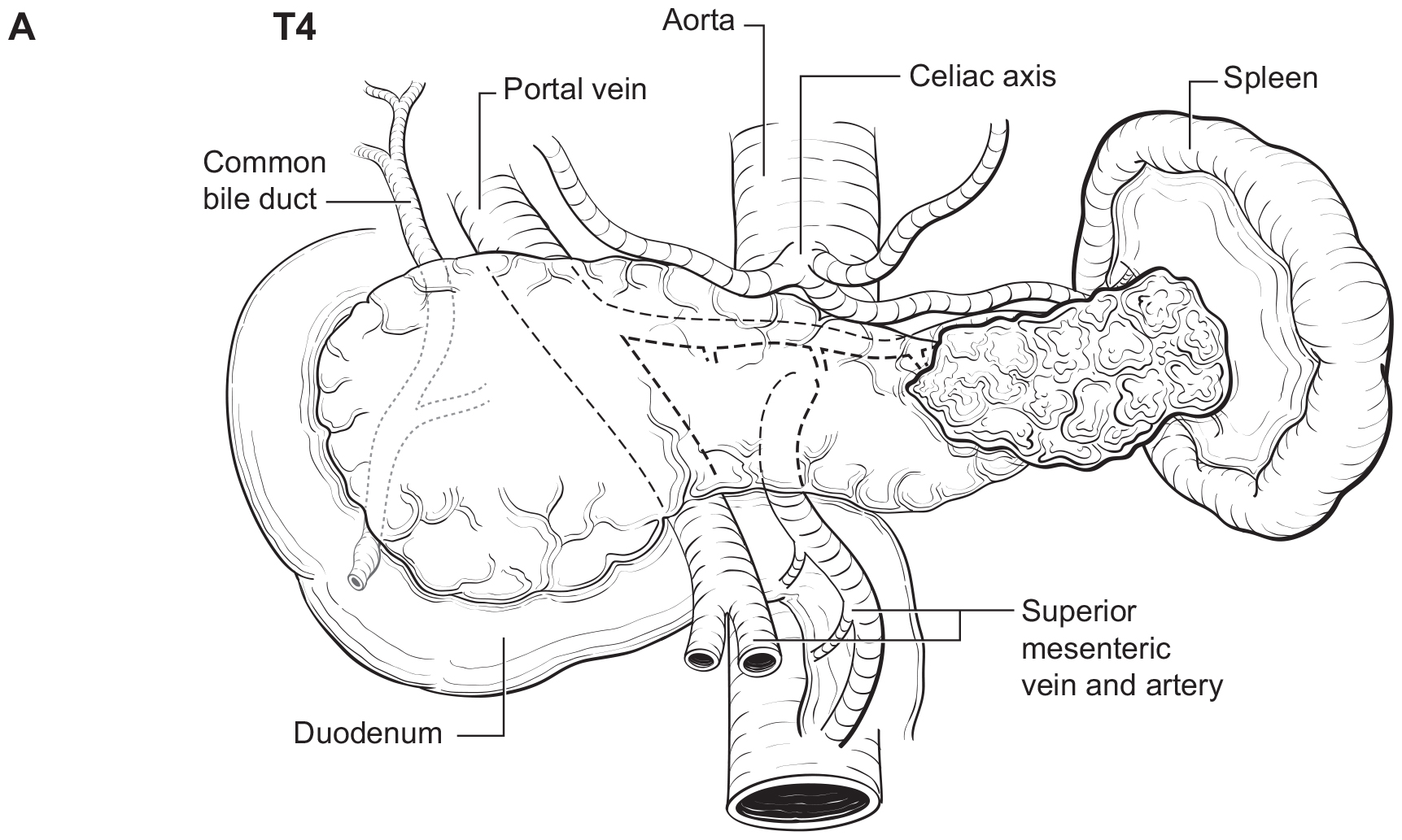
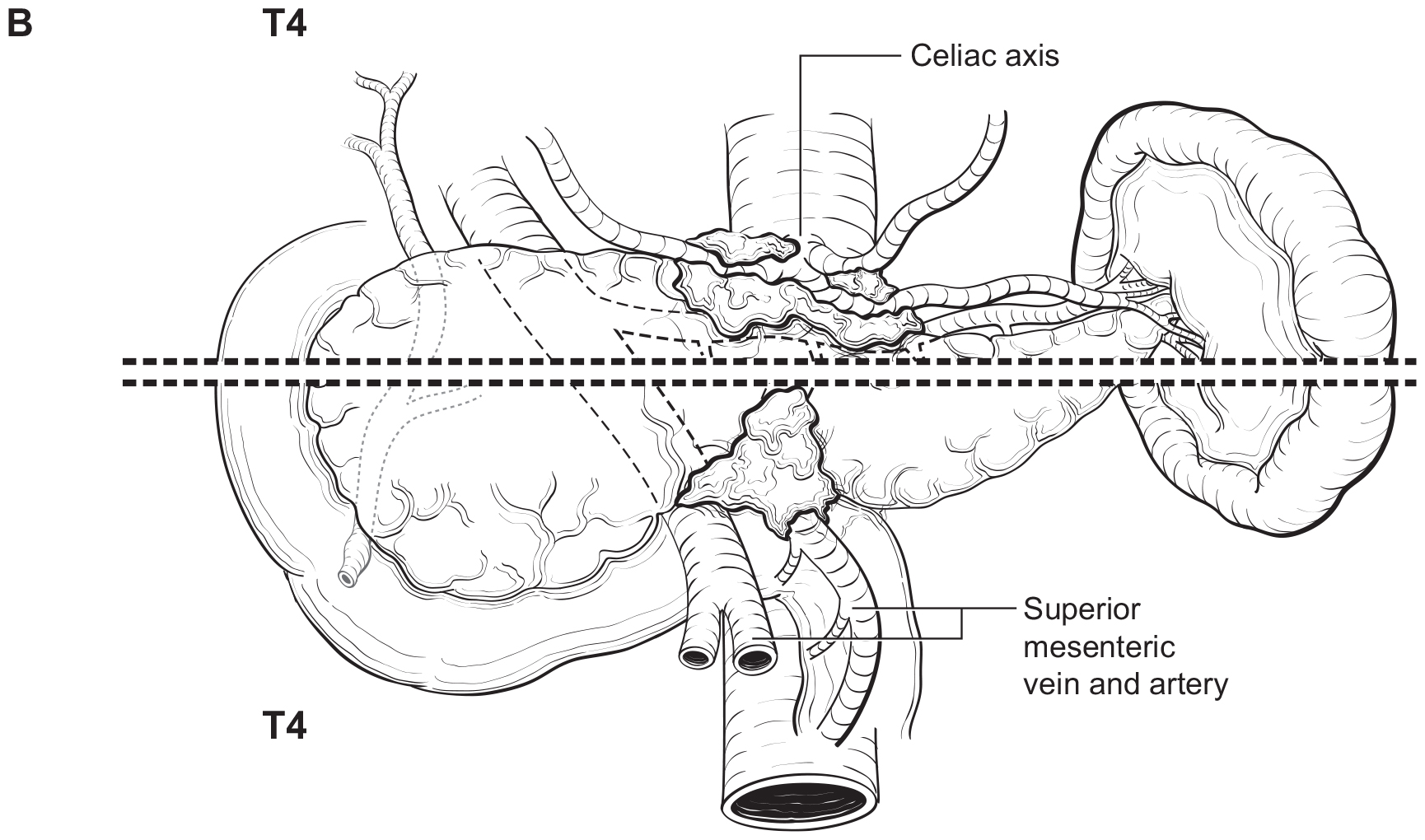
The T category definition entails recognizing the pancreas limits and tumor size for T1-T3 (Figure NET Pancreas-T1, T2 and Figure NET Pancreas-T3 (A-right side of line)). The invasion of the duodenal wall (Figure NET Pancreas-T3 (B)) including the ampulla or common bile duct (Figure NET Pancreas-T3 (A-left side of line) is T3. Invasion of nearby anatomical organs (stomach, spleen, colon, adrenal gland) or of the wall of large vessels (celiac axis, superior mesenteric artery/vein, common hepatic artery/vein, portal vein, splenic artery/vein and gastroduodenal artery/vein) is considered T4 (Figure NET Pancreas-T4).

Partial resection (pancreaticoduodenectomy or distal pancreatectomy) or total pancreatectomy, including the tumor and associated regional lymph nodes, provides the optimal information for pathological staging. In pancreaticoduodenectomy specimens, the bile duct, pancreatic duct, and superior mesenteric artery margins should be evaluated grossly and microscopically. The superior mesenteric artery margin also has been termed the retroperitoneal, vascular, or uncinate margin. In total pancreatectomy specimens, the bile duct and retroperitoneal margins should be assessed. Duodenal (with pylorus-preserving pancreaticoduodenectomy) and gastric (with standard pancreaticoduodenectomy) margins rarely are involved, but their status should be included in the surgical pathology report. Reporting of margins may be facilitated by ensuring documentation of the pertinent margins: 1) common bile (hepatic) duct, 2) pancreatic neck, 3) superior mesenteric artery, 4) other soft tissue margins (i.e., posterior pancreatic, duodenum, and stomach).

42.1 FIGURE NET PANCREAS-T1, T2. T1 (left of dotted line) is defined as tumor limited to the pancreas 2 cm in greatest dimension. T2 (right of dotted line) is defined as tumor limited to the pancreas > 2 cm but 4 cm in greatest dimension.

42.1 FIGURE NET PANCREAS-T3. T3 is defined as tumor limited to the pancreas, > 4 cm in greatest dimension (A-right of the dotted line) or tumor invading the duodenum (B), ampulla of Vater or common bile duct (A-left of the dotted line).
42.1 FIGURE NET PANCREAS-T4. T4 is defined as tumor invading adjacent organs (stomach, spleen, colon, adrenal gland) (A) or the wall of large vessels (celiac axis, superior mesenteric artery/vein, splenic artery/vein, gastroduodenal artery/ vein, portal vein) (B).
Regional Lymph Nodes

A rich lymphatic network surrounds the pancreas, and accurate tumor staging requires analysis of regional lymph nodes. Optimal histologic examination of a pancreaticoduodenectomy specimen should include analysis of 11-15 lymph nodes.31 For left sided tumors, en-bloc splenectomy may facilitate margin clearance and adequate lymph node retrieval. For patients with PanNETs at low risk for malignant behavior (i.e. low risk sporadic tumors, or insulinoma) the need for splenectomy should be balanced against the benefit of splenic preservation. Similarly, parenchymal sparing procedures such as central pancreatectomy or enucleation may be considered in highly selected low risk cases; the lymph node yield in these cases may be limited.

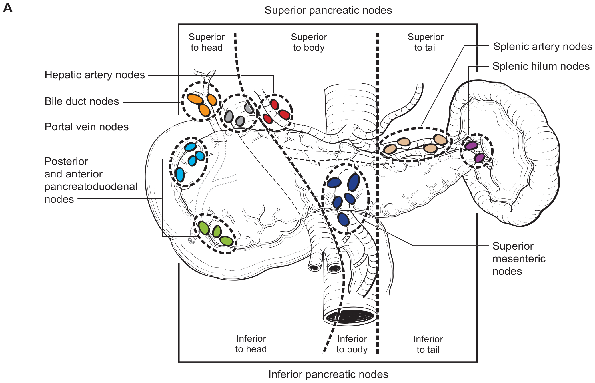
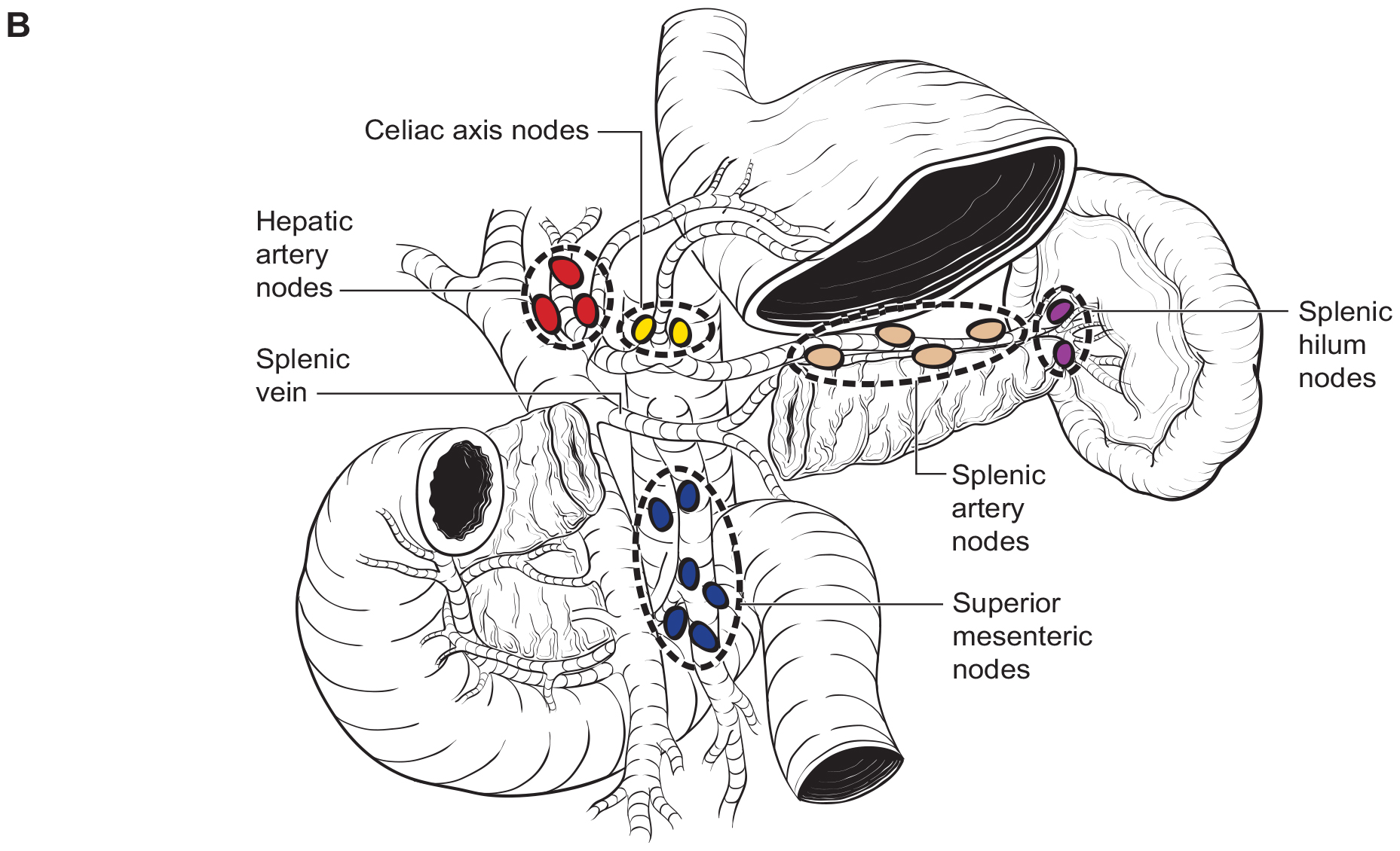
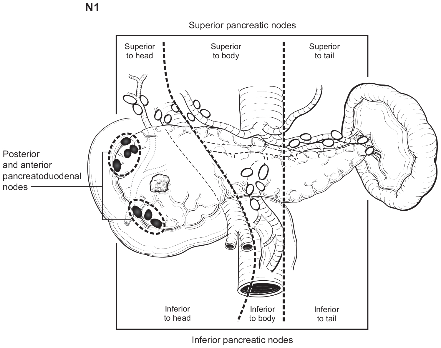
The number of lymph nodes examined and the number of positive lymph nodes should be specified in the pathology report. Anatomic division of regional lymph nodes is not necessary; however, separately submitted lymph nodes should be reported as labeled by the surgeon. An N category (N0 or N1) should be assigned as long as at least one lymph node has been assessed, even if the optimal number of lymph nodes have not been examined. NX should be applied only if no lymph nodes were assessed (e.g., if enucleation was performed). Positive peritoneal cytology is considered M1. The standard regional lymph node basins and soft tissues resected for tumors located in the head and neck of the pancreas include lymph nodes along the common bile duct, common hepatic artery, portal vein, posterior and anterior pancreatoduodenal arcades, and the superior mesenteric vein and right lateral wall of the superior mesenteric artery (Figure NET Pancreas- Nodal Map-A). For cancers located in the body and tail, regional lymph node basins include lymph nodes along the common hepatic artery, celiac axis, splenic artery, and splenic hilum (Figure NET Pancreas- Nodal Map-B). Involvement of peripancreatic lymph nodes is considered regional disease and classified as N1 (Figure NET Pancreas- N1).

42.1 FIGURE NET PANCREAS-NODAL MAP. Regional lymph nodes for Neuroendocrine Tumors of the Pancreas.
42.1 FIGURE NET PANCREAS-N1. N1 is defined as tumor involvement of regional lymph node(s).
Metastatic Sites

The M category is assigned based on the method of assessment, not the classification. The options are cM0, cM1, and pM1. cM0 indicates no distant metastasis by signs/symptoms, imaging, etc.; no evidence of tumor in distant sites or organs. This category is not assigned by pathologists. cM1 indicates the distant metastasis are identified through physical exam, signs or symptoms, any imaging results, or direct visualization during procedures. pM1 indicates at least one site of distant metastasis has been confirmed microscopically; such microscopic evidence includes tumor identified in biopsy, resection, or cytology from fine needle aspiration. Not all sites of distant metastasis must be confirmed microscopically in order to assign pM1. pM1 is further subdivided into three subcategories: M1a-metastasis confined to the liver, M1b-metastasis in at least one extrahepatic site and M1c-both hepatic and extrahepatic metastases.

The terms pM0, cMX and pMX are not valid categories.

Distant spread is common on presentation and most commonly involves the liver. Metastases to other sites, such as lung, bones, and peritoneum, also may occur. Involvement of the para-aortic or other distant lymph nodes (i.e., retroperitoneal, retrocrural, and mesenteric lymph nodes) is considered M1 disease. Seeding of the peritoneum (even if limited to the lesser sac region) is considered M1.

Additional Factors Relevant for Clinical

Additional data elements that are clinically significant but not required for staging are identified with a dagger symbol.(†)

Prognostic Tumor Characteristics

  1. †Mitotic count
  2. †Ki-67 index
  3. †Associated genetic syndrome
  4. †Chomogranin A (CgA)
  5. †Functionality
  6. †DAXX/ATRXst
  7. †ARX, PDX1 expression

Operative FactorsResidual Tumor (R)

RR Definition
R0Complete resection, margins histologically negative, no residual tumor left after resection
R1Incomplete resection, margins histologically involved, microscopic tumor remains after resection of gross disease (relevant to resection margins that are microscopically involved by tumor)
R2Incomplete resection, margins involved, or gross disease remains
Non-Tumor Factors
  1. †Age
  2. †Family history of cancer
  3. †Smoking
  4. †Alcohol consumption
  5. †Increased body mass index
  6. †Diabetes
  7. †MEN1 and other rarer heritable gene conditions (neurofibromatosis type 1, Cowden syndrome, tuberous sclerosis, von Hippel-Lindau syndrome)

Note AF: Additional Factors Impacting Treatment Decisions During First Treatment Phase

Prognostic Tumor Characteristics

Mitotic Count

Tumor grade is determined by mitotic count and Ki-67 index and correlates with progression-free survival, overall survival, and lymph node status in PanNETs.24,62,63 Mitotic count should be assessed as the number of mitoses per 2 mm2 (usually equalizing 10 high-power fields (HPF)); at least 10 mm2 equalizing 50 HPF should be evaluated if possible.

AJCC Level of Evidence: I

Mitotic count, # of mitoses per 2 mm2 (specify: )

  • < 2
  • 2 to 20>
  • 20%
  • Other (specify):
  • Not performed

Ki-67 Index

The Ki-67 index typically is measured using the MIB1 antibody, by counting the number of immunolabeled tumor cells per 500 to 2,000 cells in areas of highest nuclear labeling; the Ki-67 index is expressed as a percentage. AJCC Level of Evidence: I

Ki-67 index (specify:)

  • < 3%
  • 3% to 20%
  • >20%
  • Other (specify):
  • Not performed

Associated Genetic Syndrome

PanNETs may arise in the setting of an inherited cancer syndrome characterized by a germline mutation. Tumors arising in the setting of an inherited cancer syndrome may be multiple and appear to be associated with a better prognosis than sporadic tumors, at least in the setting of MEN1.24 AJCC Level of Evidence: II

This factor should be recorded as follows:

  • Familial syndrome
    • Multiple endocrine neoplasia type 1 (alteration in MEN1)
    • Von Hippel-Lindau disease (mutation in VHL gene)
    • Neurofibromatosis type 1 (mutation in Nf1)
    • Tuberous sclerosis complex (mutation in TSC1 or TSC2)
    • Mahvash disease (pancreatic NET caused by inactivating glucagon receptor mutation)64
    • Other syndrome
  • Sporadic tumor
  • Unknown/unable to assessChomogranin A (CgA)CgA is a 49-kDa acidic polypeptide present in the secretory granules of all neuroendocrine cells. CgA is a general NET marker, and plasma or serum CgA may be used as a marker in patients with either F- or NF-pancreatic NETs.13,65-67 CgA has prognostic significance, with higher levels indicating a worse prognosis.68 Changes over time may be useful in assessing for recurrence after surgery or response to therapy in patients with metastatic disease.65,66,69,70Despite the potential merits of monitoring CgA levels, the clinical utility of CgA is limited by the fact that it is falsely elevated in the setting of PPI use, chronic atrophic gastritis, renal failure, severe hypertension, and other conditions.8 Moreover, levels may fluctuate based on time of collection and fasting versus non-fasting states. Also, the upper limit of normal (ULN) varies widely depending on the assay used and whether plasma or serum is assessed; thus, both the assay and type of sample should be considered when comparing CgA values over time.71 As a result, routine measurementof CgA is not a consensus National Comprehensive Cancer Network (NCCN) recommendation. Multiple Clinical Laboratory Improvement Amendments (CLIA)-licensed and College of American Pathology (CAP)-accredited reference laboratories in the United States can measure CgA levels. AJCC Level of Evidence: IIFunctionalityTumors with hormone expression by tissue immunohistochemistry but not associated with a clinically relevant syndrome or signs should be recorded as nonfunctional. Similarly, tumors associated with elevated blood levels of hormones that are not associated with clinical symptoms also should be recorded as nonfunctional. Insulinomas typically have a low risk of metastasis and thus carry a good prognosis; the outcome of other F-PanNETs appears to be similar to that ofnonfunctional tumors in most studies.24,72 Importantly, the clinical manifestations and morbidity of F-PanNETs may differ, and in some cases, mortality may relate to the hormonal syndrome rather than to the extent of the neoplasm. AJCC Level of Evidence: IIIFunctionality should be characterized as follows:
  • Functional
    • Insulinoma
    • Gastrinoma (ZES)
    • Glucagonoma
    • VIPoma (Verner-Morrison syndrome)
    • Somatostatinoma
    • ACTHoma
    • PanNET causing carcinoid syndrome (5-HIAA, serotonin excess)
    • PanNET causing hypercalcemia (PTHrp or other)
    • Other
  • Nonfunctional
  • Unknown/unable to assess

DAXX/ATRX

Altered telomere length (ALT) status associate with poor prognosis in PanNET.73 DAXX and ATRX loss in turn associate with ALT status predicting metastasis and poor prognosis.18,74 The mutually exclusive loss of DAXX or ATRX can be investigated in tumor samples by specific immunohistochemistry as effective surrogate for mutational analysis.75-77 AJCC Level of Evidence: II

ARX, PDX1 Expression

ARX and PDX1 gene expression may stratify non-functioning PanNET based on cell type subtyping and with relevant prognostic differences and namely high metastatic potential (doi 10.3390/cancers1206146120). AJCC Level of Evidence: II

Non-Tumor Factors

Similar to other cancers, non-tumor factors may associate with higher risk of PanNET and include age (older age), family history of cancer (any type), smoking habits, alcohol consumption, increased body mass index and diabetes.8 AJCC Level of Evidence: II

Rationale for Changes and Future Directions

There is evidence suggesting, similar to pancreas adenocarcinoma, the number of positive lymph-nodes may be associated with patient survival for PanNETs.27,78,79 This information may be achieved by subdividing the N risk category in sub-categories (N0, N1a and N1b), where N1a may identify 1-3 positive lymph nodes and N1b more than 3 positive lymph nodes.27 Preliminary data also suggest that MGMT methylation status may be a predictive marker for temozolomide based therapies. As discussed, molecular profiling, and neuroendocrine neoplasms test (NETest)50 may provide prognostic information as well.

Registry Data Collection Variables
  1. †Size of tumor (value, unknown)
  2. †Presence of invasion into adjacent organs/structures (Y/N)
    1. If yes, which ones (pick all that apply):
      • Stomach (Y/N)
      • Duodenum (Y/N)
      • Spleen (Y/N)
      • Colon (Y/N)
      • Other:
    2. If yes, were multiple adjacent organs involved (Y/N)
  3. †Presence of necrosis
  4. †Number of tumors (multicentric disease at primary site)
  5. †Lymph node status (including number of lymph nodes assessed and number of positive nodes)
  6. †Grade (based on Ki-67 and/or mitotic count; G1, G2, G3, unknown)
  7. †Mitotic count (value, unknown)
  8. †Ki-67 index (value, unknown)
  9. †Perineural invasion (Y/N)
  10. †Lymphovascular invasion (Y/N)
  11. †Margin status (+/-)
  12. †Functional status (Y/N, type of syndrome)
  13. †Genetic syndrome (Y/N, type of syndrome)
  14. †Location in pancreas (head, tail, body, junction body/tail, junction body/head, unknown)
  15. †Type of surgery (enucleation, distal pancreatectomy with or without splenectomy, central pancreatectomy, pancreaticoduodenectomy-Whipple procedure, unknown, other)
  16. †Preoperative CgA level (absolute value with ULN; unknown)
  17. †Age of patient

Prognostic Factors

Prognostic Factors Required for Stage Grouping

Beyond the factors used to assign T, N, or M categories, no additional prognostic factors are required for stage grouping.

AJCC Level of Evidence:

Level I

The available evidence includes consistent results from multiple large, well-designed, and well-conducted national and international studies in appropriate patient populations, with appropriate endpoints and appropriate treatments.

Both prospective studies and retrospective population-based registry studies are acceptable; studies should be evaluated on the basis of methodology rather than chronology.

Level II

The available evidence is obtained from at least 1 large, well-designed, and well-conducted study in appropriate patient populations with appropriate endpoints and with external validation.

Level III

The available evidence is somewhat problematic because of a factor such as the number, size, or quality of individual studies; inconsistency of results across individual studies; appropriateness of patient population used in 1 or more studies; or the appropriateness of outcomes used in 1 or more studies.

Level IV

The available evidence is insufficient because appropriate studies have not yet been performed.

Table adapted from Amin et al.52

TNM Definitions

Assignment of AJCC TNM

AJCC data elements required for staging are identified with an asterisk (*).

*Stage classification based on time frame and criteria(see Rules for Classification)

The same classification should be used throughout the assignment of TNM and stage group.

  • c (clinical)
  • p (pathological)
  • yc (posttherapy clinical)
  • yp (posttherapy pathological)
T CategoryT Criteria
TXTumor cannot be assessed
T1Tumor limited to the pancreas, 2 cm in greatest dimension
T2Tumor limited to the pancreas, > 2 cm but 4 cm in greatest dimension
T3Tumor limited to the pancreas, > 4 cm in greatest dimension; or tumor invading the duodenum, ampulla of Vater, or common bile duct
T4Tumor invading adjacent organs (stomach, spleen, colon, adrenal gland) or the wall of large vessels (celiac axis, superior mesenteric artery/vein, splenic artery/vein, gastroduodenal artery/vein, portal vein)

Limited to the pancreas means there is no invasion of adjacent organs (stomach, spleen, colon, adrenal gland) or the wall of large vessels (celiac axis or the superior mesenteric artery). Extension of tumor into peripancreatic adipose tissue is NOT a basis for staging.

Note: Multiple tumors should be designated as such (the largest tumor should be used to assign T category):

  • Use T(#); e.g., pT3(4) N0 M0, or
  • Use the m suffix, T(m); e.g., pT3(m) N0 M0

Primary Tumor Suffix

(m) Multiple synchronous primary tumors

N CategoryN Criteria
NXRegional lymph nodes cannot be assessed
N0No tumor involvement of regional lymph node(s)
N1Tumor involvement of regional lymph node(s)

Regional Lymph Nodes Suffix

(f) FNA or core needle biopsy

M CategoryM Criteria
cM0No distant metastasis
cM1Distant metastasis
cM1aMetastasis confined to liver
cM1bMetastases in at least one extrahepatic site (e.g., lung, ovary, nonregional lymph node, peritoneum, bone)
cM1cBoth hepatic and extrahepatic metastases
pM1Microscopic confirmation of distant metastasis
pM1aMicroscopic confirmation of metastasis confined to liver
pM1bMicroscopic confirmation of metastases in at least one extrahepatic site (e.g., lung, ovary, nonregional lymph node, peritoneum, bone)
pM1cMicroscopic confirmation of both hepatic and extrahepatic metastases

Stage Prognostic

!!Calculator!!

AJCC Prognostic Stage Groups (Note PSG)

AJCC data elements required for staying are identified with an asterisk (*).

*AJCC prognostic stage group is assigned based on the stage classification and categories chosen.

When T is…and N is…and M is…Then the stage group is…
T1N0M0I
T2, T3N0M0II
T4N0M0III
Any TN1M0III
Any TAny NM1IV

Note PSG: AJCC Prognostic Stage Groups

The managing physician alone is responsible for assigning the patient's stage, because only (s)he routinely has access to all the pertinent information from physical examination, imaging studies, biopsies, diagnostic procedures, surgical findings, and pathology reports. Although the pathologist and the radiologist provide important staging information, and may provide important T-, N-, and/or M-related information, stage is defined ultimately from the synthesis of an array of patient history and physical examination findings supplemented by imaging and pathology data.

Histologic grade

HISTOLOGIC GRADE (G)

Histologic Grade (G) (Note G)

Additional data elements that are clinically significant but not required for staging are identified with a dagger symbol (†).

†Grade is assigned based on histopathologic assessment.

GG Definition
GXGrade cannot be assessed
G1Mitotic count (per 2 mm2)§ < 2 and Ki-67 index (%)§§ < 3
G2Mitotic count (per 2 mm2)§ = 2-20 or Ki-67 index (%)§§ = 3-20
G3Mitotic count (per 2 mm2)§> 20 or Ki-67 index (%)§§> 20

§2 mm2 equals 10 high-power fields at 40x magnification and an ocular field diameter of 0.5 mm; the number of high-power field for 10 mm2 is different using microscopes with different field diameter; at least 10 mm2 must be evaluated in areas of highest mitotic density.

§§MIB1 antibody; % of 500-2000 cells in areas of highest nuclear labeling.

The final grade is determined by whichever (mitotic count and Ki-67 index) places the tumor in higher grade category.

Note G: Grade (G)

Grading of PanNETs is required for prognostic stratification and should be performed on all resection specimens and on biopsy specimens containing sufficient tumor tissue to allow accurate measurement of proliferation (mitotic count per 2 mm2 (must equalize 10HPF) to determine the mitotic index and Ki-67 % in a minimum of 500 cells to determine the Ki-67 index). If multiple PanNETs or PanNET sites are sampled (e.g., a primary tumor as well as a metastasis), the grade of each primary and each site should be recorded separately. If multiple foci are sampled within a single anatomic site (e.g., multiple liver metastases), the highest grade may be recorded. The grading scheme is currently endorsed by WHO 2019 and 2022 for gastrointestinal and pancreatic neuroendocrine neoplasms.

MIB1 antibody is recommended for Ki-67 assessment. The Ki-67 index is done on the region with the highest labeling rate (“hot spot”), determined by examining the Ki-67 stain at low magnification. In the event of discordance between the grade indicated by the mitotic count and that suggested by the Ki-67 index, the higher grade should be assigned. Nuclear pleomorphism is not a useful feature for grading neuroendocrine neoplasms. Although necrosis has been regarded as a prognostic factor in some studies, its presence is not part of the grading scheme.

Bibliography

  1. Xu Z, Wang L, Dai S, et al. Epidemiologic Trends of and Factors Associated With Overall Survival for Patients With Yao JC, Eisner MP, Leary C, et al. Population-based study of islet cell carcinoma. Annals of surgical oncology. 2007;14(12):3492-3500.
  2. Dasari A, Shen C, Halperin D, et al. Trends in the Incidence, Prevalence, and Survival Outcomes in Patients With Neuroendocrine Tumors in the United States. JAMA Oncol. 2017;3(10):1335-1342.
  3. Boyar Cetinkaya R, Aagnes B, Thiis-Evensen E, Tretli S, Bergestuen DS, Hansen S. Trends in Incidence of Neuroendocrine Neoplasms in Norway: A Report of 16,075 Cases from 1993 through 2010. Neuroendocrinology. 2015.
  4. Das S, Dasari A. Epidemiology, Incidence, and Prevalence of Neuroendocrine Neoplasms: Are There Global Differences? Curr Oncol Rep. 2021;23(4):43.
  5. Capurso G, Falconi M, Panzuto F, et al. Risk factors for sporadic pancreatic endocrine tumors: a case-control study of prospectively evaluated patients. Am J Gastroenterol. 2009;104(12):3034-3041.
  6. Halfdanarson TR, Bamlet WR, McWilliams RR, et al. Risk factors for pancreatic neuroendocrine tumors: a clinic-based case-control study. Pancreas. 2014;43(8):1219-1222.
  7. Hassan MM, Phan A, Li D, Dagohoy CG, Leary C, Yao JC. Risk factors associated with neuroendocrine tumors: A U.S.-based case-control study. Int J Cancer. 2008;123(4):867-873.
  8. Leoncini E, Carioli G, La Vecchia C, Boccia S, Rindi G. Risk factors for neuroendocrine neoplasms: a systematic review and meta-analysis. Ann Oncol. 2016;27(1):68-81.
  9. Board WCTE. WHO Classification of Tumours. Digestive System Tumours: WHO Classification of Tumours, Volume 1. World Health Organization; 2019.
  10. Rindi G, Klersy C, Albarello L, et al. Competitive Testing of the WHO 2010 versus the WHO 2017 Grading of Pancreatic Neuroendocrine Neoplasms: Data from a Large International Cohort Study. Neuroendocrinology. 2018;107(4):375-386.
  11. Vinik AI, Woltering EA, Warner RR, et al. NANETS consensus guidelines for the diagnosis of neuroendocrine tumor. Pancreas. 2010;39(6):713-734.
  12. Falconi M, Eriksson B, Kaltsas G, et al. ENETS Consensus Guidelines Update for the Management of Patients with Functional Pancreatic Neuroendocrine Tumors and Non-Functional Pancreatic Neuroendocrine Tumors. Neuroendocrinology. 2016;103(2):153-171.
  13. Metz DC, Jensen RT. Gastrointestinal neuroendocrine tumors: pancreatic endocrine tumors. Gastroenterology. 2008;135(5):1469-1492.
  14. Vinik AI, Chaya C. Clinical Presentation and Diagnosis of Neuroendocrine Tumors. Hematology/oncology clinics of North America. 2016;30(1):21-48.
  15. Nahmias A, Grozinsky-Glasberg S, Salmon A, Gross DJ. Pancreatic neuroendocrine tumors with transformation to insulinoma: an unusual presentation of a rare disease. Endocrinology, diabetes & metabolism case reports. 2015;2015:150032.
  16. Jensen RT, Cadiot G, Brandi ML, et al. ENETS Consensus Guidelines for the management of patients with digestive neuroendocrine neoplasms: functional pancreatic endocrine tumor syndromes. Neuroendocrinology. 2012;95(2):98-119.
  17. Scarpa A, Chang DK, Nones K, et al. Whole-genome landscape of pancreatic neuroendocrine tumours. Nature. 2017;543(7643):65-71.
  18. Hackeng WM, Brosens LAA, Kim JY, et al. Non-functional pancreatic neuroendocrine tumours: ATRX/DAXX and alternative lengthening of telomeres (ALT) are prognostically independent from ARX/PDX1 expression and tumour size. Gut. 2022;71(5):961-973.
  19. Cejas P, Drier Y, Dreijerink KMA, et al. Enhancer signatures stratify and predict outcomes of non-functional pancreatic neuroendocrine tumors. Nat Med. 2019;25(8):1260-1265.
  20. Dreijerink KM, Hackeng WM, Singhi AD, Heaphy CM, Brosens LA. Clinical implications of cell-of-origin epigenetic characteristics in non-functional pancreatic neuroendocrine tumors. J Pathol. 2022;256(2):143-148.
  21. Hackeng WM, Schelhaas W, Morsink FHM, et al. Alternative Lengthening of Telomeres and Differential Expression of Endocrine Transcription Factors Distinguish Metastatic and Non-metastatic Insulinomas. Endocr Pathol. 2020;31(2):108-118.
  22. Oberg K. The genetics of neuroendocrine tumors. Seminars in oncology. 2013;40(1):37-44.
  23. Raj N, Shah R, Stadler Z, et al. Real-Time Genomic Characterization of Metastatic Pancreatic Neuroendocrine Tumors Has Prognostic Implications and Identifies Potential Germline Actionability. JCO Precis Oncol. 2018;2018.
  24. Rindi G, Falconi M, Klersy C, et al. TNM staging of neoplasms of the endocrine pancreas: results from a large international cohort study. Journal of the National Cancer Institute. 2012;104(10):764-777.
  25. Parekh JR, Wang SC, Bergsland EK, et al. Lymph node sampling rates and predictors of nodal metastasis in pancreatic neuroendocrine tumor resections: the UCSF experience with 149 patients. Pancreas. 2012;41(6):840-844.
  26. Chiramel J, Almond R, Slagter A, et al. Prognostic importance of lymph node yield after curative resection of gastroenteropancreatic neuroendocrine tumours. World J Clin Oncol. 2020;11(4):205-216.
  27. Guarneri G, de Mestier L, Landoni L, et al. Prognostic Role of Examined and Positive Lymph Nodes after Distal Pancreatectomy for Non-Functioning Neuroendocrine Neoplasms. Neuroendocrinology. 2021;111(8):728-738.
  28. Zhang Z, Liu M, Ji S, et al. Prognostic Value and Clinical Predictors of Lymph Node Metastases in Pancreatic Neuroendocrine Tumors. Pancreas. 2020;49(3):381-386.
  29. Kulke MH, Shah MH, Benson AB, 3rd, et al. Neuroendocrine tumors, version 1.2015. Journal of the National Comprehensive Cancer Network : JNCCN. 2015;13(1):78-108.
  30. Shah MH, Goldner WS, Benson AB, et al. Neuroendocrine and Adrenal Tumors, Version 2.2021, NCCN Clinical Practice Guidelines in Oncology. Journal of the National Comprehensive Cancer Network : JNCCN. 2021;19(7):839-868.
  31. Howe JR, Merchant NB, Conrad C, et al. The North American Neuroendocrine Tumor Society Consensus Paper on the Surgical Management of Pancreatic Neuroendocrine Tumors. Pancreas. 2020;49(1):1-33.
  32. Bolm L, Nebbia M, Wei AC, et al. Long-term Outcomes of Parenchyma-sparing and Oncologic Resections in Patients With Nonfunctional Pancreatic Neuroendocrine Tumors <3 cm in a Large Multicenter Cohort. Ann Surg. 2022;276(3):522-531.
  33. Chamberlain RS, Canes D, Brown KT, et al. Hepatic neuroendocrine metastases: does intervention alter outcomes? Journal of the American College of Surgeons. 2000;190(4):432-445.
  34. Mayo SC, de Jong MC, Pulitano C, et al. Surgical management of hepatic neuroendocrine tumor metastasis: results from an international multi-institutional analysis. Annals of surgical oncology. 2010;17(12):3129-3136.
  35. Caplin ME, Pavel M, Cwikla JB, et al. Lanreotide in metastatic enteropancreatic neuroendocrine tumors. The New England journal of medicine. 2014;371(3):224-233.
  36. Rinke A, Muller HH, Schade-Brittinger C, et al. Placebo-controlled, double-blind, prospective, randomized study on the effect of octreotide LAR in the control of tumor growth in patients with metastatic neuroendocrine midgut tumors: a report from the PROMID Study Group. Journal of clinical oncology : official journal of the American Society of Clinical Oncology. 2009;27(28):4656-4663.
  37. Oberg K. Somatostatin analog octreotide LAR in gastro-entero-pancreatic tumors. Expert review of anticancer therapy. 2009;9(5):557-566.
  38. Strosberg JR, Fine RL, Choi J, et al. First-line chemotherapy with capecitabine and temozolomide in patients with metastatic pancreatic endocrine carcinomas. Cancer. 2011;117(2):268-275.
  39. Moertel CG, Lefkopoulo M, Lipsitz S, Hahn RG, Klaassen D. Streptozocin-doxorubicin, streptozocin-fluorouracil or chlorozotocin in the treatment of advanced islet-cell carcinoma. The New England journal of medicine. 1992;326(8):519-523.
  40. Cheng PN, Saltz LB. Failure to confirm major objective antitumor activity for streptozocin and doxorubicin in the treatment of patients with advanced islet cell carcinoma. Cancer. 1999;86(6):944-948.
  41. Kunz PL, Catalano PJ, Nimeiri H, et al. A randomized study of temozolomide or temozolomide and capecitabine in patients with advanced pancreatic neuroendocrine tumors: A trial of the ECOG-ACRIN Cancer Research Group (E2211). Journal of Clinical Oncology. 2018;36(15_suppl):4004-4004.
  42. Raymond E, Dahan L, Raoul JL, et al. Sunitinib malate for the treatment of pancreatic neuroendocrine tumors. The New England journal of medicine. 2011;364(6):501-513.
  43. Yao JC, Shah MH, Ito T, et al. Everolimus for advanced pancreatic neuroendocrine tumors. The New England journal of medicine. 2011;364(6):514-523.
  44. Kulke MH, Anthony LB, Bushnell DL, et al. NANETS treatment guidelines: well-differentiated neuroendocrine tumors of the stomach and pancreas. Pancreas. 2010;39(6):735-752.
  45. Halfdanarson TR, Strosberg JR, Tang L, et al. The North American Neuroendocrine Tumor Society Consensus Guidelines for Surveillance and Medical Management of Pancreatic Neuroendocrine Tumors. Pancreas. 2020;49(7):863-881.
  46. Ramage JK, Ahmed A, Ardill J, et al. Guidelines for the management of gastroenteropancreatic neuroendocrine (including carcinoid) tumours (NETs). Gut. 2012;61(1):6-32.
  47. anderson MA, Carpenter S, Thompson NW, Nostrant TT, Elta GH, Scheiman JM. Endoscopic ultrasound is highly accurate and directs management in patients with neuroendocrine tumors of the pancreas. The American journal of gastroenterology. 2000;95(9):2271-2277.
  48. Oberg K, Eriksson B. Nuclear medicine in the detection, staging and treatment of gastrointestinal carcinoid tumours. Best practice & research Clinical endocrinology & metabolism. 2005;19(2):265-276.
  49. Plockinger U, Rindi G, Arnold R, et al. Guidelines for the diagnosis and treatment of neuroendocrine gastrointestinal tumours. A consensus statement on behalf of the European Neuroendocrine Tumour Society (ENETS). Neuroendocrinology. 2004;80(6):394-424.
  50. Puliani G, Di Vito V, Feola T, et al. NETest: A Systematic Review Focusing on the Prognostic and Predictive Role. Neuroendocrinology. 2022;112(6):523-536.
  51. Falconi M, Bartsch DK, Eriksson B, et al. ENETS Consensus Guidelines for the management of patients with digestive neuroendocrine neoplasms of the digestive system: well-differentiated pancreatic non-functioning tumors. Neuroendocrinology. 2012;95(2):120-134.
  52. Morse B, Jeong D, Thomas K, Diallo D, Strosberg JR. Magnetic Resonance Imaging of Neuroendocrine Tumor Hepatic Metastases: Does Hepatobiliary Phase Imaging Improve Lesion Conspicuity and Interobserver Agreement of Lesion Measurements? Pancreas. 2017;46(9):1219-1224.
  53. Hope TA, Pampaloni MH, Nakakura E, et al. Simultaneous (68)Ga-DOTA-TOC PET/MRI with gadoxetate disodium in patients with neuroendocrine tumor. Abdom Imaging. 2015;40(6):1432-1440.
  54. Lebtahi R, Cadiot G, Sarda L, et al. Clinical impact of somatostatin receptor scintigraphy in the management of patients with neuroendocrine gastroenteropancreatic tumors. Journal of nuclear medicine : official publication, Society of Nuclear Medicine. 1997;38(6):853-858.
  55. Hofman MS, Lau WF, Hicks RJ. Somatostatin receptor imaging with 68Ga DOTATATE PET/CT: clinical utility, normal patterns, pearls, and pitfalls in interpretation. Radiographics : a review publication of the Radiological Society of North America, Inc. 2015;35(2):500-516.
  56. Toumpanakis C, Kim MK, Rinke A, et al. Combination of cross-sectional and molecular imaging studies in the localization of gastroenteropancreatic neuroendocrine tumors. Neuroendocrinology. 2014;99(2):63-74.
  57. Bosman FT, Carneiro F, Hruban RH, Theise ND. WHO classification of tumours of the digestive system. World Health Organization; 2010.
  58. Travis WD, Brambilla E, Muller-Hermelink HK, Harris CC. Pathology and genetics of tumours of the lung, pleura, thymus and heart. 2004.
  59. Klimstra DS, Modlin IR, Adsay NV, et al. Pathology reporting of neuroendocrine tumors: application of the Delphic consensus process to the development of a minimum pathology data set. The American journal of surgical pathology. 2010;34(3):300-313.
  60. Klimstra DS. Pathology reporting of neuroendocrine tumors: essential elements for accurate diagnosis, classification, and staging. Seminars in oncology. 2013;40(1):23-36.
  61. van Velthuysen MF, Couvelard A, Rindi G, et al. ENETS standardized (synoptic) reporting for neuroendocrine tumour pathology. J Neuroendocrinol. 2022;34(3):e13100.
  62. Panzuto F, Boninsegna L, Fazio N, et al. Metastatic and locally advanced pancreatic endocrine carcinomas: analysis of factors associated with disease progression. Journal of clinical oncology : official journal of the American Society of Clinical Oncology. 2011;29(17):2372-2377.
  63. Panzuto F, Merola E, Rinzivillo M, et al. Advanced digestive neuroendocrine tumors: metastatic pattern is an independent factor affecting clinical outcome. Pancreas. 2014;43(2):212-218.
  64. Lucas MB, Yu VEYR. Mahvash disease: pancreatic neuroendocrine tumor syndrome caused by inactivating glucagon receptor mutation. Journal of Molecular and Genetic Medicine. 2013.
  65. de Herder WW. Biochemistry of neuroendocrine tumours. Best practice & research Clinical endocrinology & metabolism. 2007;21(1):33-41.
  66. Stronge RL, Turner GB, Johnston BT, et al. A rapid rise in circulating pancreastatin in response to somatostatin analogue therapy is associated with poor survival in patients with neuroendocrine tumours. Annals of clinical biochemistry. 2008;45(Pt 6):560-566.
  67. Yang X, Yang Y, Li Z, et al. Diagnostic value of circulating chromogranin a for neuroendocrine tumors: a systematic review and meta-analysis. PloS one. 2015;10(4):e0124884.
  68. Han X, Zhang C, Tang M, et al. The value of serum chromogranin A as a predictor of tumor burden, therapeutic response, and nomogram-based survival in well-moderate nonfunctional pancreatic neuroendocrine tumors with liver metastases. European journal of gastroenterology & hepatology. 2015;27(5):527-535.
  69. Massironi S, Rossi RE, Casazza G, et al. Chromogranin A in diagnosing and monitoring patients with gastroenteropancreatic neuroendocrine neoplasms: a large series from a single institution. Neuroendocrinology. 2014;100(2-3):240-249.
  70. Yao JC, Pavel M, Phan AT, et al. Chromogranin A and neuron-specific enolase as prognostic markers in patients with advanced pNET treated with everolimus. The Journal of clinical endocrinology and metabolism. 2011;96(12):3741-3749.
  71. Glinicki P, Jeske W, Kapuscinska R, Zgliczynski W. Comparison of chromogranin A (CgA) levels in serum and plasma (EDTA2K) and the respective reference ranges in healthy males. Endokrynologia Polska. 2015;66(1):53-56.
  72. Strosberg JR, Cheema A, Weber J, Han G, Coppola D, Kvols LK. Prognostic validity of a novel American Joint Committee on Cancer Staging Classification for pancreatic neuroendocrine tumors. Journal of clinical oncology : official journal of the American Society of Clinical Oncology. 2011;29(22):3044-3049.
  73. Kim JY, Brosnan-Cashman JA, An S, et al. Alternative Lengthening of Telomeres in Primary Pancreatic Neuroendocrine Tumors Is Associated with Aggressive Clinical Behavior and Poor Survival. Clin Cancer Res. 2017;23(6):1598-1606.
  74. Singhi AD, Liu TC, Roncaioli JL, et al. Alternative Lengthening of Telomeres and Loss of DAXX/ATRX Expression Predicts Metastatic Disease and Poor Survival in Patients with Pancreatic Neuroendocrine Tumors. Clin Cancer Res. 2017;23(2):600-609.
  75. Heaphy CM, Bi WL, Coy S, et al. Telomere length alterations and ATRX/DAXX loss in pituitary adenomas. Mod Pathol. 2020;33(8):1475-1481.
  76. Heaphy CM, Singhi AD. The diagnostic and prognostic utility of incorporating DAXX, ATRX, and alternative lengthening of telomeres to the evaluation of pancreatic neuroendocrine tumors. Hum Pathol. 2022;129:11-20.
  77. Hechtman JF, Klimstra DS, Nanjangud G, Frosina D, Shia J, Jungbluth AA. Performance of DAXX Immunohistochemistry as a Screen for DAXX Mutations in Pancreatic Neuroendocrine Tumors. Pancreas. 2019;48(3):396-399.
  78. Partelli S, Javed AA, andreasi V, et al. The number of positive nodes accurately predicts recurrence after pancreaticoduodenectomy for nonfunctioning neuroendocrine neoplasms. Eur J Surg Oncol. 2018;44(6):778-783.
  79. Zhang P, Li YL, Qiu XD, et al. Clinicopathological characteristics and risk factors for recurrence of well-differentiated pancreatic neuroendocrine tumors after radical surgery: a case-control study. World J Surg Oncol. 2019;17(1):66.