Cancers Staged Using This Staging System
Adult Hodgkin and non-Hodgkin lymphomas
Cancers Not Staged Using This Staging System
These histopathologic types of cancer | Are staged according to the classification for | and can be found in chapter |
---|---|---|
Ocular adnexal lymphoma | Ocular adnexal lymphoma | 71 |
Pediatric lymphoma | Pediatric lymphoma | 80 |
Primary cutaneous Lymphoma | Primary cutaneous lymphoma | 81 |
Multiple myeloma | Plasma cell myeloma | 82 |
Summary of Changes
Change | Details of Change | Level of Evidence |
---|---|---|
Ann Arbor staging | The Cotswold modification1 of the Ann Arbor staging system2,3 has been updated to the Lugano classification4 | I |
A and B Classification (Symptoms) | B symptoms were eliminated for non-Hodgkin lymphoma (retained for Hodgkin lymphoma). | I |
X subscript | X subscript for bulk was eliminated. The diameter of the largest mass must be recorded. | I |
Stage III | The extension of disease into extralymphatic sites (E lesions) was eliminated from Stage III; any extralymphatic involvement with nodal disease above and below the diaphragm is Stage IV. | I |
Stage IIIS | Involvement of the spleen no longer part of stage grouping. | I |
Stage II | Although four staging categories are retained, the concept of the Lugano classification is to divide patients into limited and advanced stages. Stage II bulky is variably categorized as limited- or advanced-stage based on the histology and prognostic factors. | I |
Imaging | Posteroanterior chest X-ray is no longer required for the determination of bulk in Hodgkin or non-Hodgkin lymphoma. | I |
ICD-O-3 Topography Codes
Code | Description |
---|---|
C00.0 | External upper lip |
C00.1 | External lower lip |
C00.2 | External lip, NOS |
C00.3 | Mucosa of upper lip |
C00.4 | Mucosa of lower lip |
C00.5 | Mucosa of lip, NOS |
C00.6 | Commissure of lip |
C00.8 | Overlapping lesion of lip |
C00.9 | Lip, NOS |
C01.9 | Base of tongue, NOS |
C02.0 | Dorsal surface of tongue, NOS |
C02.1 | Border of tongue |
C02.2 | Ventral surface of tongue, NOS |
C02.3 | Anterior 2/3 of tongue, NOS |
C02.4 | Lingual tonsil |
C02.8 | Overlapping lesion of tongue |
C02.9 | Tongue, NOS |
C03.0 | Upper gum |
C03.1 | Lower gum |
C03.9 | Gum, NOS |
C04.0 | Anterior floor of mouth |
C04.1 | Lateral floor of mouth |
C04.8 | Overlapping lesion of floor of mouth |
C04.9 | Floor of mouth, NOS |
C05.0 | Hard palate |
C05.1 | Soft palate, NOS |
C05.2 | Uvula |
C05.8 | Overlapping lesion of palate |
C05.9 | Palate, NOS |
C06.0 | Cheek mucosa |
C06.1 | Vestibule of mouth |
C06.2 | Retromolar area |
C06.8 | Overlapping lesion of other and unspecified parts of mouth |
C06.9 | Mouth, NOS |
C07.9 | Parotid gland |
C08.0 | Submandibular gland |
C08.1 | Sublingual gland |
C08.1 | Sublingual gland duct |
C08.8 | Overlapping lesion of major salivary glands |
C08.9 | Major salivary gland, NOS |
C09.0 | Tonsillar fossa |
C09.1 | Tonsillar pillar |
C09.8 | Overlapping lesion of tonsil |
C09.9 | Tonsil, NOS |
C10.0 | Vallecula |
C10.1 | Anterior surface of epiglottis |
C10.2 | Lateral wall of oropharynx |
C10.3 | Posterior wall of oropharynx |
C10.4 | Branchial cleft |
C10.8 | Overlapping lesion of oropharynx |
C10.9 | Oropharynx, NOS |
C11.0 | Superior wall of nasopharynx |
C11.1 | Posterior wall of nasopharynx |
C11.2 | Lateral wall of nasopharynx |
C11.3 | Anterior wall of nasopharynx |
C11.8 | Overlapping lesion of nasopharynx |
C11.9 | Nasopharynx, NOS |
C12.9 | Pyriform sinus |
C13.0 | Postcricoid region |
C13.1 | Hypopharyngeal aspect of aryepiglottic fold |
C13.2 | Posterior wall of hypopharynx |
C13.8 | Overlapping lesion of hypopharynx |
C13.9 | Hypopharynx, NOS |
C14.0 | Pharynx, NOS |
C14.2 | Waldeyer ring |
C14.8 | Overlapping lesion of lip, oral cavity and pharynx |
C15.0 | Cervical esophagus |
C15.1 | Thoracic esophagus |
C15.2 | Abdominal esophagus |
C15.3 | Upper third of esophagus |
C15.4 | Middle third of esophagus |
C15.5 | Lower third of esophagus |
C15.8 | Overlapping lesion of esophagus |
C15.9 | Esophagus, NOS |
C16.0 | Cardia, NOS |
C16.1 | Fundus of stomach |
C16.2 | Body of stomach |
C16.3 | Gastric antrum |
C16.4 | Pylorus |
C16.5 | Lesser curvature of stomach, NOS |
C16.6 | Greater curvature of stomach, NOS |
C16.8 | Overlapping lesion of stomach |
C16.9 | Stomach, NOS |
C17.0 | Duodenum |
C17.1 | Jejunum |
C17.2 | Ileum |
C17.3 | Meckel diverticulum |
C17.8 | Overlapping lesion of small intestine |
C17.9 | Small intestine, NOS |
C18.0 | Cecum |
C18.1 | Appendix |
C18.2 | Ascending colon |
C18.3 | Hepatic flexure of colon |
C18.4 | Transverse colon |
C18.5 | Splenic flexure of colon |
C18.6 | Descending colon |
C18.7 | Sigmoid colon |
C18.8 | Overlapping lesion of colon |
C18.9 | Colon, NOS |
C19.9 | Rectosigmoid junction |
C20.9 | Rectum, NOS |
C21.0 | Anus, NOS |
C21.1 | Anal canal |
C21.2 | Cloacogenic zone |
C21.8 | Overlapping lesion of rectum, anus and anal canal |
C22.0 | Liver |
C22.1 | Intrahepatic bile duct |
C23.9 | Gallbladder |
C24.0 | Extrahepatic bile duct |
C24.1 | Ampulla of Vater |
C24.8 | Overlapping lesion of biliary tract |
C24.9 | Biliary tract, NOS |
C25.0 | Head of pancreas |
C25.1 | Body of pancreas |
C25.2 | Tail of pancreas |
C25.3 | Pancreatic duct |
C25.4 | Islets of Langerhans |
C25.7 | Other specified parts of pancreas |
C25.8 | Overlapping lesion of pancreas |
C25.9 | Pancreas, NOS |
C26.0 | Intestinal tract, NOS |
C26.8 | Overlapping lesion of digestive system |
C26.9 | Gastrointestinal tract, NOS |
C30.0 | Nasal cavity |
C30.1 | Middle ear |
C31.0 | Maxillary sinus |
C31.1 | Ethmoid sinus |
C31.2 | Frontal sinus |
C31.3 | Sphenoid sinus |
C31.8 | Overlapping lesion of accessory sinuses |
C31.9 | Accessory sinus, NOS |
C32.0 | Glottis |
C32.1 | Supraglottis |
C32.2 | Subglottis |
C32.3 | Laryngeal cartilage |
C32.8 | Overlapping lesion of larynx |
C32.9 | Larynx, NOS |
C33.9 | Trachea |
C34.0 | Main bronchus |
C34.1 | Upper lobe, lung |
C34.2 | Middle lobe, lung |
C34.3 | Lower lobe, lung |
C34.8 | Overlapping lesion of lung |
C34.9 | Lung, NOS |
C37.9 | Thymus |
C38.0 | Heart |
C38.1 | Anterior mediastinum |
C38.2 | Posterior mediastinum |
C38.3 | Mediastinum, NOS |
C38.4 | Pleura, NOS |
C38.8 | Overlapping lesion of heart, mediastinum and pleura |
C39.0 | Upper respiratory tract, NOS |
C39.8 | Overlapping lesion of respiratory system and intrathoracic organs |
C39.9 | Ill-defined sites within respiratory system |
C40.0 | Long bones of upper limb, scapula and associated joints |
C40.1 | Short bones of upper limb and associated joints |
C40.2 | Long bones of lower limb and associated joints |
C40.3 | Short bones of lower limb and associated joints |
C40.8 | Overlapping lesion of bones, joints and articular cartilage of limbs |
C40.9 | Bone of limb, NOS |
C41.0 | Bones of skull and face and associated joints |
C41.1 | Mandible |
C41.2 | Vertebral column |
C41.3 | Rib, sternum, clavicle and associated joints |
C41.4 | Pelvic bones, sacrum, coccyx and associated joints |
C41.8 | Overlapping lesion of bones, joints and articular cartilage |
C41.9 | Bone, NOS |
C42.0 | Blood |
C42.1 | Bone marrow |
C42.2 | Spleen |
C42.3 | Reticuloendothelial system, NOS |
C42.4 | Hematopoietic system, NOS |
C47.0 | Peripheral nerves and autonomic nervous system of head, face, and neck |
C47.1 | Peripheral nerves and autonomic nervous system of upper limb and shoulder |
C47.2 | Peripheral nerves and autonomic nervous system of lower limb and hip |
C47.3 | Peripheral nerves and autonomic nervous system of thorax |
C47.4 | Peripheral nerves and autonomic nervous system of abdomen |
C47.5 | Peripheral nerves and autonomic nervous system of pelvis |
C47.6 | Peripheral nerves and autonomic nervous system of trunk, unspecified |
C47.8 | Overlapping lesion of peripheral nerves and autonomic nervous system |
C47.9 | Autonomic nervous system, NOS |
C48.0 | Retroperitoneum |
C48.1 | Specified parts of peritoneum |
C48.2 | Peritoneum, NOS |
C48.8 | Overlapping lesion of retroperitoneum and peritoneum |
C49.0 | Connective, subcutaneous and other soft tissues of head, face, and neck |
C49.1 | Connective, subcutaneous and other soft tissues of upper limb and shoulder |
C49.2 | Connective, subcutaneous and other soft tissues of lower limb and hip |
C49.3 | Connective, subcutaneous and other soft tissues of thorax |
C49.4 | Connective, subcutaneous and other soft tissues of abdomen |
C49.5 | Connective, subcutaneous and other soft tissues of pelvis |
C49.6 | Connective, subcutaneous and other soft tissues of trunk NOS |
C49.8 | Overlapping lesion of connective, subcutaneous and other soft tissues |
C49.9 | Connective, subcutaneous and other soft tissues, NOS |
C50.0 | Nipple |
C50.1 | Central portion of breast |
C50.2 | Upper-inner quadrant of breast |
C50.3 | Lower-inner quadrant of breast |
C50.4 | Upper-outer quadrant of breast |
C50.5 | Lower-outer quadrant of breast |
C50.6 | Axillary tail of breast |
C50.8 | Overlapping lesion of breast |
C50.9 | Breast, NOS |
C51.1 | Labium minus |
C51.2 | Clitoris |
C51.8 | Overlapping lesion of vulva |
C51.9 | Vulva, NOS |
C52.9 | Vagina, NOS |
C53.0 | Endocervix |
C53.1 | Exocervix |
C53.8 | Overlapping lesion of cervix uteri |
C53.9 | Cervix uteri |
C54.0 | Isthmus uteri |
C54.1 | Endometrium |
C54.2 | Myometrium |
C54.3 | Fundus uteri |
C54.8 | Overlapping lesion of corpus uteri |
C54.9 | Corpus uteri |
C55.9 | Uterus, NOS |
C56.9 | Ovary |
C57.0 | Fallopian tube |
C57.1 | Broad ligament |
C57.2 | Round ligament |
C57.3 | Parametrium |
C57.4 | Uterine adnexa |
C57.7 | Other specified parts of female genital organs |
C57.8 | Overlapping lesion of female genital organs |
C57.9 | Female genital tract, NOS |
C58.9 | Placenta |
C60.0 | Prepuce |
C60.1 | Glans penis |
C60.2 | Body of penis |
C60.8 | Overlapping lesion of penis |
C61.9 | Prostate gland |
C62.0 | Undescended testis |
C62.1 | Descended testis |
C62.9 | Testis, NOS |
C63.0 | Epididymis |
C63.1 | Spermatic cord |
C63.7 | Other specified parts of male genital organs |
C63.8 | Overlapping lesion of male genital organs |
C63.9 | Male genital organs, NOS |
C64.9 | Kidney, NOS |
C65.9 | Renal pelvis |
C66.9 | Ureter |
C67.0 | Trigone of bladder |
C67.1 | Dome of bladder |
C67.2 | Lateral wall of bladder |
C67.3 | Anterior wall of bladder |
C67.4 | Posterior wall of bladder |
C67.5 | Bladder neck |
C67.6 | Ureteric orifice |
C67.7 | Urachus |
C67.8 | Overlapping lesion of bladder |
C67.9 | Bladder, NOS |
C68.0 | Urethra |
C68.1 | Paraurethral gland |
C68.8 | Overlapping lesion of urinary organs |
C68.9 | Urinary system, NOS |
C69.1 | Cornea, NOS |
C69.2 | Retina |
C69.3 | Choroid |
C69.4 | Ciliary body |
C73.9 | Thyroid gland |
C74.0 | Cortex of adrenal gland |
C74.1 | Medulla of adrenal gland |
C74.9 | Adrenal gland, NOS |
C76.0 | Head, face or neck, NOS |
C76.1 | Thorax, NOS |
C76.2 | Abdomen, NOS |
C76.3 | Pelvis, NOS |
C76.4 | Upper limb, NOS |
C76.5 | Lower limb, NOS |
C76.7 | Other ill-defined sites |
C76.8 | Overlapping lesion of ill-defined sites |
C77.0 | Lymph nodes of head, face, and neck |
C77.1 | Intrathoracic lymph nodes |
C77.2 | Intra-abdominal lymph nodes |
C77.3 | Lymph nodes of axilla or arm |
C77.4 | Lymph nodes of inguinal region or leg |
C77.5 | Pelvic lymph nodes |
C77.8 | Lymph nodes of multiple regions |
C77.9 | Lymph node, NOS |
C80.9 | Unknown primary site |
WHO Classification of Tumors
This list includes histology codes and preferred terms from the WHO Classification of Tumors and the International Classification of Diseases for Oncology (ICD-O). Most of the terms in this list represent malignant behavior. For cancer reporting purposes, behavior codes /3 (denoting malignant neoplasms), /2 (denoting in situ neoplasms), and in some cases /1 (denoting neoplasms with uncertain and unknown behavior) may be appended to the 4-digit histology codes to create a complete morphology code.
Code | Description |
---|---|
9675 | Malignant lymphoma, mixed small and large cell, diffuse [obs] |
9678 | Primary effusion lymphoma |
9679 | Mediastinal (thymic) large B-cell lymphoma |
9680 | Malignant lymphoma, large B-cell, diffuse, NOS |
9684 | Malignant lymphoma, large B-cell, diffuse, immunoblastic, NOS |
9688 | T-cell rich large B-cell lymphoma |
9712 | Intravascular large B-cell lymphoma |
9735 | Plasmablastic lymphoma |
9737 | ALK-positive large B-cell lymphoma |
9738 | Large B-cell lymphoma arising in human herpesvirus 8 (HHV8)-associated multicentric Castleman disease |
International Agency for Research on Cancer, World Health Organization. International Classification of Diseases for Oncology. ICD-O-3-Online.http://codes.iarc.fr/home. Accessed August 16, 2017. Used with permission.
All newly diagnosed patients with malignant lymphomas should have formal documentation of the anatomic disease extent before the initial therapeutic intervention; that is, clinical stage must be assigned and recorded. Although patients with recurrent disease generally do not have a new clinical stage assigned at the time of relapse, some prognostic models include stage at the time of second-line therapy, particularly in Hodgkin lymphoma (HL) and diffuse large B-cell lymphoma (DLBCL), with intent to proceed with high-dose therapy and autologous stem cell rescue.5-8 In all cases, recording of the anatomic disease extent at the time of relapse is recommended.
Lugano Classification Modification of the Ann Arbor Staging System
Anatomic staging of lymphomas traditionally has been based on the Ann Arbor classification system, which was originally developed more than 30 years ago for HL. It was based on the relatively predictable pattern of spread of HL and improved the ability to determine which patients might be suitable candidates for radiation therapy.2 It was updated as the Cotswold system to address some of the issues present in the original staging system and to accommodate newer diagnostic techniques, including computed tomography (CT) scan.1 It subsequently was applied to non-Hodgkin lymphoma (NHL) as well, despite the fact that the pattern of spread is less predictable than that of HL. The Ann Arbor classification has been accepted as the best means of describing the anatomic disease extent and has been useful as a universal system for a variety of lymphomas; therefore, it was adopted by the AJCC and the Union for International Cancer Control (UICC) as the official system for classifying the anatomic extent of disease in HL and NHL, with the exception of cutaneous lymphomas (e.g., mycosis fungoides), which are discussed later in this chapter. However, advances in diagnostics and therapy provided the impetus to review and modernize the evaluation and staging of lymphoma. Workshops were held at the 11th and 12th International Conference on Malignant Lymphoma to study areas in need of clarification or updating and then to review the proposed changes. The Lugano classification was published and forms the basis for revised recommendations regarding anatomic staging and evaluation of disease before and after therapy.4 This staging system is adopted by the AJCC.
For the purposes of coding and staging, lymph nodes, Waldeyer's ring, thymus, and spleen are considered nodal or lymphatic sites. Extranodal or extralymphatic sites include the adrenal glands, blood, bone, bone marrow, central nervous system (CNS; leptomeningeal and parenchymal brain disease), gastrointestinal (GI) tract, gonads, kidneys, liver, lungs, skin, ocular adnexae (conjunctiva, lacrimal glands, and orbital soft tissue), skin, uterus, and others. HL rarely presents in an extranodal site alone, but about 25% of NHLs are extranodal at presentation. The frequency of extranodal presentation varies dramatically among different lymphomas, however, with some (e.g., mycosis fungoides and mucosa-associated lymphoid tissue [MALT] lymphomas) being virtually always extranodal, except in advanced stages of the diseases, and some (e.g., follicular lymphoma) seldom being extranodal, except for bone marrow involvement.
The Lugano classification includes an E suffix for lymphomas with either localized extralymphatic presentation (Stage IE) or by contiguous spread from nodal disease (Stage IIE). For example, lymphoma presenting in the thyroid gland with cervical lymph node involvement should be staged as IIE. However, in a change from the Cotswold modification of the Ann Arbor staging system, E lesions do not apply to patients with Stage III nodal disease; any patient with nodal disease above and below the diaphragm with concurrent contiguous extralymphatic involvement is Stage IV (previously Stage IIIE). Frequently, extensive lymph node involvement is associated with extranodal extension of disease that also may directly invade other organs. Such extension should be described with the E suffix if the nodal disease is on one side of the diaphragm. For example, mediastinal lymph nodes with adjacent lung extension should be classified as Stage IIE disease. Other examples of Stage IIE diseases include extension into the anterior chest wall and into the pericardium from a large mediastinal mass (two areas of extralymphatic involvement) and no nodal involvement below the diaphragm; involvement of the iliac bone in the presence of adjacent iliac lymph node involvement and no nodal involvement above the diaphragm; involvement of a lumbar vertebral body in conjunction with para-aortic lymph node involvement and no nodal involvement above the diaphragm; and involvement of the pleura or chest wall as an extension from adjacent internal mammary nodes. A pleural or pericardial effusion with negative (or unknown) cytology is not an E lesion. Liver involvement is an exception; any liver involvement by contiguous or noncontiguous spread should be recorded as Stage IV.
The definition of disease bulk varies according to lymphoma histology. In HL, the extent of mediastinal disease is defined as the ratio between the maximum diameter of the mediastinal mass and maximal intrathoracic diameter based on CT imaging in the Lugano classification. In HL, bulk at other sites is defined as a mass >10 cm. A recent analysis has suggested that in early stage disease, masses > 7 cm (at any site) may dictate the inclusion of radiation to provide optimal outcomes.9 For NHL, the recommended definitions of bulk vary by lymphoma subtype. In follicular lymphoma, 6 cm has been suggested based on the Follicular Lymphoma International Prognostic Index, version 2 (FLIPI-2) and its validation.10,11 In DLBCL, cutoffs ranging from 5 to 10 cm have been used, although 10 cm is recommended.12
Lymph Node Regions
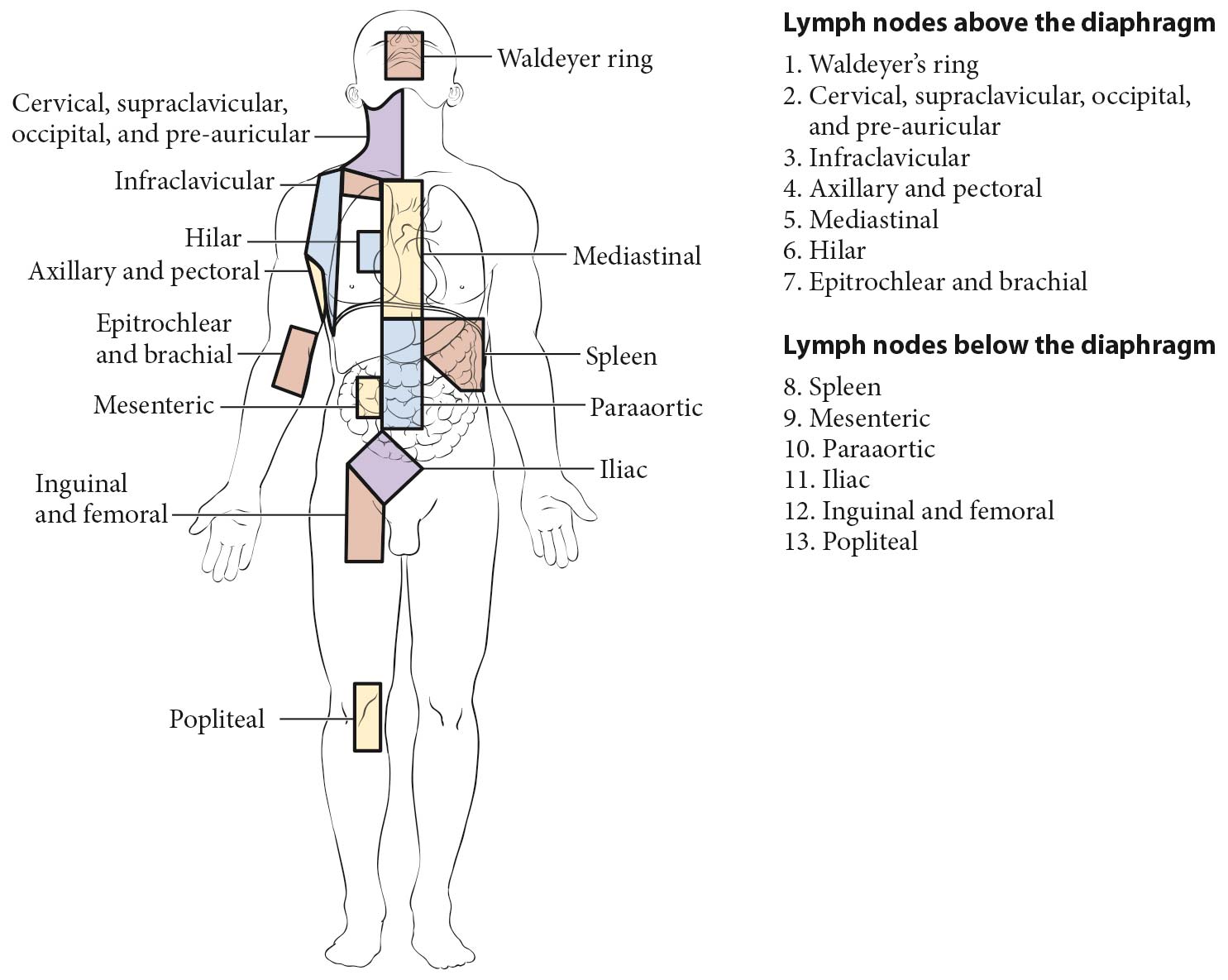
The staging classification for lymphoma uses the term lymph node region. The lymph node regions were defined at the Rye Symposium in 1965 and have been used in the Ann Arbor classification; this is unchanged in the Lugano classification (Figure 105.1). They are not based on any physiologic principles but rather have been agreed upon by convention. The currently accepted classification of core nodal regions is as follows:
In addition to these core regions, HL and NHLs may involve epitrochlear lymph nodes, popliteal lymph nodes, internal mammary lymph nodes (considered mediastinal by convention), occipital lymph nodes, submental lymph nodes, preauricular lymph nodes (all considered cervical, Figure 105.1), and many other small nodal areas. Clinical prognostic models may include specific definitions of nodal regions. For example, in follicular lymphoma, the FLIPI-2 uses a different definition of nodal regions (see prognostic factors for follicular lymphoma). This is also the case in determination of favorable versus unfavorable early-stage HL as proposed by the German Hodgkin Study Group (GHSG) and the European Organisation for Research and Treatment of Cancer (EORTC; see prognostic factors for HL).
A and B Classification (Symptoms)
For HL, each stage should be classified as either A or B according to the absence or presence of defined constitutional symptoms. The designation A or B is not included in the revised staging of NHL,4 although clinicians are encouraged to record the presence of these symptoms in the medical record. The symptoms are as follows:
Other symptoms, such as chills, pruritus, alcohol-induced pain, and fatigue, are not included in the A or B designation but are recorded in the medical record, as the reappearance of these symptoms may be a harbinger of recurrence.
Criteria for Organ Involvement
Lymph Node Involvement
Lymph node involvement is demonstrated by enlargement of a node detected clinically or by imaging when alternative pathology may reasonably be ruled out. Imaging criteria include demonstration of fludeoxyglucose (FDG) avidity on FDG positron emission tomography (FDG-PET) or unexplained node enlargement on CT. Suspicious nodes should always be biopsied if treatment decisions are based on their involvement, preferably with an excisional biopsy; fine-needle aspirations are strongly discouraged because of the potential for false negatives or misdiagnosis because of loss of lymph node architecture. Core needle biopsy may be able to provide adequate material for diagnosis, particularly of a secondary site.
Spleen Involvement
Spleen involvement is suggested by unequivocal palpable splenomegaly and demonstrated by radiologic confirmation (FDG-PET or CT). Positive findings on FDG-PET include diffuse uptake, a solitary mass, miliary lesions, or nodules and those on CT include enlargement of >13 cm in cranial-caudal dimension, a mass, or nodules that are neither cystic nor vascular.
Liver Involvement
Liver involvement is demonstrated on FDG-PET by diffuse uptake or mass lesions and on CT by nodules that are neither cystic nor vascular. Clinical enlargement alone, with or without abnormalities of liver function tests, is not adequate. Liver biopsy may be used to confirm the presence of liver involvement in a patient with abnormal liver function tests or when imaging assessment is equivocal, if treatment will be altered on the basis of those results.
Lung Involvement
Lung involvement is demonstrated by FDG-avid pulmonary nodules on FDG-PET and evidence of parenchymal involvement on CT in the absence of other likely causes, especially infection. Lung biopsy may be required to clarify equivocal cases.
Bone Involvement
Bone involvement is demonstrated in FDG-avid lymphoma by avid lesions on FDG-PET. It is quite common for FDG-PET to demonstrate more sites of bone involvement than CT imaging. A bone biopsy from an involved area of bone may be necessary for a precise diagnosis, if treatment decisions depend on the findings.
CNS Involvement
CNS involvement often is heralded by symptoms and is demonstrated by (a) a spinal intradural deposit or spinal cord or meningeal involvement, which may be diagnosed on the basis of the clinical history and findings supported by plain radiology, cerebrospinal fluid (CSF) examination with flow cytometry, CT, and/or magnetic resonance (MR) imaging (spinal extradural deposits should be carefully assessed because they may be the result of soft tissue disease that represents extension from bone metastasis or disseminated disease) and (b) parenchymal brain disease demonstrated on CT and/or MR imaging, which may be confirmed by biopsy.
Bone Marrow Involvement
Bone marrow involvement is assessed by an aspiration and bone marrow biopsy. In HL, it is rare to have bone marrow involvement in the absence FDG-avid bone site. Therefore, if FDG-PET/CT is performed as part of the staging evaluation, routine bone marrow aspiration and biopsy is not required for staging of HL. In DLBCL, the presence of FDG-avid skeletal lesions precludes the need for a bone marrow aspiration and biopsy. However, the procedure generally should be done in the absence of FDG-avid bone disease because of the risk of identifying discordant bone marrow involvement by a small cell lymphoma. For the indolent B-cell lymphoma, bone marrow aspiration and biopsy remains the standard for evaluation; however, it may be deferred in patients who are candidates for initial observation. Immunohistochemistry (IHC) and/or flow cytometry may be useful adjuncts to histologic interpretation to determine whether a lymphocytic infiltrate is malignant.
Clinical staging includes careful recording of medical history and physical examination; imaging of chest, abdomen, and pelvis; blood chemistry determination; complete blood count; erythrocyte sedimentation rate (ESR; in HL); and bone marrow biopsy (if indicated) (Table 105.1).
The basic staging investigation in HL and NHLs includes physical examination; complete blood count; lactate dehydrogenase (LDH) measurement; liver function tests; FDG-PET (for FDG-avid lymphomas); contrast-enhanced CT scan of the neck, chest, abdomen, and pelvis; and bone marrow aspiration and biopsy in selected cases. In the Lugano classification, both FDG-PET and diagnostic contrast-enhanced CT scanning are recommended. However, in clinical practice, FDG-PET (for FDG-avid lymphomas) may suffice for diagnosis and response evaluation; however, at the end of treatment, contrast-enhanced CT may be useful if imaging is planned during follow up. In patients presenting with extranodal lymphoma, imaging of the presenting area with either CT or MR is required to define local disease extent. In patients at high risk for occult CNS involvement (see Table 105.1), CSF cytology is performed, preferably with flow cytometry. Biopsies of any suspicious lesions also may be conducted as part of the initial clinical staging, especially if this would alter stage assignment. Bone marrow aspiration and biopsy is recommended in indolent lymphoma and aggressive NHL if there is no FDG-avid skeletal disease. It is not routinely necessary for staging of HL. Liver biopsy is not required as part of clinical staging, unless abnormal liver function occurs in the presence of otherwise limited-stage disease. Clinical staging is repeated at the end of therapy and forms the basis for defining response.
Additionally, baseline evaluation of HIV status should be done in all cases, as this may have an impact on treatment. Evaluation of hepatitis B serology is essential if anti-CD20 therapy is contemplated and is recommended in all cases because of the risk of reactivation of occult hepatitis B with chemotherapy alone. In patients receiving anti-CD20 therapy and for patients with small lymphocytic lymphoma (SLL)/chronic lymphocytic leukemia (CLL), baseline quantitative immunoglobulins are helpful to evaluate for the presence of hypogammaglobulinemia.
The use of the term pathological staging is reserved for patients who undergo staging laparotomy with an explicit intent to assess the presence of abdominal disease or to define histologic microscopic disease extent in the abdomen. As a result of improved diagnostic imaging, staging laparotomy and pathological staging generally are no longer performed.
|
|
*CNS risk score: 1 point for each of the following risk factors: age >60 years; performance status (PS) ≤2; LDH greater than upper limit of normal; two or more extralymphatic sites; Stage III/IV; adrenal or renal involvement.
Prognostic Factors Required for Stage Grouping
Stage is only one component in clinical prognostication. Clinical prognostic models have become the cornerstone for categorization of patients into various risk groups and in some cases, to guide therapy. However, the clinical prognostic models vary by lymphoma histology. In addition, important insights have been gained into the biology of the lymphoid neoplasms that also have had a profound impact on outcome and an emerging impact on therapy. These important factors for determination of clinical risk are discussed by disease entity.
The Lugano classification includes no prognostic variables required for determination of stage.
Additional Factors Recommended for Clinical Care
Several factors have emerged that have a reproducible impact on the outcome of patients with DLBCL.
International Prognostic Index (IPI) and Related Indices. The International Non-Hodgkin Lymphoma Prognostic Factors Project used pretreatment prognostic factors in a sample of several thousand patients with aggressive lymphomas treated with doxorubicin-based combination chemotherapy to develop a predictive model of outcome for aggressive NHL.17 A multivariate analysis identified five pretreatment characteristics as independent, statistically significant prognostic factors: age in years (≤60 vs. >60); Stage I or II (localized) versus III or IV (advanced); number of extranodal sites of involvement (0-1 vs. >=2); patient's performance status (Eastern Cooperative Oncology Group [ECOG] 0 or 1 vs. >=2); and serum LDH level (normal vs. abnormal). With the use of these five pretreatment risk factors, patients could be assigned to one of the four risk groups on the basis of the number of presenting risk factors: low (0 or 1), low intermediate (2), high intermediate (3), and high (4 or 5). When patients were analyzed by risk factors, they were found to have very different outcomes with regard to complete response (CR), relapse-free survival (RFS), and overall survival (OS). The outcomes indicated that the low-risk patients had an 87% CR rate and an OS rate of 73% at 5 years, in contrast to a 44% CR rate and 26% 5-year survival in patients in the high-risk group. A similar pattern of decreasing survival with several adverse factors was observed when only younger patients were considered (age-adjusted IPI), as well as when only limited stage (I-II) patients were considered (stage-modified IPI).
The IPI has remained reliable in the rituximab era; however, the delineation among risk groups is not as robust. This has led to suggestions to modify the grouping in the IPI. The revised IPI (R-IPI) restratified the same five clinical factors18 into three categories: good risk, zero factors; intermediate risk, one to two factors; and poor risk (three to five factors). This risk stratification performed better in patients treated in the rituximab era but has not been universally reproduced or adopted.
The National Comprehensive Cancer Network IPI (NCCN-IPI)19 uses the same five clinical factors; however, the key difference is the recognition that age and LDH are continuous variables for which simple dichotomization is inappropriate. Age is divided into three groups: 41-60 years, 1 point; 61-75 years, 2 points, and >75 years, 3 points. LDH is divided into three groups: within normal limits, 0 points; greater than one to three times the upper limit of normal (ULN), 1 point; and greater than three times the ULN, 2 points. A point is given for any extralymphatic involvement of bone marrow, CNS, liver, lung, and/or GI tract. The NCCN-IPI has a total of 8 points, and the groups are stratified as follows: low risk, 0-1 points; low-intermediate risk, 2-3 points; intermediate-high risk, 4-5 points; and high risk, >=6 points. Compared with the IPI, the NCCN-IPI had better discrimination between the low- and high-risk groups with respect to 5-year OS: NCCN-IPI low 96% versus high 33%; IPI low 90% versus high 54%. This index has been validated independently, albeit with the suggestion that addition of albumin and β2-microglobulin may further improve prognostication.20
Cell of Origin (COO). The heterogeneity of DLBCL has been appreciated on morphologic grounds since the introduction of the REAL and subsequent World Health Organization (WHO) classifications.21,22 Gene expression profiling confirmed this heterogeneity and found that DLBCL, not otherwise specified, in large part could be divided into tumors derived from germinal center B cells (GC-DLBCL) or activated B cells (ABC-DLBCL).23 COO is a strong correlate of outcome in retrospective analyses in the rituximab era, with ABC-DLBCL having an inferior outcome compared with GC-DLBCL.24 Furthermore, several novel therapies, including ibrutinib and lenalidomide, have differential activity based on COO; both these agents are more active in ABC-DLBCL.
Distinction of DLBCL based on COO was incorporated into the 4th edition of the WHO classification, but its use in clinical practice was optional.13 In the upcoming revision, the recommendation is that COO be determined by the best available means. Although the Hans algorithm25 using IHC for CD10, BCL6, and MUM1/IRF4 has been the most commonly used method for determining COO, its limitations have been widely reported. The gold standard, gene expression profiling of tumor RNA on high-density gene arrays, was still a discovery tool unsuitable for clinical application. Recently, several assays were developed that use RNA extracted from formalin-fixed paraffin-embedded tissue and provide reproducible information about the COO that correlates with the gold standard and robustly predicts clinical outcome; however, these test have not yet been approved by the Clinical Laboratory Improvement Amendments (CLIA).26,27 As of this writing, IHC is still the most commonly available tool for determining COO, and this needs to be recorded by the registrar as reflected in the pathology report.
Double-hit and Double-expressor DLBCL. The double/triple-hit lymphomas (simultaneous translocation of BCL2 or BCL6 and MYC or translocation of BCL2/BCL6 and MYC) have been reported to have very poor outcomes. Biologically these tumors are GC-DLBCL but have outcomes inferior to those that do not have the dual translocations.4,28-32 It may be impractical to perform fluorescence in situ hybridization (FISH) on all cases of GC-DLBCL. IHC for MYC expression may serve as a screening tool to identify cases in which FISH should be performed. IHC for BCL2 or BCL6 is not useful in predicting translocations in these genes, however.
All cases of DLBCL should be evaluated by IHC for expression of BCL2 (>=70% of neoplastic cells is positive), BCL6 (>=50% is positive), and MYC (>=40% is positive) because patients with double expression (BCL2/MYC or BCL6/MYC) or triple expression (BCL2/BCL6/MYC) have an inferior outcome.33-36 Compared with patients with double or triple translocations, expression (without translocation) carries an intermediate prognosis. Unlike the double/triple-hit lymphomas, double expression is not restricted to GC-DLBCL, and double expression of BCL2 and MYC in the absence of translocation is more often seen in ABC-DLBCL.35
Special Clinical Considerations Affecting Therapy. Several relatively common clinical scenarios have an impact on therapeutic decisions, namely primary mediastinal large B cell lymphoma (PMBL); testicular lymphoma; and DLBCL associated with HIV. PMBL is a clinicopathologic diagnosis facilitated by communication of the clinical picture by the clinician to the pathologist. This diagnosis may have an impact on treatment choice. In the case of testicular lymphoma, there is a risk of disease in the contralateral testis, necessitating inclusion of scrotal radiation into the treatment plan. Furthermore, there is a risk of late parenchymal brain involvement, which may be affected by prophylactic CNS-directed therapy during initial treatment. Association of DLBCL with HIV infection also affects the choice of therapy and the decision to include antiviral therapy.
Definition: Five clinical factors predict outcome (0-1, low risk; 2, low-intermediate risk; 3, intermediate-high risk; 4-5, high risk): age (>60 years), PS (>=2), LDH (>ULN), extranodal sites (>=2), and CS (III/IV) are adverse. aaIPI does not include age; risk groups are 0-1, 2, 3, and 4 risk factors (RFs).
Clinical Significance: Robust predictor of outcome, even in the rituximab era.
Definition: IPI risk factors (RFs) are grouped into three risk groups:good, 0 RF; intermediate, 1-2 RFs; poor, 3-5 RFs.
Clinical Significance: R-IPIhas better performance than IPI in rituximab era (although IPI continues to work).
Definition: IPI risk factors (RFs) restratified:age (41-60, 1 point; 61-75, 2 points; >75, 3 points); stage (III/IV, 1 point); PS (2-4, 1 point); extranodal sites (bone marrow, CNS, liver, lung, GI-yes, 1 point); LDH (>1-3 × ULN, 1 point; >3 × ULN, 2 points). Total 8 points: low risk, 0-1; low-intermediate risk, 2-3; intermediate-high risk, 4-5; high risk, >=6.
Clinical Significance: Better discrimination of low- and high-risk patients than IPI in rituximab era.
Definition: Stage-modified IPI risk factors (RFs):age >60; LDH > ; PS (2-4); Stage II.Adverse risk: >=1 RF.
Clinical Significance: Stage-modified IPI discriminates RFs for patients with early-stage DLBCL (Stage I-II).
Definition: Determine the COO of DLBCL by best available means* (minimally, IHC for CD10, BCL6, IRF4/MUM1) .
Clinical Significance: In retrospective studies, differential outcomes with standard therapy and targeted therapy (e.g., ibrutinib and lenalidomide).38-42
*Determination of COO is evolving. There are several emerging platforms that are superior to IHC, including the Lymph2CX (NanoString Technologies, Seattle, WA) 26 and Fluidigm (South San Francisco, CA) assays.27
Definition: Restricted to GC-DLBCL phenotype: FISH for translocations of MYC with BCL2 and/or BCL6 translocation.
Clinical Significance: Double-/triple-hit lymphoma has a very poor outcome with conventional therapy.
Numerous factors are being evaluated to help determine prognosis and guide therapy in DLBCL.
Tumor Somatic Mutations. One of the factors that might become important is determination of tumor-specific somatic mutations. Its value recently was demonstrated in elucidating our understanding of the response of ABC-DLBCL to ibrutinib.41 Activating mutation of CARD11, which is downstream of BTK, the target of ibrutinib, predicted lack of activity, whereas activating mutation of CD79b, upstream of BTK, was associated with enhanced activity of ibrutinib as a single agent. However, evaluation of somatic mutation cannot be recommended as a necessary component of care without additional data.
Definition: Identification of specific gene mutations by targeted sequencing.
Clinical Significance: Mutations in specific genes may provide basis for sensitivity or resistance to treatment: e.g., CARD11 mutation in ABC-DLBCL predicts resistance to ibrutinib.
Risk Assesment Models
Risk assessment models and prognostic tools play an important role in cancer medicine because they provide a mechanism to integrate disparate data elements into a process that leads to decreased prognostic heterogeneity. Such processes are useful for (1) identifying and characterizing important prognostic factors, (2) improving prognostic predictions for individual patients, and (3) designing, conducting, and analyzing clinical trials.43 The most common type of prognostic tool is a prognostic calculator that provides time-specific outcome (e.g., 5-year OS) probability predictions for individual patients based on their demographic, clinical, and tumor characteristics. The prognostic nomogram developed by Yang et al44 is an example of a risk calculator. Another type of prognostic tool is a prognostic classifier that places patients into ordered prognostic risk classes (either directly or based on cutoffs for individual probability estimates). The remaining tools referenced in this chapter (e.g., IPI, MIPI, FLIPI, and CLL-IPI) are prognostic classifiers. The AJCC Precision Medicine Core (PMC) developed and published criteria for critical evaluation of prognostic calculators,45 which are presented and discussed in Chapter 4. The prognostic nomogram developed by Yang et al44 meets all but one of the AJCC PMC criteria because it lacks discussion of how missing data were treated.
Stage | Stage description |
---|---|
Limited stage | |
I | Involvement of a single lymphatic site (i.e., nodal region, Waldeyer's ring, thymus, or spleen) |
IE | Single extralymphatic site in the absence of nodal involvement (rare in HL) |
II | Involvement of two or more lymph node regions on the same side of the diaphragm |
IIE | Contiguous extralymphatic extension from a nodal site with or without involvement of other lymph node regions on the same side of the diaphragm |
II bulky* | Stage II with disease bulk** |
Advanced stage | |
III | Involvement of lymph node regions on both sides of the diaphragm; nodes above the diaphragm with spleen involvement |
IV | Diffuse or disseminated involvement of one or more extralymphatic organs, with or without associated lymph node involvement or noncontiguous extralymphatic organ involvement in conjunction with nodal Stage II disease or any extralymphatic organ involvement in nodal Stage III disease Stage IV includes any involvement of the CSF, bone marrow, liver, or multiple lung lesions (other than by direct extension in IIE disease). |
*Stage II bulky may be considered either early- or advanced-stage disease based on lymphoma histology and prognostic factors (see discussion of HL prognostic factors).
**The definition of disease bulk varies according to the lymphoma histology. In the Lugano classification,4 bulk in HL is defined as a mass greater than one third of the thoracic diameter on CT of the chest or a mass greater than 10 cm. For NHL, the recommended definitions of bulk vary by lymphoma histology. In follicular lymphoma, 6 cm has been suggested based on the FLIPI-2 and its validation.10,11 In DLBCL, cutoffs ranging from 5 to 10 cm have been used, although 10 cm is recommended.12
HL uses an A or B designation with stage group. A/B is no longer used in NHL.