Cancers Staged Using This Staging System
Malignant melanoma of the iris
Cancers Not Staged Using This Staging System
These histopathologic types of cancer | Are staged according to the classification for | and can be found in chapter |
---|---|---|
Malignant melanoma of the ciliary body and choroid | Uvea: Choroidal and Ciliary Body Melanomas | 67.2 |
Cutaneous melanoma metastatic to the iris, ciliary body, and choroid | Melanoma of the skin | 47 |
Secondary intraocular extension of conjunctival melanoma | Conjunctival melanoma | 66 |
Summary of Changes
Change | Details of Change | Level of Evidence |
---|---|---|
Definition of Primary Tumor (T) | T2a-b: Iris melanomas without secondary glaucoma are assigned to subcategories T2a and T2b so that they may be distinguished from those with glaucoma. | III |
Definition of Primary Tumor (T) | New category T2a for iris melanoma: tumor confluent with or extending into the ciliary body but not into the choroid, without secondary glaucoma | III |
Definition of Primary Tumor (T) | New category T2b for iris melanoma: tumor confluent with or extending into the ciliary body and choroid, without secondary glaucoma | III |
Definition of Primary Tumor (T) | Changed T2c category for iris melanoma: tumor confluent with or extending into the ciliary body, choroid, or both, with secondary glaucoma | III |
Definition of Primary Tumor (T) | Deleted T3a for iris melanoma: Subcategory T3a (with secondary glaucoma) was removed because of the very small number of T3 iris melanomas. | III |
ICD-O-3 Topography Codes
Code | Description |
---|---|
C69.4 | Ciliary body and iris |
WHO Classification of Tumors
This list includes histology codes and preferred terms from the WHO Classification of Tumors and the International Classification of Diseases for Oncology (ICD-O). Most of the terms in this list represent malignant behavior. For cancer reporting purposes, behavior codes /3 (denoting malignant neoplasms), /2 (denoting in situ neoplasms), and in some cases /1 (denoting neoplasms with uncertain and unknown behavior) may be appended to the 4-digit histology codes to create a complete morphology code.
Code | Description |
---|---|
8720 | Malignant melanoma, NOS |
8730 | Amelanotic melanoma |
8770 | Mixed epithelioid and spindle cell melanoma |
8771 | Epithelioid cell melanoma |
8772 | Spindle cell melanoma, NOS |
8773 | Spindle cell melanoma, type A |
8774 | Spindle cell melanoma, type B |
Histology is not ideal for clinical use in patient care, as it describes an unspecified or outdated diagnosis. Data collectors may use this code only if there is not enough information in the medical record to document a more specific diagnosis.
International Agency for Research on Cancer, World Health Organization. International Classification of Diseases for Oncology. ICD-O-3-Online.http://codes.iarc.fr/home. Accessed August 16, 2017. Used with permission.
This chapter addresses malignant melanoma of the uvea, the most common intraocular cancer in adults. It predominantly affects white Caucasians and Hispanic people, as compared with Asian and African populations. It is a cancer that shows a high propensity to metastasize hematogenously to the liver. Staging of uveal melanoma is divided in two systems, one for anteriorly located iris melanomas and the other for posteriorly located ciliary body and choroidal melanomas, because these two types differ not only in anatomic location but also in prognosis. Both systems are based on assessment of the anatomic extent of the tumor. The system for ciliary body and choroidal melanoma was revised extensively for the AJCC Cancer Staging Manual, 7th Edition; it is evidence-based and has since been externally validated. Only minor adjustments are introduced in the AJCC Cancer Staging Manual, 8th Edition. Most uveal melanomas are managed conservatively, mainly with radiotherapy, although select large tumors continue to be treated with enucleation.1 Prognostic biopsies of conservatively treated uveal melanomas that allow analysis of their cytogenetic, gene expression, and molecular genetic features are increasingly common. However, evidence for a long-term association between these characteristics and survival according to the anatomic extent of the tumor is still incomplete.
Uveal melanoma may occur in the iris, ciliary body, or choroid. Of these sites, choroidal melanoma is the most common, with an estimated 8,000 new cases per year. The original AJCC Cancer Staging Manual, 2nd to 5th Edition staging systems for uveal melanoma was based on definitions from one epidemiological study.2 In 2003, in an effort to be relevant to the size categories widely in use at that time, the AJCC Cancer Staging Manual, 6th Edition staging was reconciled with that of the Collaborative Ocular Melanoma Study (COMS).2 For these editions, there was no foundation of clinical evidence available from which to create an accurate system.
In contrast, the 7th Edition AJCC staging system for uveal melanoma was evidence based; it was empirically derived from a collaborative database of 7,369 patients.3 The previous T categories, based on cut points of tumor thickness and largest tumor basal diameter, were replaced by redefined T categories based on blocks representing 3x3-mm size fractions (Figure 86.1). In addition, this edition took into account involvement of the ciliary body and extrascleral tissues, the other predominant and independent clinical predictors of survival in uveal melanoma. For the first time, staging was defined so that categories of anatomic extent with mutually similar survival were assigned to the same stage. Empirical data were insufficient to propose a major revision of the AJCC staging system for iris melanomas.
During the past 5 years, the 7th Edition staging system for choroidal and ciliary body melanomas has been independently validated. Ten-year survival rates for the seven stagesI, IIA-B, IIIA-C, and IVin the 5,403 patients in the original European dataset used to formulate the stages,3 and in a later international validation study of 3,217 patients,4 are clearly distinct between stages and consistent in these two large studies, especially with regard to 5-year survival (Table 86.1). Follow-up was shorter in the validation study, which likely explains the moderate differences in 10-year survival rates, especially in higher-stage categories, which contained fewer patients; however, the confidence intervals overlap. Several single-center studies and series of specific groups of patients, such as children and young adults, also have supported the validity of staging on the basis of anatomic extent.5-7
5-Year survival rate | 10-Year survival rate | |||
---|---|---|---|---|
Stage | Original study (%) | Validation study (%) | Original study (%) | Validation study (%) |
I | 96 (94-97) | 97 (95-98) | 88 (84-91) | 94 (91-96) |
IIA | 89 (87-91) | 89 (86-91) | 80 (76-83) | 84 (80-88) |
IIB | 81 (78-84) | 79 (75-83) | 67 (62-71) | 70 (62-76) |
IIIA | 66 (62-70) | 67 (59-73) | 45 (39-51) | 60 (51-68) |
IIIB | 45 (39-52) | 50 (33-65) | 27 (19-36) | 50 (33-65) |
IIIC | 26 (13-40) | 25 (4-53) | N/A | N/A |
N/A, not available because of a very small number of patients surviving; numbers in parentheses are 95% confidence intervals. |
Although it would be ideal to implement cytogenetic, GEP, and molecular genetic prognosticators in the AJCC staging system for uveal melanoma, the aforementioned observations are too recent to allow that with confidence. Specifically, the average follow-up in patients with identified changes is less than 5 years, and the number of patients in many AJCC T size categories is still very small. Preliminary observations suggest that most patients with disomy 3 and probably those with the most favorable GEP class1Ahave a very small risk of metastasis, one that approaches the risk in Stage I or less.6 Conversely, patients in AJCC Stages IIA to IIIC have increasing proportions of uveal melanomas that show monosomy 3 and chromosome 8 changes, or represent GEP class 2. These patients likely are responsible for most of the variation seen in survival, by anatomic extent, in AJCC Stages IIA to IIIC.6 The question of how patients with a genetic signature of intermediate risk fit into this scheme is still open. A genetic amendment to the AJCC staging system eventually will reassign patients to stages according to a combination of genetic data and anatomic extent. As in the 7th and 8th Editions, such changes must be evidence based. Time will allow for the emergence of the most reliable and cost-effective genetic biomarkers to be included in the AJCC staging system for uveal melanoma. Lastly, it is important to note that AJCC anatomic staging continues to be invaluable when cytogenetic prognostication is unavailable or not offered to the patient.
Primary Site(s)
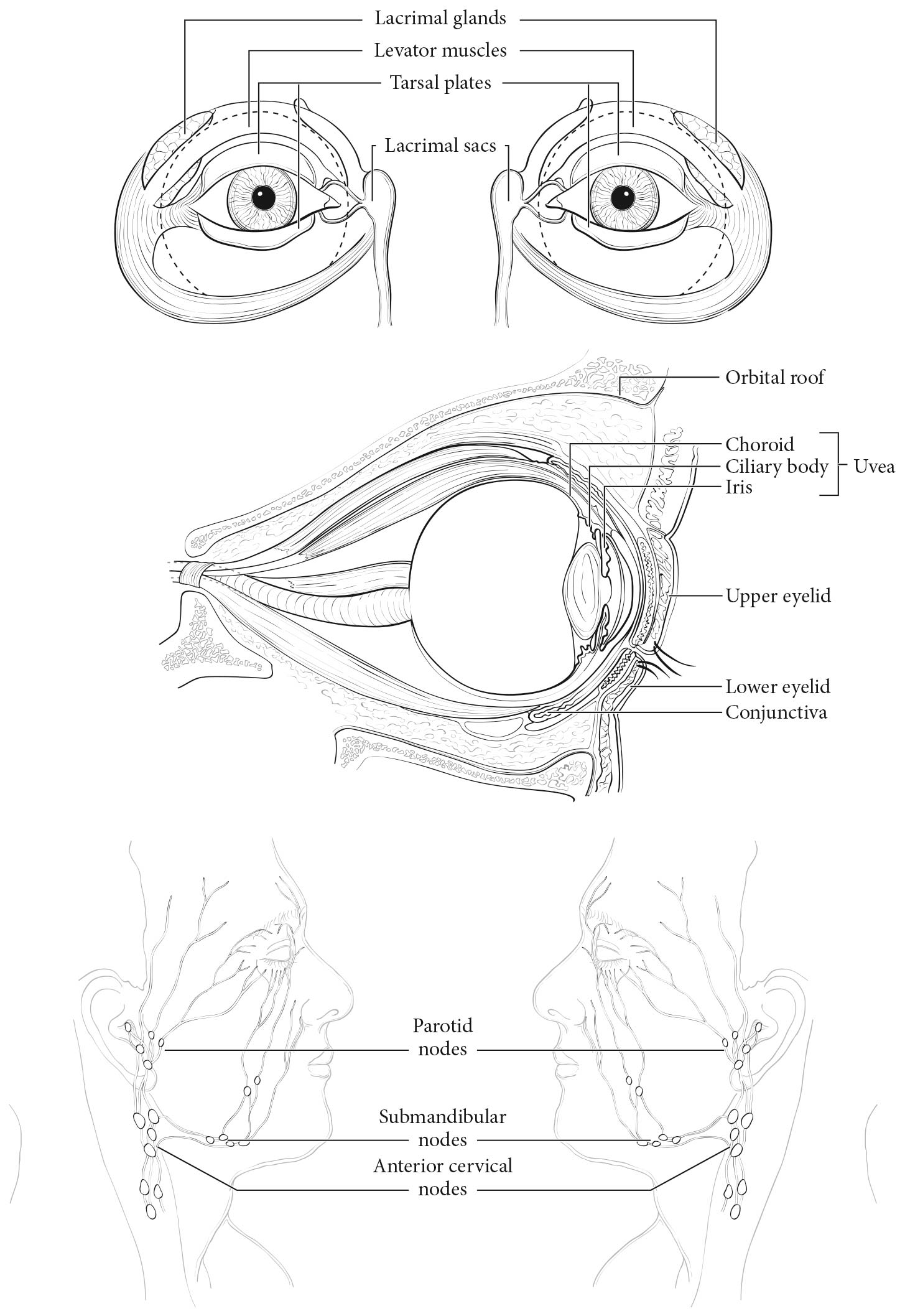
The uveal tract is the middle layer of the eye, situated between the sclera externally and the retina and analogous neuroepithelial tissues internally. The uveal tract is divided into three regions: the iris, ciliary body, and choroid. Uveal melanomas arise most commonly in the choroid, less frequently in the ciliary body, and least often in the iris.4
The uveal tract is a highly vascular structure that comprises blood vessels and intervening stroma. The stroma contains variable numbers of melanocytes of neural crest origin, from which uveal melanomas arise. The uvea has no basal membrane comparable with that of the skin to be breached; therefore, uveal melanoma is immediately in contact with blood vessels.19 Moreover, because there is no traditional lymphatic drainage within the eye and orbit, uveal melanomas metastasize almost exclusively hematogenously. Most uveal melanomas are slow-growing tumors,20,21 so clinical metastases also may grow slowly, only to appear decades after successful treatment of the primary tumor.22,23
Choroidal melanomas extend commonly into the subretinal space through Bruch's membrane, which separates the choroid from the retina. More rarely, they further extend through the retina to reach the vitreous cavity. Choroidal melanomas also may extend through the sclera into the orbit, either by growing along intrascleral blood vessels and nerves or by directly invading the sclera, whereas iris and ciliary body melanomas may extend to the conjunctiva, either along aqueous outflow channels or by direct invasion.24 Choroidal melanomas occasionally invade the optic nerve by direct extension,25 and rare retinoinvasive melanomas26 may invade it by disseminating along the retina or through the vitreous.
In the rare event that uveal melanoma metastasizes to the regional lymph nodes, it occurs after extraocular spread and invasion of conjunctival or adnexal lymphatics.27 This category consequently applies only to uveal melanomas with anterior extrascleral extension.
The relevant regional lymph nodes include the following:
Clinical Classification
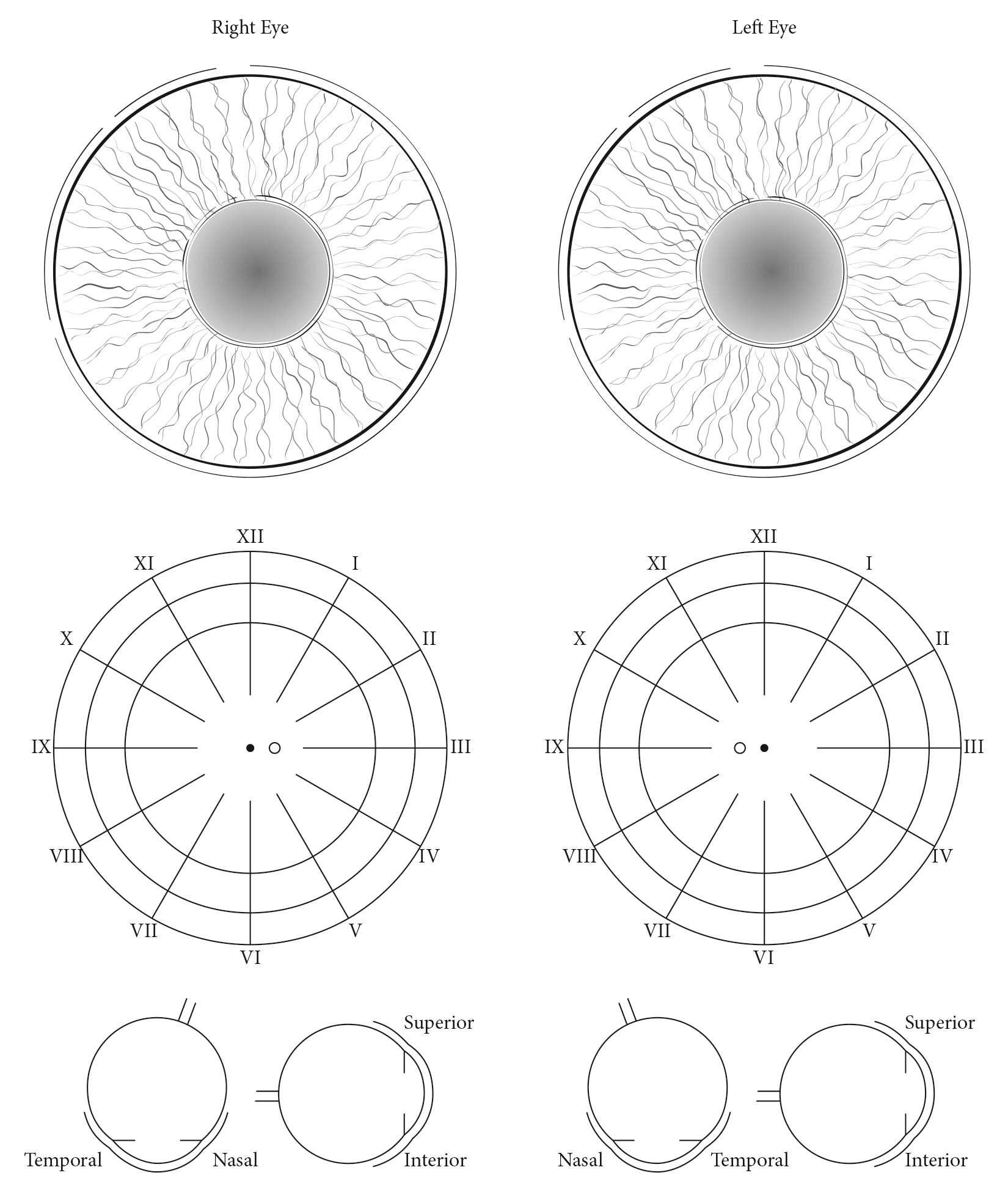
Iris MelanomasIris melanomas typically are visible through the cornea. They originate from, and are predominantly located in, this region of the uvea. If less than half the tumor volume is located within the iris and it involves the chamber angle, the tumor likely originated in the ciliary body and should be classified accordingly.
Ciliary body involvement may be evaluated and measured by slit-lamp examination, ophthalmoscopy, gonioscopy, transillumination, and anterior segment optical coherence tomography. However, calipers and anterior segment ultrasound imaging are used for more accurate assessment. Extension through the sclera is evaluated visually before and during surgery and preoperatively with ultrasonography, computed tomography (CT), or magnetic resonance (MR) imaging.
Pigmented iris tumors that demonstrate intrinsic vascularity; measure greater than 3 clock hours, are greater than 1 mm in thickness, and are associated with sentinel vessels, sector cataract, dispersion of melanocytic tumor cells, secondary glaucoma, and extrascleral extension are more likely iris melanomas than benign melanocytic proliferations.35
Of iris melanomas, 56% are classified as T1, 34% as T2, 2% as T3, and 1% as T4. Kaplan-Meier estimates of survival at 5 years for categories T1, T2, and higher were about 100%, 80%, and 50%, respectively.35
Choroidal and Ciliary Body MelanomasChoroidal and ciliary body melanomas commonly are diagnosed, measured, and assessed based on clinical examination, including slit-lamp examination, direct and indirect ophthalmoscopy, and ultrasonography. Additional methods, such as anterior segment ultrasound imaging, optical coherence tomography, fundus autofluorescence, standard and wide-field fundus photography, fluorescein and indocyanine green angiography, CT, positron emission tomography (PET)/CT, and MR imaging may enhance the accuracy of appraisal, especially in atypical cases.
The large prospective randomized COMS demonstrated 99.6% accuracy in the clinical diagnosis of medium-sized and large choroidal melanomas. However, smaller melanomas typically exhibit fewer diagnostic characteristics, making them more difficult to diagnose.36-38 A multitude of clinical findings have been used to predict the risk of growth of small choroidal melanocytic lesions. Choroidal melanocytic tumors with orange pigment, subretinal fluid, and thickness greater than 2 mm should always be referred for evaluation. A low internal reflectivity on ultrasound examination also suggests the diagnosis of uveal melanoma. Currently, small uveal melanocytic lesions often are either observed for growth or biopsied before being defined and treated as uveal melanomas. When a needle aspiration or vitreous cutter biopsy is pursued, a nonmalignant result will not exclude the possibility of uveal melanoma because of potential sampling error, technical error, or tumor heterogeneity.39,40
The T categories describe the anatomic extent of choroidal and ciliary body melanomas, expressed as tumor size and involvement of the ciliary body and extrascleral tissues.3 The category thresholds are defined in a nonrectangular, tabular format to achieve homogeneous survival within each category (Figure 86.1). Category T4 also includes larger extrascleral extensions. Survival worsens not only with increasing T category,7 but also with the subcategories within each T category.3 Staging takes this variation into account and should always be used for reporting survival. Stages I to IIIC are confined to uveal melanoma patients who have no evidence of metastases, either at regional or distant sites, based on clinical, radiologic, and laboratory evaluation. Stage IV indicates metastatic disease or noncontiguous intraorbital invasion. Staging is not implemented for iris melanomas.
MetastasesAt the time of diagnosis of the intraocular tumor, lymph node metastases are found in fewer than 1% of patients and systemic metastases in only 2-3% of patients.41 Liver imaging and chest radiography are recommended to exclude both hepatic metastasis and a nonocular primary tumor metastatic to the uvea.31,41 Local tumor recurrence is associated with a higher risk of metastasis.42 Sentinel lymph node biopsy is not practiced.
No international consensus has been reached regarding posttreatment review guidelines and surveillance techniques for early detection of metastatic uveal melanoma. Although national guidelines exist,43 there is no existing objective evidence that any surveillance improves prognosis.44Available evidence includes the COMS finding that physical examination and liver function tests are inadequate,45 although the latter are useful for excluding diffuse hepatic metastasis. Largely dependent on customs and practice, as well as knowledge that metastases typically present in the liver, it is common to examine patients one to two times per year with abdominal imaging (e.g., ultrasound, MR imaging, and CT).46 Some eye cancer centers use PET/CT for initial staging and subsequent surveillance of patients at high risk for metastasis, in part because whole-body scanning is more likely to detect nonhepatic metastases (e.g., to skin and bone) as well as second, nonocular primary cancers.41
M1 was divided into three subcategories in the previous edition based on the largest diameter of the largest metastasis. This measure has been shown to correlate strongly with survival after diagnosis of metastases. The divisions were based on a European collaborative dataset of 249 patients with metastatic uveal melanoma. Median survival times for subcategories M1a to M1c were 17.5 months, 9.6 months, and 5.0 months, respectively, and they included 47%, 45%, and 8% of patients, respectively. These subcategories are retained in the 8th Edition.
Patients with Stage IV uveal melanoma have clinical or radiologic evidence of regional (N1a-b) or systemic (M1a-c) metastases, or positive diagnostic biopsies of these regional nodes or distant metastases. Suspected orbital invasion, regional lymph node involvement, and systemic metastasis are confirmed by needle biopsy or resection.
Because staging of metastatic uveal melanoma is evolving and depends on several factors in addition to diameter of the largest metastasis, especially liver enzyme levels and performance status,28,47 no substaging currently is proposed.
Pathological Classification
Resection of the primary tumor by iridectomy, iridocyclectomy, local resection, or enucleation is required for complete pathological staging; then, the extent of the tumor measured in clock hours of involvement, basal dimensions, tumor thickness, and margins of resection may be assessed. It is uncommon in current practice to resect small choroidal melanomas because these tumors may be controlled effectively with nonsurgical treatment, such as plaque brachytherapy or proton beam therapy.
The T categories of anatomic extent are the same for clinical and pathological classification. When histopathologic measurements are recorded after fixation, however, tumor diameter and thickness may be underestimated because of tissue shrinkage. On the other hand, assessment of ciliary body involvement and extrascleral extension often is more accurate if a resection specimen is available for histopathologic evaluation.
Suspected orbital invasion, regional lymph node involvement, and systemic metastasis are confirmed by needle biopsy or resection.
Uveal melanomas exhibit marked variation in cytologic composition. They display a spectrum of cell types ranging from slender spindle cells through plump spindle cells to epithelioid cells. Many tumors contain some admixture of these different types.
Today, DNA- or RNA-based genetic profiling and GEP, if available and combined with data on tumor size and histopathology, may enhance accuracy in providing a prognosis for an individual patient as compared with clinical and histopathologic data alone. Consideration should be given to harvesting tumor tissue for this purpose during the histopathologic examination.
Regional lymphadenectomy ordinarily includes six or more regional lymph nodes. Because of the rarity of regional lymph node metastasis, sentinel lymph node biopsy is not practiced.
Prognostic Factors Required for Stage Grouping
Beyond the factors used to assign T, N, or M categories, no additional prognostic factors are required for stage grouping.
Additional Factors Recommended for Clinical Care
The size of uveal melanoma is strongly associated with risk for metastasis.3,4,48,49Although it generally is accepted that largest basal tumor diameter is the predominant clinical predictor of prognosis, tumor thickness is an independent prognostic indicator, even when ciliary body involvement and extrascleral extension are taken into account.3 The largest tumor basal diameter may be estimated in optic disk diameters (DD; average: 1 DD = 1.5 mm), and tumor thickness may be estimated in diopters (average: 2.5 diopters = 1 mm). Evaluation and comparison of fundus photography and ultrasonography provide more accurate measurements.
Intraocular location of a uveal melanoma relative to the ciliary body is independently associated with metastatic risk.3,50 Ciliary body involvement is evaluated by slit-lamp examination, ophthalmoscopy, gonioscopy, transillumination, and anterior segment optical coherence tomography. Anterior segment ultrasound imaging provides a more accurate assessment. Tumors confined to the iris carry the most favorable prognosis, followed by those confined in the choroid; ciliary body involvement carries the least favorable prognosis.
The presence of extraocular extension is independently associated with higher metastatic risk.3,51 Anterior extension through the sclera is evaluated visually, measured, and photographed. Posterior extension may be measured with ultrasonography, CT, or MR imaging.
Cell type is independently associated with metastatic risk.49,52 Spindle cells have ovoid nuclei and tend to grow in a compact cohesive fashion. Epithelioid cells are larger, more irregularly contoured, and pleomorphic, and contain abundant, typically eosinophilic cytoplasm. Their nuclei and nucleoli are larger, and they grow less cohesively than spindle cells. No consensus has been reached regarding what proportions of spindle and epithelioid cells qualify a uveal melanoma as being of mixed and epithelioid type. Some ophthalmic pathologists primarily record the presence or absence of epithelioid cells and do not try to subclassify uveal melanomas with epithelioid cells into mixed or epithelioid categories. Spindle cell melanomas predict the longest and epithelioid cell melanomas the shortest survival times.
Monosomy 3, especially if combined with a frequently coexisting gain in chromosome 8q, is independently associated with metastatic risk.10,11,18,51,53-56 The clinical prognostic significance of other recurring chromosomal changes is not as firmly established. Chromosome 3 and 8 status may be determined with karyotyping or fluorescent in situ hybridization, but these studies do not identify isodisomy 3. Array comparative genomic hybridization, DNA polymorphism analysis (e.g., single-nucleotide polymorphism and microsatellite), and multiplex ligation-dependent probe amplification provide more reliable results. Droplet polymerase chain reaction allows copy number assessment of small tissue samples. Disomy 3 predicts the longest (partial) survival, monosomy 3 or chromosome 8q gain alone predicts an intermediate survival, and the combination of monosomy 3 and chromosome 8q gain predicts the shortest survival time. However, determination of chromosome status may be affected by prior irradiation.57
GEP class 2 (high grade) or equivalent is independently associated with higher metastatic risk.13,14 The profile is determined by using RNA extracted from the tumor and a gene chip. Class 1A predicts the longest survival, class 1B an intermediate survival, and class 2 the shortest survival time.
Mitotic count is independently associated with metastatic risk.55 The number of mitotic figures is counted under light microscopy per 40 high-power fields (HPF; 40x), which typically have a field area of 0.15-0.19 mm2. The College of American Pathologists Protocol for the Examination of Specimens from Patients with Uveal Melanoma (www.cap.org) recommends using a field area of 0.152 mm2 for standardization of reporting. Higher counts are associated with shorter survival. Mitotic cells also may be identified by immunohistochemical methods.58
The presence of certain types of extravascular matrix patterns is independently associated with risk of metastasis.52,59,60 This is documented conclusively for individual loops and for loops forming networks consisting of at least three back-to-back loops. The patterns are assessed with light microscopy under a dark green filter after staining with periodic acid-Schiff stain without counterstain. Absence of both loops and networks is associated with the longest survival and presence of loops forming networks is associated with the shortest survival time.
Microvascular density is independently associated with metastatic risk.59-61 The number of immunopositive elements is labeled with a marker for vascular endothelial cells (e.g., CD34 epitope, CD31 epitope, factor VIII-related antigen) and counted from areas of densest vascularization (typical field area, 0.31 mm2). Higher counts are associated with shorter survival.
The number of tumor-infiltrating macrophages is independently associated with metastatic risk.54,62,63 The number of immunopositive elements labeled with a marker for macrophages (e.g., CD68 epitope, CD163 epitope) is semiquantitatively graded as few, moderate, or many, for example, against published standard photographs.62 Higher numbers are associated with shorter survival.
Preliminary results suggest that identification of mutations in BAP1, EIF1AX, and SF3B1 may further enhance prognostication in uveal melanoma, especially if the BAP1 gene is not mutated and the tumor has disomy 3.15-18,64 Mutations in these genes may be assessed from either a biopsy sample of the tumor or a resection specimen using sequencing (e.g., Sanger sequencing). Expression of BAP1 protein also may be assessed immunohistochemically.65-67 Mutation leads to the absence of normally occurring nuclear immunoreaction. Currently, it is estimated that BAP1 is mutated in 45-55%, EIF1AX in 10-20%, and SF3B1 in 10-20% of all primary uveal melanomas, the first predominantly in those with monosomy 3 and the second two in those with disomy 3. EIF1AX is mutated in about 6% and 48% of monosomy 3 and disomy 3 primary uveal melanomas, and SF3B1 (usually in codon 625) in 3-7% and 27-29% of tumors, respectively. Mutations in BAP1 have been found in 80-85% and those in SF3B1 in about 5% of metastatic uveal melanomas, according to preliminary studies.68BAP1 mutations also may occur in the germline, and these inherited mutations predispose to familial uveal melanoma and other cancers, especially cutaneous melanoma, mesothelioma, and renal cell cancer.69 If EIF1AX is mutated in a disomy 3 melanoma (typically with wild-type BAP1), the risk of metastasis is preliminarily estimated to be 6 to 10 times lower than if EIF1AX is wild type.18 When it rarely is mutated in a monosomy 3 melanoma (typically with mutated BAP1), the risk of metastasis is estimated to be three or four times lower than if EIF1AX is wild type. Patients with SF3B1 mutations also carry a lower risk of metastasis than those with wild-type SF3B1 in their tumor.16,17
Mutations in GNAQ are found in 40-45% and those in QNA11 in 30-35% of primary uveal melanomas, usually in codon 626, and are considered early or initiating events.70-72 These mutations may be assessed from a biopsy sample, from a resection specimen, or potentially, from circulating DNA by using sequencing (e.g., Sanger sequencing) or related techniques. GNA11 mutations are more frequent than GNAQ mutations in uveal melanomas with BAP1 mutations, and the former may mark a higher risk of metastasis and, in metastatic uveal melanoma, shorter survival.68,73
The number of tumor-infiltrating lymphocytes is associated with metastatic risk and may be relevant with regard to immunotherapy.54,74-76 The presence of lymphocytes may be assessed semiquantitatively or quantitativelyfor example, by confocal imaging of cells stained by immunohistochemistry or immunofluorescence. Higher numbers are associated with shorter survival.
The level of expression of human leukocyte antigen (HLA) molecules is associated with metastatic risk and may be relevant with regard to immunotherapy.77,78 The level of expression is determined by assessing the percentage of immunopositive tumor cells. Higher expression is related to shorter survival.
The AJCC recently established guidelines that will be used to evaluate published statistical prediction models for the purpose of granting endorsement for clinical use.79 Although this is a monumental step toward the goal of precision medicine, this work was published only very recently. Therefore, the existing models that have been published or may be in clinical use have not yet been evaluated for this cancer site by the Precision Medicine Core of the AJCC. In the future, the statistical prediction models for this cancer site will be evaluated, and those that meet all AJCC criteria will be endorsed.
Clinical trials addressing treatments for metastatic uveal melanoma are recommended to stratify patients on the basis of the M categories (M1a-c).
Definition of Primary Tumor (T)
T Category | T Criteria |
---|---|
TX | Primary tumor cannot be assessed |
T0 | No evidence of primary tumor |
T1 | Tumor limited to the iris |
T1a | Tumor limited to the iris, not more than 3 clock hours in size |
T1b | Tumor limited to the iris, more than 3 clock hours in size |
T1c | Tumor limited to the iris with secondary glaucoma |
T2 | Tumor confluent with or extending into the ciliary body, choroid, or both |
T2a | Tumor confluent with or extending into the ciliary body, without secondary glaucoma |
T2b | Tumor confluent with or extending into the ciliary body and choroid, without secondary glaucoma |
T2c | Tumor confluent with or extending into the ciliary body, choroid, or both, with secondary glaucoma |
T3 | Tumor confluent with or extending into the ciliary body, choroid, or both, with scleral extension |
T4 | Tumor with extrascleral extension |
T4a | Tumor with extrascleral extension less than or equal to 5 mm in largest diameter |
T4b | Tumor with extrascleral extension greater than 5 mm in largest diameter |
Iris melanomas originate from, and are predominantly located in, this region of the uvea. If less than half the tumor volume is located within the iris, the tumor may have originated in the ciliary body, and consideration should be given to classifying it accordingly.
Definition of Regional Lymph Node (N)
N Category | N Criteria |
---|---|
NX | Regional lymph nodes cannot be assessed |
N0 | No regional lymph node involvement |
N1 | Regional lymph node metastases or discrete tumor deposits in the orbit |
N1a | Metastasis in one or more regional lymph node(s) |
Definition of Distant Metastasis (M)
M Category | M Criteria |
---|---|
M0 | No distant metastasis by clinical classification |
M1 | Distant metastasis |
M1a | Largest diameter of the largest metastasis less than or equal to 3.0 cm |
M1b | Largest diameter of the largest metastasis 3.1-8.0 cm |
M1c | Largest diameter of the largest metastasis greater than or equal to 8.1 cm |
HISTOLOGIC GRADE (G)
G | G Definition |
---|---|
GX | Grade cannot be assessed |
G1 | Spindle cell melanoma (greater than 90% spindle cells) |
G2 | Mixed cell melanoma (greater than 10% epithelioid cells and less than 90% spindle cells) |
G3 | Epithelioid cell melanoma (greater than 90% epithelioid cells) |
Because of the lack of universal agreement regarding which proportion of epithelioid cells classifies a tumor as mixed or epithelioid, some ophthalmic pathologists currently combine grades 2 and 3 (nonspindle, i.e. epithelioid cells detected) and contrast them with grade 1 (spindle, i.e. no epithelioid cells detected).