section name header

Authors

Chapter Summary

Cancers Staged Using This Staging System

Parathyroid carcinoma

Summary of Changes

This is a new chapter for the AJCC Cancer Staging Manual.

ICD-O-3 Topography Codes

CodeDescription
C75.0Parathyroid gland

WHO Classification of Tumors

This list includes histology codes and preferred terms from the WHO Classification of Tumors and the International Classification of Diseases for Oncology (ICD-O). Most of the terms in this list represent malignant behavior. For cancer reporting purposes, behavior codes /3 (denoting malignant neoplasms), /2 (denoting in situ neoplasms), and in some cases /1 (denoting neoplasms with uncertain and unknown behavior) may be appended to the 4-digit histology codes to create a complete morphology code.

CodeDescription
8000Neoplasm, malignant
8001Tumor cells, malignant
8005Malignant tumor, clear cell type
8010Carcinoma, NOS
8140Parathyroid carcinoma
8290Oxyphilic adenocarcinoma
8310Clear cell adenocarcinoma, NOS
8322Water-clear cell adenocarcinoma

Lloyd RV, Osamura RY, Klöppel G, Rosai J, eds. World Health Organization Classification of Tumours of Endocrine Organs. Lyon: IARC; 2017. Used with permission.

International Agency for Research on Cancer, World Health Organization. International Classification of Diseases for Oncology. ICD-O-3-Online.http://codes.iarc.fr/home. Accessed August 16, 2017. Used with permission.

Introduction

Parathyroid carcinoma accounts for fewer than 1% of cases of primary hyperparathyroidism, and data are limited. Few studies have been published on parathyroid carcinoma, and most are retrospective reviews from individual institutions or large databases such as the Surveillance, Epidemiology, and End Results (SEER) database and the National Cancer Data Base (NCDB). There is no generally accepted staging system, and identification of significant prognostic factors has been challenging because of the wide variation among existing studies. As a result, the panel feels that proposing a staging system at this time would be premature. Instead, this chapter includes recommendations for recording specific variables in the cancer registry to be used to develop a formal staging system in future AJCC manuals.

Classification Rules

Primary Site(s)

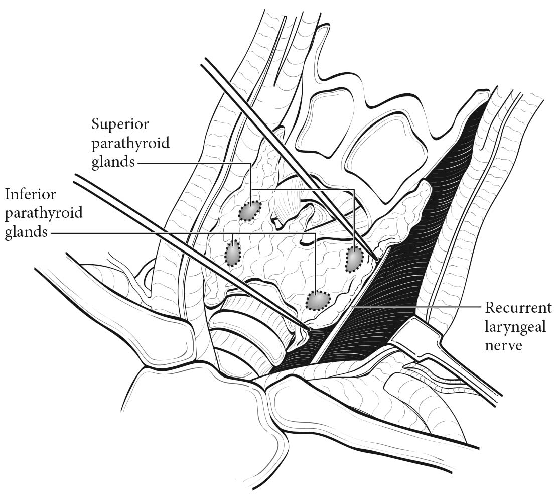
The parathyroid glands are composed of chief cells, oxyphil cells, and clear cells, and are usually located in the neck, adjacent to the thyroid gland. Because the parathyroid glands descend during embryologic development, the location of each gland varies. The superior parathyroid glands, derived from the fourth branchial pouch, descend with the thyroid gland, and are commonly located posterior to the upper third of the thyroid lobe.1 The inferior parathyroid glands descend with the thymus from the third branchial pouch, and may be located anywhere from the superior thyroidal poles to the anterior mediastinum.1,2 Parathyroid glands also may be located within the thyroid gland.3 Although the exact number of parathyroid glands varies among individuals, most people have two superior and two inferior parathyroid glands. (Figure 95.1)

95.1 Anatomy of the parathyroid gland.

The average weight of an individual gland is 30 to 50 mg.1,2 These glands are usually contained within a thin connective tissue capsule.1 If the capsule is absent, there may be nests of ectopic epithelial parathyroid cells mixed with fatty tissue near the gland.1

Parathyroid carcinoma develops within the parathyroid gland itself, and the location of the carcinoma is dependent on the embryologic descent and location of the affected gland.

Regional Lymph Nodes

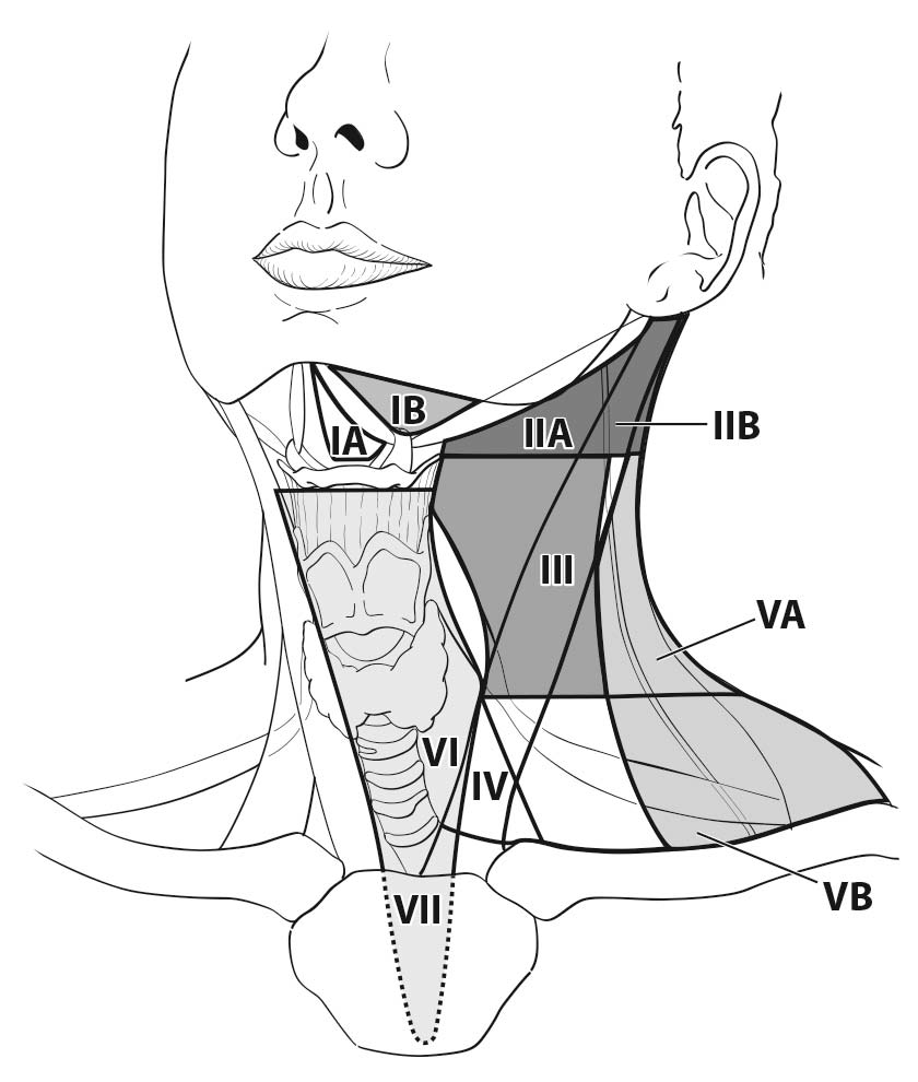
Parathyroid carcinoma has been shown to metastasize to locoregional lymph nodes. Occasionally, parathyroid cancer metastasizes to lymph nodes in the central compartment of the neck (level VI or VII) near the primary tumor.4 Rarely, parathyroid carcinoma metastasizes to lymph nodes in the lateral neck (levels II, III, IV, and V) (Figure 95.2).

95.2 Lymph node levels in the neck.

Metastatic Sites

Evidence of disease beyond the neck should be considered distant metastasis. Parathyroid carcinoma metastasizes to the lung in most cases.5-7 Among patients who develop metastatic disease, as many as 15% have bony metastases.7-9 Other described sites of metastases include the brain, skin, liver, mediastinum, adrenal glands, and pancreas. 2,5,7

Clinical Classification

Patients who present with a palpable neck mass, a serum calcium level greater than 14 mg/dL, and significantly elevated parathyroid hormone (PTH) levels should be suspected of harboring parathyroid carcinoma. Symptoms are similar to those in patients with severe primary hyperparathyroidism, including fatigue, cognitive deficits (difficulty with sleep, concentration, memory, multitasking, depression), bone/joint pain, fragility fractures, osteoporosis, pancreatitis, and kidney stones.5,6,8,10 Some patients with parathyroid carcinoma may develop weight loss and thromboembolic disease.2 Rarely, patients may present with neck pain.5 Some parathyroid carcinomas do not overproduce PTH, and these patients may be asymptomatic, with normal calcium levels.11

The diagnosis of parathyroid carcinoma should be considered in any patient with significantly elevated calcium and PTH levels. Preoperative biopsy of patients with suspected parathyroid carcinoma is not recommended because of the risk of seeding the needle track with tumor. Because patients with parathyroid carcinoma often present with the same symptoms as patients with benign, sporadic primary hyperparathyroidism, the diagnosis is often assigned intraoperatively.10 Unlike a parathyroid adenoma, a parathyroid carcinoma appears as a firm, white-gray mass often adhering to surrounding tissues.

Consideration should be given to en bloc resection of the parathyroid with the ipsilateral thyroid lobe and/or locoregional lymph nodes if necessary for complete tumor resection.

Often, parathyroid carcinoma is not recognized at the first operation, and scar tissue at the second operation may increase the difficulty of recognizing parathyroid cancer. In these cases, persistent disease (defined as biochemical evidence of inappropriately elevated calcium 6 months after the first operation) and time to recurrence should be considered.

Microscopic and macroscopic classifications of the primary tumor have not been standardized. Whether size or extent of invasion affects overall survival remains controversial. Until more is known about prognostic features associated with the primary tumor in parathyroid carcinoma, the expert panel recommends classifying the primary tumor according to both size and extent of invasion.

The prognostic significance of regional lymph node involvement is unclear, and studies are conflicting.7,8,11-15 Until more is known about the prognostic significance of regional lymph node metastasis, the panel recommends collecting the type of lymph node dissection, the number of lymph nodes removed, and the number of lymph nodes with metastatic disease.

Parathyroid carcinoma is usually sporadic and may be associated with primary, secondary, or tertiary hyperparathyroidism. Patients with previous neck irradiation or certain hereditary syndromes may be at increased risk for developing parathyroid carcinoma. For instance, hyperparathyroidism-jaw tumor syndrome (HPT-JT) is a rare autosomal dominant disease caused by a mutation in the CDC73 (HRPT2) gene located on chromosome 1q25-32.16 Patients with this syndrome have ossifying fibromas of the jaw and various tumors/cysts of the kidneys, as well as Mullerian tract tumors in females. While 90% of patients develop primary hyperparathyroidism during their lifetime, as many as 15% develop parathyroid carcinoma.2

Familial isolated primary hyperparathyroidism is a rare autosomal dominant disease thought to be a variant of multiple endocrine neoplasia type I. This disease is also associated with an increased risk of developing parathyroid carcinoma.17

Imaging

Patients with parathyroid carcinoma often have imaging characteristics similar to those of patients with parathyroid adenomas. However, patients who are found to have a large mass, complex cystic lesions with internal septations, central necrosis, and/or compression of adjacent tissues should raise the preoperative suspicion of parathyroid carcinoma.10

Preoperative imaging studies may be performed in patients with a biochemical diagnosis of primary hyperparathyroidism to facilitate performance of a minimally invasive parathyroidectomy. This approach involves a focused resection of an abnormal parathyroid gland in conjunction with intraoperative PTH monitoring.18,19 If there is clinical suspicion for parathyroid carcinoma at operation, then en bloc resection of adjacent tissue (i.e. thyroid) and/or adjacent lymph nodes should be performed. Preoperative imaging is also essential in patients undergoing reoperative parathyroidectomy.20-24

The most commonly used imaging modalities include ultrasonography, which allows concurrent examination of the thyroid; technetium-99 sestamibi scanning (in conjunction with single-photon emission computed tomography [CT] and/or X-ray based CT); and four-dimensional CT, which uses thin-section dynamic scanning techniques with multiplanar reconstruction capabilities.25-29 In addition, fludeoxyglucose positron emission tomography may be helpful for the detection of parathyroid carcinoma, particularly for recurrence of distant metastatic disease; however, current data on this imaging modality are limited. Several preoperative imaging algorithms have been proposed; the optimal algorithm is informed by institutional capabilities and knowledge of one's institutional resources, capabilities, reported sensitivities, and operative outcomes.30

Pathological Classification

The diagnosis of parathyroid carcinoma is based on a combination of clinical and histologic findings on the resected parathyroid gland. The most reliable criteria for the diagnosis of parathyroid carcinoma are the presence of vascular or perineural invasion, invasion into adjacent soft tissues, and/or regional and distant metastasis.1,2,31 Supportive findings include the presence of broad fibrous bands, necrosis, mitotic figures, and trabecular growth; although without the criteria noted earlier, these features are insufficient for a diagnosis of carcinoma. 1,2 Similarly, although the Ki-67 proliferation index (PI) is often elevated (>5%) in parathyroid carcinoma, this finding is not always conclusive because it may be elevated in benign disease.1

Registry Data Collection Variables
  1. Age at diagnosis
  2. Gender
  3. Race
  4. Size of primary tumor in millimeters
  5. Location of primary tumor: left or right and superior (upper) or inferior (lower)
  6. Invasion into surrounding tissue (present or absent)
  7. Distant metastasis
  8. Number of lymph nodes removed (by level)
  9. Number of lymph nodes positive (by level)
  10. Highest preoperative calcium (number in tenths in milligrams per deciliter [e.g., 11.5 mg/dL])
  11. Highest preoperative PTH (whole number in picograms per milliliter [e.g., 350 pg/mL])
  12. Lymphovascular invasion (present or absent)
  13. Grade (LG or HG)
  14. Weight of primary tumor (in milligrams)
  15. Mitotic rate
  16. Time to recurrence (months)

Prognostic Factors

Prognostic Factors Required for Stage Grouping

Beyond the factors used to assign T, N, or M categories, no additional prognostic factors are required for stage grouping.

Additional Factors Recommended for Clinical Care

Age

Defined as age at initial diagnosis. Among nine retrospective studies, three identified older age as a risk factor for decreased overall survival.5-8,11-14,32 Most of the studies showing that age is not predictive of survival were limited in statistical value because of small sample sizes.

AJCC Level of Evidence: III

Gender

Defined as male or female. Most of the studies evaluating gender as a prognostic factor showed that it was not predictive of survival.6,8,11,12,14 However, most these studies were limited because of small sample sizes. Studies from large databases or meta-analyses suggest that men have a worse prognosis.7,13,32

AJCC Level of Evidence: III

Size of Primary Tumor

Defined as measurement of the longest axis of the primary tumor in millimeters. Studies evaluating size as a prognostic factor are conflicting.5,7,8,12-14,32 Likewise, no analysis has been performed to determine whether there is a significant cutoff value in terms of prognosis.

AJCC Level of Evidence: III

Extent of Invasion of Primary Tumor

Few studies have been published regarding whether the extent of invasion of the primary tumor affects overall prognosis, and the results are conflicting.7,8 Comparison among studies is difficult, because classification according to extent of the primary tumor was not standardized before publication of this chapter.

AJCC Level of Evidence: III

Location of Primary Tumor

Defined as left or right and superior (upper) or inferior (lower)

AJCC Level of Evidence: Not identified

Lymph Node Metastasis

Several analyses showed lymph node metastases to be predictive of outcome, but some of the large database studies did not identify lymph node metastasis as a prognostic factor.7,8,11-14,32 This discrepancy may be related to how the survival analysis was performed (e.g., disease-specific vs. overall survival, exclusion of patients with more than one primary tumor).

AJCC Level of Evidence: III

Distant Metastasis

Distant metastasis is defined as evidence of disease beyond the central and lateral neck. Several published studies demonstrated that the presence of distant metastasis portends a worse survival.8,11-13 In fact, the presence of distant metastasis is the only consistent factor across the literature that is predictive of overall survival.

AJCC Level of Evidence: I

Preoperative Calcium at Diagnosis

Defined as highest preoperative serum calcium level at diagnosis before the first operation. Few studies evaluated preoperative calcium as a predictive factor, and the results are conflicting.5,7,8,11 Likewise, these analyses were limited because of small sample sizes.

AJCC Level of Evidence: III

Preoperative PTH Level

Defined as highest PTH level at diagnosis before the first operation. Two studies concluded that preoperative PTH is not predictive of survival, but both were limited because of small sample sizes.7,8 Recorded PTH level should include the assay's limits of normal.

AJCC Level of Evidence: III

Lymphovascular Invasion

Defined as tumor in a lymphatic or vascular space. Few studies evaluated this factor as a prognostic variable, and the results are conflicting.7,8

AJCC Level of Evidence: III

Mitotic Rate

Defined as the number of mitoses per high-power field.2 Few institutions have evaluated the number of mitotic figures as a prognostic factor, and the results are limited because of small sample sizes and a lack of uniform methodology for assessment.7,8,33

AJCC Level of Evidence: III

Weight of Primary Tumor

Defined as the weight of the primary tumor in milligrams.

AJCC Level of Evidence: IV

Cytonuclear Grade

Defined as low grade or high grade. Only a few studies attempted to define grade in parathyroid carcinomas.33,34 Low-grade tumors consist of round monomorphic nuclei with only mild to moderate nuclear size variation, indistinct nucleoli, and chromatin characteristics resembling those of normal parathyroid or of adenoma. High-grade tumors have more pleomorphism, with a nuclear size variation greater than 4:1; prominent nuclear membrane irregularities; chromatin alterations, including hyperchromasia or margination of chromatin; and prominent nucleoli. Unlike the random “endocrine atypia” seen even in normal parathyroid glands, high-grade tumors show several discrete confluent areas with nuclear changes. Few data exist regarding grade as a prognostic factor for parathyroid cancer.14

AJCC Level of Evidence: III

Time to Recurrence

Defined as time after first operation to time of first recurrence in months. There is not sufficient evidence that time to recurrence is predictive of outcome.11

AJCC Level of Evidence: III

Ki-67 Index

Defined as <5% versus >=5%. Ki-67 PI typically is elevated in carcinomas. However, in primary tumors, an elevated Ki-67 PI is not sufficiently sensitive or specific for distinction from benign disease, because not all parathyroid carcinomas have an elevated Ki-67 PI and Ki-67 may be increased in parathyromatosis (seeding of parathyroid cells into surrounding tissue via rupture of capsule or cystic component) and some parathyroid adenomas.35 However, elevated Ki-67 is noted more frequently (up to 80%) in metastatic disease.35 One small study did not find Ki-67 to be predictive of survival.6

AJCC Level of Evidence: III

Growth Pattern

Solid Growth Pattern

Defined as present or not present. Solid is best defined as a confluent proliferation of tumor cells without intervening stroma.

Trabecular Growth Pattern

Defined as present or not present. A trabecular growth pattern is composed of long ribbons of tumor cells with intervening fibrosis. Peripheral palisading often is present.

AJCC Level of Evidence: IV

Genetic Mutations

Defined as presence of germline or somatic mutation.

Germline Mutations

  • Hereditary HPT-JT
  • Familial isolated primary hyperparathyroidism

Somatic Mutations

Sporadic parathyroid carcinomas are often associated with a somatic mutation of the RB and CDC73 genes and cyclin D1 overexpression, although some may have a p53 mutation.1

AJCC Level of Evidence: IV

Intraoperative Tumor Rupture

Defined as rupture of the primary tumor during the first operation. One study demonstrated that intraoperative tumor rupture is predictive of worse survival.8

AJCC Level of Evidence: III

Presence of Fibrous Bands

Defined as present or not present. There is conflicting and limited evidence showing that the presence of fibrous bands is predictive of overall survival.7,8

AJCC Level of Evidence: III

Parafibromin Staining

Defined as present or not present. Parafibromin, the protein encoded by CDC73, is retained in parathyromatosis, adenomas, hyperplasias, and most atypical parathyroid adenomas.35,36 This marker is lost in up to two thirds of parathyroid carcinomas and thus may be diagnostically useful.35,37 However, some studies suggest that loss of parafibromin may occur in some atypical parathyroid adenomas, especially if associated with HPT-JT syndrome.10,37,38 Furthermore, the rare parathyroid carcinomas arising in the setting of secondary/tertiary hyperparathyroidism do not have CDC73 alterations and will not show parafibromin loss.39

AJCC Level of Evidence: IV

Perineural Invasion

Defined as present/not present.

AJCC Level of Evidence: IV

Necrosis

Defined as present/not present.

AJCC Level of Evidence: IV

Type of Hyperparathyroidism

Defined as primary versus secondary versus tertiary hyperparathyroidism

AJCC Level of Evidence: Not identified

Risk Assessment

Risk Assesment Models

The AJCC recently established guidelines that will be used to evaluate published statistical prediction models for the purpose of granting endorsement for clinical use.40 Although this is a monumental step toward the goal of precision medicine, this work was published only very recently. Therefore, the existing models that have been published or may be in clinical use have not yet been evaluated for this cancer site by the Precision Medicine Core of the AJCC. In the future, the statistical prediction models for this cancer site will be evaluated, and those that meet all AJCC criteria will be endorsed.

TNM Definitions

Clinical T (cT)

cT CategorycT Criteria
cTXPrimary tumor cannot be assessed
cT0No evidence of primary tumor
cTisAtypical parathyroid neoplasm (neoplasm of uncertain malignant potential)
cT1Localized to the parathyroid gland with extension limited to soft tissue
cT2Direct invasion into the thyroid gland
cT3Direct invasion into recurrent laryngeal nerve, esophagus, trachea, skeletal muscle, adjacent lymph nodes, or thymus
cT4Direct invasion into major blood vessel or spine

*Defined as tumors that are histologically or clinically worrisome but do not fulfill the more robust criteria (i.e., invasion, metastasis) for carcinoma. They generally include tumors that have two or more concerning features, such as fibrous bands, mitotic figures, necrosis, trabecular growth, or adherence to surrounding tissues intraoperatively.31,41 Atypical parathyroid neoplasms usually have a smaller dimension, weight, and volume than carcinomas and are less likely to have coagulative tumor necrosis.10

Pathological T (pT)

pT CategorypT Criteria
pTXPrimary tumor cannot be assessed
pT0No evidence of primary tumor
pTisAtypical parathyroid neoplasm (neoplasm of uncertain malignant potential)
pT1Localized to the parathyroid gland with extension limited to soft tissue
pT2Direct invasion into the thyroid gland
pT3Direct invasion into recurrent laryngeal nerve, esophagus, trachea, skeletal muscle, adjacent lymph nodes, or thymus
pT4Direct invasion into major blood vessel or spine
cTXPrimary tumor cannot be assessed
cT0No evidence of primary tumor
cTisAtypical parathyroid neoplasm (neoplasm of uncertain malignant potential)
cT1Localized to the parathyroid gland with extension limited to soft tissue
cT2Direct invasion into the thyroid gland
cT3Direct invasion into recurrent laryngeal nerve, esophagus, trachea, skeletal muscle, adjacent lymph nodes, or thymus
cT4Direct invasion into major blood vessel or spine

*Defined as tumors that are histologically or clinically worrisome but do not fulfill the more robust criteria (i.e., invasion, metastasis) for carcinoma. They generally include tumors that have two or more concerning features, such as fibrous bands, mitotic figures, necrosis, trabecular growth, or adherence to surrounding tissues intraoperatively.31,41 Atypical parathyroid neoplasms usually have a smaller dimension, weight, and volume than carcinomas and are less likely to have coagulative tumor necrosis.10

Neoadjuvant Clinical T (yT)

ycT CategoryycT Criteria
ycTXPrimary tumor cannot be assessed
ycT0No evidence of primary tumor
ycTisAtypical parathyroid neoplasm (neoplasm of uncertain malignant potential)
ycT1Localized to the parathyroid gland with extension limited to soft tissue
ycT2Direct invasion into the thyroid gland
ycT3Direct invasion into recurrent laryngeal nerve, esophagus, trachea, skeletal muscle, adjacent lymph nodes, or thymus
ycT4Direct invasion into major blood vessel or spine

*Defined as tumors that are histologically or clinically worrisome but do not fulfill the more robust criteria (i.e., invasion, metastasis) for carcinoma. They generally include tumors that have two or more concerning features, such as fibrous bands, mitotic figures, necrosis, trabecular growth, or adherence to surrounding tissues intraoperatively.31,41 Atypical parathyroid neoplasms usually have a smaller dimension, weight, and volume than carcinomas and are less likely to have coagulative tumor necrosis.10

Neoadjuvant Pathological T (yT)

ypT CategoryypT Criteria
ypTXPrimary tumor cannot be assessed
ypT0No evidence of primary tumor
ypTisAtypical parathyroid neoplasm (neoplasm of uncertain malignant potential)
ypT1Localized to the parathyroid gland with extension limited to soft tissue
ypT2Direct invasion into the thyroid gland
ypT3Direct invasion into recurrent laryngeal nerve, esophagus, trachea, skeletal muscle, adjacent lymph nodes, or thymus
ypT4Direct invasion into major blood vessel or spine

*Defined as tumors that are histologically or clinically worrisome but do not fulfill the more robust criteria (i.e., invasion, metastasis) for carcinoma. They generally include tumors that have two or more concerning features, such as fibrous bands, mitotic figures, necrosis, trabecular growth, or adherence to surrounding tissues intraoperatively.31,41 Atypical parathyroid neoplasms usually have a smaller dimension, weight, and volume than carcinomas and are less likely to have coagulative tumor necrosis.10

Definition of Regional Lymph Node (N)

Clinical N (cN)
cN CategorycN Criteria
cNXRegional nodes cannot be assessed
cN0No regional lymph node metastasis
cN1Regional lymph node metastasis
cN1aMetastasis to level VI (pretracheal, paratracheal, and prelaryngeal/Delphian lymph nodes) or superior mediastinal lymph nodes (level VII)
cN1bMetastasis to unilateral, bilateral, or contralateral cervical (level I, II, III, IV, or V) or retropharyngeal nodes
Pathological N (pN)
pN CategorypN Criteria
pNXRegional nodes cannot be assessed
pN0No regional lymph node metastasis
pN1Regional lymph node metastasis
pN1aMetastasis to level VI (pretracheal, paratracheal, and prelaryngeal/Delphian lymph nodes) or superior mediastinal lymph nodes (level VII)
pN1bMetastasis to unilateral, bilateral, or contralateral cervical (level I, II, III, IV, or V) or retropharyngeal nodes
cNXRegional nodes cannot be assessed
cN0No regional lymph node metastasis
cN1Regional lymph node metastasis
cN1aMetastasis to level VI (pretracheal, paratracheal, and prelaryngeal/Delphian lymph nodes) or superior mediastinal lymph nodes (level VII)
cN1bMetastasis to unilateral, bilateral, or contralateral cervical (level I, II, III, IV, or V) or retropharyngeal nodes
Neoadjuvant Clinical N (pY)
ycN CategoryycN Criteria
ycNXRegional nodes cannot be assessed
ycN0No regional lymph node metastasis
ycN1Regional lymph node metastasis
ycN1aMetastasis to level VI (pretracheal, paratracheal, and prelaryngeal/Delphian lymph nodes) or superior mediastinal lymph nodes (level VII)
ycN1bMetastasis to unilateral, bilateral, or contralateral cervical (level I, II, III, IV, or V) or retropharyngeal nodes
Neoadjuvant Pathological N (pY)
ypN CategoryypN Criteria
ypNXRegional nodes cannot be assessed
ypN0No regional lymph node metastasis
ypN1Regional lymph node metastasis
ypN1aMetastasis to level VI (pretracheal, paratracheal, and prelaryngeal/Delphian lymph nodes) or superior mediastinal lymph nodes (level VII)
ypN1bMetastasis to unilateral, bilateral, or contralateral cervical (level I, II, III, IV, or V) or retropharyngeal nodes

Definition of Distant Metastasis (M)- Clinical M (cN)

cM CategorycM Criteria
cM0No distant metastasis
cM1Distant metastasis
pM1Microscopic evidence of distant metastasis

Definition of Distant Metastasis (M)- Pathological M (pN)

pM CategorypM Criteria
cM0No distant metastasis
cM1Distant metastasis
pM1Microscopic evidence of distant metastasis

Definition of Distant Metastasis (M)- Neoadjuvant Clinical M (pY)

ycM CategoryycM Criteria
cM0No distant metastasis
cM1Distant metastasis
pM1Microscopic evidence of distant metastasis

Definition of Distant Metastasis (M)- Neoadjuvant Pathological M (pY)

ypM CategoryypM Criteria
cM0No distant metastasis
cM1Distant metastasis
pM1Microscopic evidence of distant metastasis

Stage Prognostic

Clinical

There is no Clinical (cTNM) stage group available at this time.

Pathological

There is no Pathological (pTNM) stage group available at this time.

Neoadjuvant Clinical

There is no Postneoadjuvant Clinical Therapy (ycTNM) stage group available at this time.

Neoadjuvant Pathological

There is no Postneoadjuvant Pathological Therapy (ypTNM) stage group available at this time.

Histopathologic type

Tumor cells may be arranged in trabecular, sheet-like, or rosette-like patterns. Occasionally the nodular structures have central calcifications with necrosis. Nuclear morphology varies between pleomorphism with clumped chromatin and enlarged nucleoli. Proof of vascular invasion or invasion into adjacent organs is a defining hallmark. Terms that may be used include invasive parathyroid neoplasm, neoplasm of undetermined malignant significance, parathyroid neoplasm with locally invasive or atypical features, and parathyroid neoplasm with invasion into connective tissue.

Histologic grade

HISTOLOGIC GRADE (G)

Cytonuclear grade is defined as low grade or high grade. Low-grade tumors consist of round monomorphic nuclei with only mild to moderate nuclear size variation, indistinct nucleoli, and chromatin characteristics resembling those of normal parathyroid or of adenoma. High-grade tumors have more pleomorphism, with a nuclear size variation greater than 4:1; prominent nuclear membrane irregularities; chromatin alterations, including hyperchromasia or margination of chromatin; and prominent nucleoli. Unlike the random “endocrine atypia” seen even in normal parathyroid glands, high-grade tumors show several discrete confluent areas with nuclear changes.

GG Definition
LGLow grade: round monomorphic nuclei with only mild to moderate nuclear size variation, indistinct nucleoli, and chromatin characteristics resembling those of normal parathyroid or of adenoma
HGHigh grade: more pleomorphism, with a nuclear size variation greater than 4:1; prominent nuclear membrane irregularities; chromatin alterations, including hyperchromasia or margination of chromatin; and prominent nucleoli. High-grade tumors show several discrete confluent areas with nuclear changes.

Survival

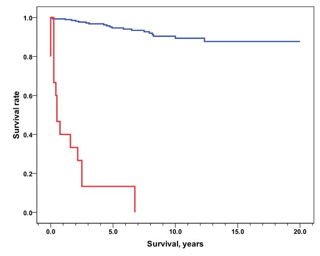
95.3 Cancer-specific survival comparing patients with disease localized to the neck versus patients with distant metastasis. Data was obtained from the SEER program for patients diagnosed from 1973 to 2012 (patients with multiple primary tumors excluded).

Bibliography

  1. Lloyd RV. Endocrine Pathology:: Differential Diagnosis and Molecular Advances. Springer Science & Business Media; 2010.
  2. V-SR HM. Atlas of Endocrine Neoplasia. Houston: The University of Texas M.D. anderson Cancer Center. 2006.
  3. Mazeh H, Kouniavsky G, Schneider DF, et al. Intrathyroidal parathyroid glands: small, but mighty (a Napoleon phenomenon). Surgery. Dec 2012;152(6):1193-1200.
  4. Schulte KM, Talat N, Miell J, Moniz C, Sinha P, Diaz-Cano S. Lymph node involvement and surgical approach in parathyroid cancer. World journal of surgery. Nov 2010;34(11):2611-2620.
  5. Busaidy NL, Jimenez C, Habra MA, et al. Parathyroid carcinoma: a 22-year experience. Head & neck. Aug 2004;26(8):716-726.
  6. Iihara M, Okamoto T, Suzuki R, et al. Functional parathyroid carcinoma: Long-term treatment outcome and risk factor analysis. Surgery. Dec 2007;142(6):936-943; discussion 943 e931.
  7. Talat N, Schulte KM. Clinical presentation, staging and long-term evolution of parathyroid cancer. Annals of surgical oncology. Aug 2010;17(8):2156-2174.
  8. Villar-del-Moral J, Jimenez-Garcia A, Salvador-Egea P, et al. Prognostic factors and staging systems in parathyroid cancer: a multicenter cohort study. Surgery. Nov 2014;156(5):1132-1144.
  9. Li M, Lu H, Gao Y. FDG-anorectic parathyroid carcinoma with FDG-avid bone metastasis on PET/CT images. Clinical nuclear medicine. Nov 2013;38(11):916-918.
  10. Quinn CE, Healy J, Lebastchi AH, et al. Modern experience with aggressive parathyroid tumors in a high-volume New England referral center. Journal of the American College of Surgeons. Jun 2015;220(6):1054-1062.
  11. Harari A, Waring A, Fernandez-Ranvier G, et al. Parathyroid carcinoma: a 43-year outcome and survival analysis. The Journal of clinical endocrinology and metabolism. Dec 2011;96(12):3679-3686.
  12. Hsu K-T, Sippel RS, Chen H, Schneider DF. Is central lymph node dissection necessary for parathyroid carcinoma? Surgery. 2014;156(6):1336-1341.
  13. Lee PK, Jarosek SL, Virnig BA, Evasovich M, Tuttle TM. Trends in the incidence and treatment of parathyroid cancer in the United States. Cancer. May 1 2007;109(9):1736-1741.
  14. Sadler C, Gow KW, Beierle EA, et al. Parathyroid carcinoma in more than 1,000 patients: A population-level analysis. Surgery. Dec 2014;156(6):1622-1629; discussion 1629-1630.
  15. Hundahl SA, Fleming ID, Fremgen AM, Menck HR. Two hundred eighty-six cases of parathyroid carcinoma treated in the U.S. between 1985-1995: a National Cancer Data Base Report. The American College of Surgeons Commission on Cancer and the American Cancer Society. Cancer. Aug 1 1999;86(3):538-544.
  16. Carpten JD, Robbins CM, Villablanca A, et al. HRPT2, encoding parafibromin, is mutated in hyperparathyroidism-jaw tumor syndrome. Nature genetics. Dec 2002;32(4):676-680.
  17. Udelsman R, Akerstrom G, Biagini C, et al. The surgical management of asymptomatic primary hyperparathyroidism: proceedings of the Fourth International Workshop. The Journal of clinical endocrinology and metabolism. Oct 2014;99(10):3595-3606.
  18. Siperstein A, Berber E, Barbosa GF, et al. Predicting the success of limited exploration for primary hyperparathyroidism using ultrasound, sestamibi, and intraoperative parathyroid hormone: analysis of 1158 cases. Annals of surgery. Sep 2008;248(3):420-428.
  19. Udelsman R, Lin Z, Donovan P. The superiority of minimally invasive parathyroidectomy based on 1650 consecutive patients with primary hyperparathyroidism. Annals of surgery. Mar 2011;253(3):585-591.
  20. Chen H, Wang TS, Yen TW, et al. Operative failures after parathyroidectomy for hyperparathyroidism: the influence of surgical volume. Annals of surgery. Oct 2010;252(4):691-695.
  21. Hessman O, Stalberg P, Sundin A, et al. High success rate of parathyroid reoperation may be achieved with improved localization diagnosis. World journal of surgery. May 2008;32(5):774-781; discussion 782-773.
  22. Mortenson MM, Evans DB, Lee JE, et al. Parathyroid exploration in the reoperative neck: improved preoperative localization with 4D-computed tomography. Journal of the American College of Surgeons. May 2008;206(5):888-895; discussion 895-886.
  23. Udelsman R, Donovan PI. Remedial parathyroid surgery: changing trends in 130 consecutive cases. Annals of surgery. Sep 2006;244(3):471-479.
  24. Yen TW, Wang TS, Doffek KM, Krzywda EA, Wilson SD. Reoperative parathyroidectomy: an algorithm for imaging and monitoring of intraoperative parathyroid hormone levels that results in a successful focused approach. Surgery. Oct 2008;144(4):611-619; discussion 619-621.
  25. Harari A, Allendorf J, Shifrin A, DiGorgi M, Inabnet WB. Negative preoperative localization leads to greater resource use in the era of minimally invasive parathyroidectomy. American journal of surgery. Jun 2009;197(6):769-773.
  26. Lubitz CC, Hunter GJ, Hamberg LM, et al. Accuracy of 4-dimensional computed tomography in poorly localized patients with primary hyperparathyroidism. Surgery. Dec 2010;148(6):1129-1137; discussion 1137-1128.
  27. Rodgers SE, Hunter GJ, Hamberg LM, et al. Improved preoperative planning for directed parathyroidectomy with 4-dimensional computed tomography. Surgery. Dec 2006;140(6):932-940; discussion 940-931.
  28. Solorzano CC, Carneiro-Pla DM, Irvin GL, 3rd. Surgeon-performed ultrasonography as the initial and only localizing study in sporadic primary hyperparathyroidism. Journal of the American College of Surgeons. Jan 2006;202(1):18-24.
  29. Starker LF, Mahajan A, Björklund P, Sze G, Udelsman R, Carling T. 4D parathyroid CT as the initial localization study for patients with de novo primary hyperparathyroidism. Annals of surgical oncology. 2011;18(6):1723-1728.
  30. Wang TS, Cheung K, Farrokhyar F, Roman SA, Sosa JA. Would scan, but which scan? A cost-utility analysis to optimize preoperative imaging for primary hyperparathyroidism. Surgery. Dec 2011;150(6):1286-1294.
  31. Seethala RR OJ, Virji M. Pathology of the Parathyroid Glands. In: Barnes EL, ed. Surgical Pathology of the Head and Neck. New York. Informa healthcare. 2008:1429-1473.
  32. Asare EA, Sturgeon C, Winchester DJ, et al. Parathyroid Carcinoma: An Update on Treatment Outcomes and Prognostic Factors from the National Cancer Data Base (NCDB). Annals of surgical oncology. Nov 2015;22(12):3990-3995.
  33. Bondeson L, Sandelin K, Grimelius L. Histopathological variables and DNA cytometry in parathyroid carcinoma. The American journal of surgical pathology. Aug 1993;17(8):820-829.
  34. Yip L, Seethala RR, Nikiforova MN, et al. Loss of heterozygosity of selected tumor suppressor genes in parathyroid carcinoma. Surgery. Dec 2008;144(6):949-955; discussion 954-945.
  35. Fernandez-Ranvier GG, Khanafshar E, Tacha D, et al. Defining a molecular phenotype for benign and malignant parathyroid tumors. Cancer. Jan 15 2009;115(2):334-344.
  36. Tan MH, Morrison C, Wang P, et al. Loss of parafibromin immunoreactivity is a distinguishing feature of parathyroid carcinoma. Clin Cancer Res. Oct 1 2004;10(19):6629-6637.
  37. Gill AJ, Clarkson A, Gimm O, et al. Loss of nuclear expression of parafibromin distinguishes parathyroid carcinomas and hyperparathyroidism-jaw tumor (HPT-JT) syndrome-related adenomas from sporadic parathyroid adenomas and hyperplasias. The American journal of surgical pathology. Sep 2006;30(9):1140-1149.
  38. Kruijff S, Sidhu SB, Sywak MS, Gill AJ, Delbridge LW. Negative parafibromin staining predicts malignant behavior in atypical parathyroid adenomas. Annals of surgical oncology. Feb 2014;21(2):426-433.
  39. Tominaga Y, Tsuzuki T, Matsuoka S, et al. Expression of parafibromin in distant metastatic parathyroid tumors in patients with advanced secondary hyperparathyroidism due to chronic kidney disease. World journal of surgery. May 2008;32(5):815-821.
  40. Kattan MW, Hess KR, Amin MB, et al. American Joint Committee on Cancer acceptance criteria for inclusion of risk models for individualized prognosis in the practice of precision medicine. CA: a cancer journal for clinicians. Jan 19 2016.
  41. McCoy KL, Seethala RR, Armstrong MJ, et al. The clinical importance of parathyroid atypia: is long-term surveillance necessary? Surgery. Oct 2015;158(4):929-935; discussion 935-926.