- Implanted electronic device that continually monitors the heart for bradycardia, ventricular tachycardia (VT), and ventricular fibrillation (VF)
- Detects and treats VT and VF with antitachycardia pacing (ATP) or shock therapy.
- Implant is similar to a pacemaker implant, and most often takes place in the Electrophysiology Laboratory.
- System components and placement are similar to those of a permanent pacemaker, with these important differences:
- The generator is larger, with expanded battery capacity and storage capacitors that allow for the delivery of high-energy shocks
- Device memory capacity is greater, to allow for detailed recording of arrhythmias (electrograms or EGMs) and precise data regarding treated episodes
- The right ventricular pacing lead is also the defibrillation lead, and has one or two metal coils on its surface, for the purpose of delivering shock energy from the generator to the heart
- The generator is most often implanted in the left pectoral area, while pacemakers are routinely place on either the left or the right side
- All ICDs are also pacemakers and can provide single-chamber, dual-chamber, or biventricular pacing
- Implanted as primary prevention for patients at high risk for ventricular arrhythmias
- Coronary disease and low ejection fraction
- MI and impaired left ventricular systolic function
- Dilated cardiomyopathy with low ejection fraction
- Hypertrophic cardiomyopathy
- Genetic abnormalities (i.e., Long QT syndrome and Brugada syndrome)
- Implanted as secondary prevention for patients who survive life-threatening arrhythmias
- VF or hemodynamically unstable VT
- Sustained VT with structural heart disease
|
Types of ICD therapies Implantable cardioverter-defibrillators (ICDs) can deliver a range of therapies depending on the arrhythmia detected and how the device is programmed. Therapies include ATP, cardioversion, defibrillation, biventricular pacing, and bradycardia pacing. |
Patient management
- Know the device and how it's programmed, including:
- Type and model of ICD
- Detection rates for ventricular arrhythmias
- Types of therapies programmed
- Bradycardia pacing mode and rates
- Review above sections including “Evaluating pacemakers” and “Evaluating the patient,” as this information applies to the ICD patient, as well
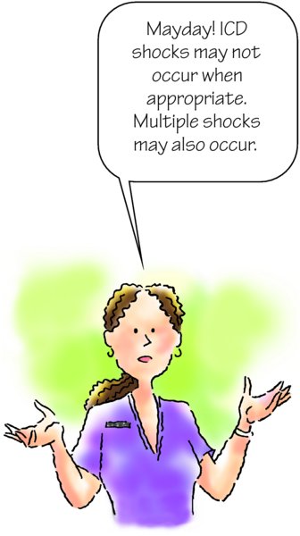
- Keep in mind that shocks may not occur despite VT or VF under certain circumstances, such as:
- If the heart rate is less than the detection rate
- If there's a lead or circuitry problem
- If therapy is suspended or turned off
- If the battery is depleted
- If cardiac arrest occurs in a patient with an ICD, CPR and advanced cardiac life support should be used immediately
- If the patient needs external defibrillation, take the following steps:
- Position the paddles about 6 inches away from the ICD
- Alternatively, use anterior–posterior position for defibrillation patches
- Anticipate that external defibrillation may result in a change in ICD function or settings
- ICD programmed parameters should be verified with a programmer, after external defibrillation is performed
Patient teaching
- Review above sections under “Pacemakers,” including “Patient teaching” and “Understanding EMI,” as these guidelines apply to the ICD patient, as well
- Risk of syncope may continue because the device treats the arrhythmia but does not prevent VT or VF
- Help the patient to understand any prescribed long-term recommendations regarding driving or other high-risk activity
- Help the patient and family formulate a plan for reporting shock therapy, by collaborating with the cardiology/electrophysiology team regarding recommendations
- Instruct the patient and family to call 911 with symptoms of concern (i.e., dyspnea, chest pain, syncope) or at any point they consider the patient to be unstable