section name header

Introduction

What Causes It

  • Digoxin toxicity (most common cause)
  • Cardiomyopathy
  • MI
  • Valvular heart disease
  • Wolff–Parkinson–White (WPW) syndrome
  • Cor pulmonale
  • Systemic hypertension
  • COPD
  • Drugs
    • Albuterol
    • Cocaine
    • Marijuana
    • Theophylline
  • Electrolyte imbalances
  • Hypoxia
  • Physical or psychological stress
  • Congenital anomalies
  • Hyperthyroidism


Identifying atrial tachycardia

This rhythm strip illustrates atrial tachycardia. Look for these distinguishing characteristics:

Rhythm

  • Regular

Rate

  • 210 beats/min

P wave

  • Almost hidden in T wave

PR interval

  • 0.12 second

QRS complex

  • 0.10 second

T wave

  • Distorted by P wave

QT interval

  • 0.20 second

Other

  • None
Memory jogger

To remember the three forms of atrial tachycardia, think of the acronym AMP:

Atrial tachycardia with block

Multifocal atrial tachycardia

Paroxysmal atrial tachycardia

Triggers

  • Alcohol
  • Caffeine
  • Nicotine

What to Look for

  • Rapid heart rate
  • Regular or irregular rhythm, depending on type of atrial tachycardia
  • Sudden feeling of palpitations, especially with PAT
  • Decreased cardiac output and possible hypotension, chest pain, and syncope from persistent tachycardia and rapid ventricular rate, which leads to decreased ventricular filling time, increased myocardial oxygen consumption, and decreased oxygen supply to the myocardium

  • Serious ventricular arrhythmias, especially if the patient has heart disease

Identifying types of atrial tachycardia

Atrial tachycardia comes in three varieties. Here's a quick rundown of each.

Atrial tachycardia with block

Atrial tachycardia with block is caused by increased automaticity of the atrial tissue. As the atrial rate speeds up and AV conduction becomes impaired, a 2:1 block typically occurs. Occasionally a type I (Wenckebach) second-degree AV block may be seen. Look for these distinguishing characteristics:
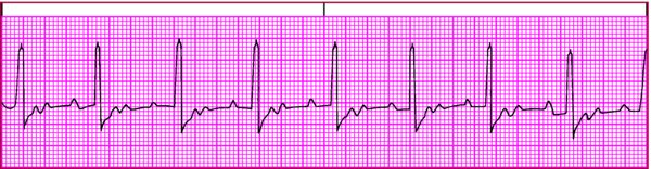
Rhythm

  • Atrialregular; ventricularregular if block is constant, irregular if block is variable

Rate

  • Atrial150 to 250 beats/min, multiple of ventricular rate; ventricularvaries with block

P wave

  • Slightly abnormal; shape depends on site of ectopic pacemaker

PR interval

  • Usually normal

QRS complex

  • Usually normal

T wave

  • Usually indistinguishable

QT interval

  • Possibly indiscernible

Other

  • More than one P wave for each QRS complex

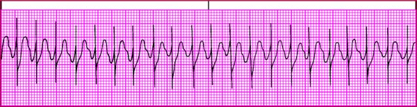
Multifocal atrial tachycardia

In MAT, atrial tachycardia occurs with numerous atrial foci firing intermittently. MAT produces varying P waves on the strip and occurs most commonly in patients with chronic pulmonary disease. The irregular baseline in this strip is caused by movement of the chest wall. Look for these distinguishing characteristics:
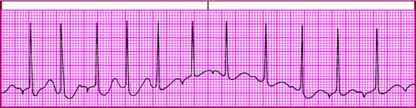
Rhythm

  • Atrial and ventricularirregular

Rate

  • Atrial100 to 250 beats/min, usually under 160; ventricular100 to 250 beats/min

P wave

  • Configuration varies; must see at least three different P wave shapes

PR interval

  • Variable

QRS complex

  • Usually normal; can be aberrant if arrhythmia persists

T wave

  • Usually distorted

QT interval

  • Possibly indiscernible

Other

  • None

Paroxysmal atrial tachycardia

A type of paroxysmal supraventricular tachycardia, PAT features brief periods of tachycardia that alternate with periods of normal sinus rhythm. PAT starts and stops suddenly as a result of rapid firing of an ectopic focus. It commonly follows frequent PACs, one of which initiates the tachycardia. Look for these distinguishing characteristics:
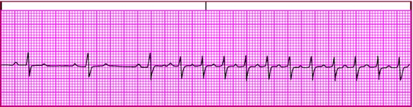
Rhythm

  • Regular

Rate

  • 150 to 250 beats/min

P wave

  • Abnormal, possibly hidden in previous T wave

PR interval

  • Identical for each cycle

QRS complex

  • Possibly aberrantly conducted

T wave

  • Usually distorted

QT interval

  • Possibly indiscernible

Other

  • One P wave for each QRS complex

How It's Treated

  • Treatment depends on the type of tachycardia and the severity of symptoms. Treatment is directed toward eliminating the cause and decreasing the ventricular rate.
  • Inquire about digoxin use, assess the patient for evidence of digoxin toxicity, and monitor digoxin blood levels.
  • Valsalva maneuver or carotid sinus massage may be used to treat PAT. If vagal maneuvers are used, keep resuscitative equipment readily available because vagal stimulation can cause bradycardia, ventricular arrhythmias, and asystole.
  • Drug therapy (pharmacologic cardioversion) may be used to increase the degree of AV block and decrease the ventricular response rate. Appropriate drugs include:
    • adenosine (Adenocard)

    • amiodarone (Cordarone)
    • beta-adrenergic blockers (metoprolol)
    • calcium channel blockers (diltiazem)
    • digoxin
  • If other treatments fail or if the patient is unstable, synchronized electrical cardioversion may be used.
  • Atrial overdrive pacing may stop the arrhythmia by suppressing spontaneous depolarization of the ectopic pacemaker with a series of paced electrical impulses.
  • If the arrhythmia is related to WPW syndrome, catheter ablation may be used to control recurrent episodes of PAT.
  • In patients with chronic pulmonary disease, treatment is directed at correcting hypoxia and electrolyte imbalances. The rhythm may not respond to antiarrhythmic drug treatment.
Through the ages

No carotid sinus massage for elderly

Because carotid bruits may be absent, even with significant disease, carotid atherosclerosis might go undiagnosed in older adults. As a result, cardiac sinus massage shouldn't be performed in late middle-aged and older patients. Embolic stroke may result if carotid massage is performed on a patient with significant atherosclerosis.