section name header

Information

  • Gather all needed supplies.
  • Explain the procedure to the patient.
  • Answer the patient's questions.
  • Ask the patient to lie in a supine position in the center of the bed with his arms at his sides.
  • If the patient can't tolerate lying flat, raise the head of the bed to semi-Fowler position.
  • Ensure privacy.
  • Expose the patient's arms, legs, and chest.
  • Drape the patient for comfort.

Selecting lead sites

  • Choose areas that are flat and fleshy, not muscular or bony.
  • As needed, take steps to enhance electrode contact with the skin:
    • Clip excessively hairy areas.
    • Remove excess oil and other substances from the skin.
  • To ensure an accurate recording, be sure to apply the electrodes correctly.
  • Keep in mind that inaccurate placement of an electrode may lead to inaccurate waveforms and incorrect ECG interpretation.
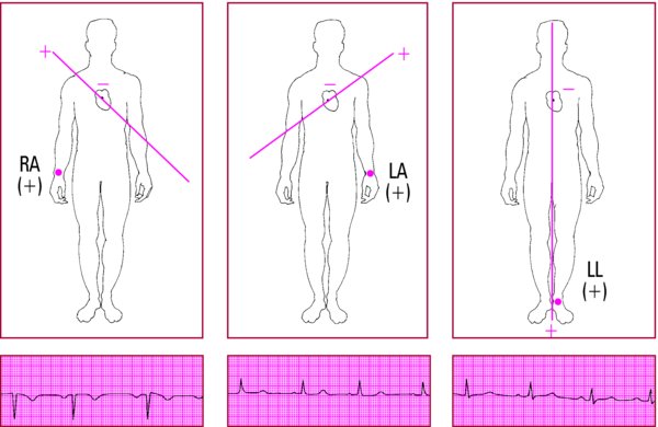
Placing the leads

Limb Leads

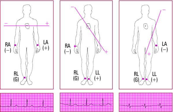
  • Place electrodes on both of the patient's arms and on the left leg.
  • Place an electrode on the right leg. (This is a ground that doesn't contribute to the waveform.)

Precordial Leads

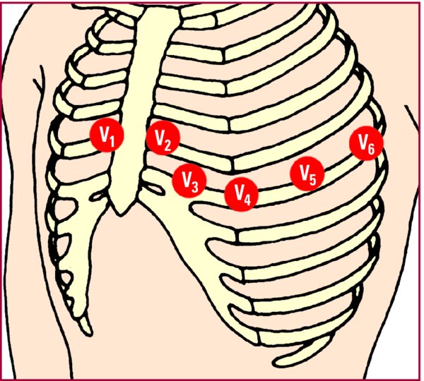
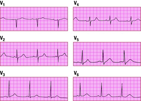
  • Place the six unipolar precordial leads (V1 through V6) in sequence across the chest.
Through the ages

Obtaining a pediatric 12-lead ECG

You'll need patience when obtaining a 12-lead ECG from a pediatric patient. With the help of the parents, if possible, try distracting the attention of a young child. If artifact from arm and leg movement is a problem, place the electrodes in a more proximal position on the limb.


Limb lead placement
Proper lead placement is critical for accurate recording of cardiac rhythms. These drawings show correct electrode placement for the six limb leads. RA stands for right arm; LA, left arm; RL, right leg; and LL, left leg. A plus sign (+) indicates a positive pole, a minus sign () indicates a negative pole, and G indicates a ground. Below each drawing is a sample ECG strip for that lead.
Lead ILead IILead III
Connects the right arm (negative pole) with the left arm (positive pole).Connects the right arm (negative pole) with the left leg (positive pole).Connects the left arm (negative pole) with the left leg (positive pole).

Lead aVRLead aVLLead aVF
Connects the right arm (positive pole) with the heart (negative pole).Connects the left arm (positive pole) with the heart (negative pole).Connects the left leg (positive pole) with the heart (negative pole).

Precordial lead placement
To record a 12-lead ECG, place electrodes on the patient's arms and left leg and place a ground lead on the patient's right leg. The three standard limb leads (I, II, and III) and the three augmented leads (aVR, aVL, and aVF) are recorded using these electrodes. Then, to record the precordial chest leads, place electrodes as follows:

  • V1fourth intercostal space (ICS), right sternal border
  • V2fourth ICS, left sternal border
  • V3midway between V2 and V4
  • V4fifth ICS, left midclavicular line
  • V5fifth ICS, left anterior axillary line
  • V6fifth ICS, left midaxillary