section name header

Introduction

Identifying atrial fibrillation

This rhythm strip illustrates atrial fibrillation. Look for these distinguishing characteristics:

Rhythm

  • Irregularly irregular

Rate

  • Atrialindiscernible; ventricular130 beats/min

P wave

  • Absent; replaced by fine fibrillatory waves

PR interval

  • Indiscernible

QRS complex

  • 0.08 second

T wave

  • Indiscernible

QT interval

  • Unmeasurable

Other

  • None

Other considerations

  • Atrial rhythm may vary between fibrillatory line and flutter waves, called atrial fib-flutter.
  • It may be difficult to differentiate atrial fibrillation from atrial flutter and MAT.


What Causes It

  • Acute MI
  • Atrial septal defect
  • Cardiomyopathy
  • Coronary artery disease
  • Hypertension
  • Pericarditis
  • Valvular heart disease (especially mitral valve disease)
  • Rheumatic heart disease
  • Cardiac surgery
  • COPD
  • Drugs such as aminophylline
  • Digoxin toxicity
  • Endogenous catecholamine released during exercise
  • Hyperthyroidism

Triggers

  • Alcohol
  • Caffeine
  • Nicotine

What to Look for

  • Irregularly irregular pulse rhythm with normal or abnormal heart rate
  • Radial pulse rate that's slower than the apical pulse rate
  • Palpable peripheral pulse only with stronger contractions, not with weaker ones that occur with atrial fibrillation
  • Evidence of decreased end-diastolic volume (by about 20%) from loss of atrial kick, decreased diastolic filling time from rapid heart rate, and decreased cardiac output:
    • Light-headedness, syncope, and hypotension with new-onset atrial fibrillation and a rapid ventricular rate
  • Possibly no symptoms with chronic atrial fibrillation, in which the patient may be able to compensate for decreased cardiac output, but an increased risk of pulmonary, cerebral, or other thromboembolic events

How It's Treated

  • Interventions aim to control the ventricular rate to less than 100 with ideal at 80 or less and establish anticoagulation. Sinus rhythm may or may not be achievable.
  • Treatment typically includes drug therapy to control the ventricular response or a combination of electrical cardioversion and drug therapy.
  • If the patient is hemodynamically unstable, synchronized electrical cardioversion should be performed immediately. It's most successful if done within 48 hours after atrial fibrillation starts.
  • If atrial fibrillation has lasted longer than 48 hours, electrical cardioversion shouldn't be performed unless the patient is adequately anticoagulated because of the risk of thromboembolism.
    • If a thrombus forms in the atria, the resumption of normal contractions can result in systemic emboli.
    • Anticoagulation therapy is crucial in reducing the risk of thromboembolism. Warfarin and heparin are used for anticoagulation before and after elective cardioversion.
    • A transesophageal echocardiogram may be obtained before cardioversion to rule out thrombi in the atria.
  • Drugs such as metoprolol (Lopressor), diltiazem (Cardizem), verapamil (Calan), and amiodarone (Cardarone) can be given after successful cardioversion to maintain normal sinus rhythm and to control the ventricular rate in chronic atrial fibrillation.
  • Symptomatic atrial fibrillation that doesn't respond to standard treatment may be treated with radio-frequency ablation or surgical removal of the left atrial appendage


  • Monitor the apical and peripheral pulses; watch for evidence of decreased cardiac output and heart failure. If the patient isn't on a cardiac monitor, stay alert for an irregular pulse and differences in the radial and apical pulse rates.
  • If drug therapy is used, monitor serum drug levels and watch for evidence of toxicity.
  • Tell the patient to report changes in pulse rate, dizziness, faintness, chest pain, and signs of heart failure, such as dyspnea and peripheral edema.

Distinguishing atrial fibrillation from MAT

On an ECG, atrial fibrillation may look a lot like MAT. To determine whether a rhythm is atrial fibrillation or the similar MAT, focus on the P waves and the atrial and ventricular rhythms. You may find it helpful to look at a rhythm strip that's longer than 6 seconds.

Atrial fibrillation

  • Carefully look for discernible P waves before each QRS complex.
  • If you can't clearly identify P waves and if fibrillatory waves appear in place of P waves, then the rhythm is probably atrial fibrillation.
  • Carefully look at the rhythm, focusing on the R-R intervals. One of the hallmarks of atrial fibrillation is an irregularly irregular rhythm.

MAT

  • P waves are present in MAT. Keep in mind, though, that at least three different P-wave shapes are visible in a single rhythm strip.
  • You should be able to see most or all of the P-wave shapes repeat.
  • Although the atrial and ventricular rhythms are irregular, the irregularity usually isn't as pronounced as it is in atrial fibrillation.

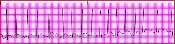
Distinguishing atrial fibrillation from atrial flutter

It isn't uncommon for atrial flutter to have an irregular pattern of impulse conduction to the ventricles. In some leads, this pattern may be confused with atrial fibrillation. Here's how to tell the two arrhythmias apart.

Atrial fibrillation

  • Remember that fibrillatory waves (f waves) occur in an irregular pattern, making the atrial rhythm irregular.
  • If you see atrial activity on the rhythm strip that, in some places, looks like flutter waves and seems to be regular for a short time and, in other places, looks like fibrillatory waves, interpret the rhythm as atrial fibrillation. Coarse fibrillatory waves sometimes have the characteristic sawtooth appearance of flutter waves.
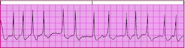
Atrial flutter

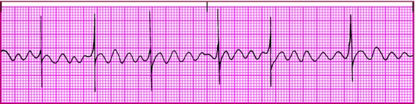
  • Look for characteristic abnormal P waves that produce a sawtooth appearance, known as flutter waves (F waves). These can be identified most easily in leads I, II, and V1.
  • Remember that the atrial rhythm is regular. You should be able to map the F waves across the rhythm strip. Although some F waves may occur within the QRS or T waves, other F waves are visible and occur on time.

Risk of restoring sinus rhythm

A patient with atrial fibrillation is at increased risk for developing an atrial thrombus and a systemic arterial embolism. Because the atria don't contract together in atrial fibrillation, blood may pool on the atrial wall and mural thrombi can form. Thrombus formation places the patient at higher risk for emboli and stroke.

When normal sinus rhythm is restored and the atria contract normally, clots can break away and travel through the pulmonary or systemic circulation, resulting in stroke or arterial occlusion.