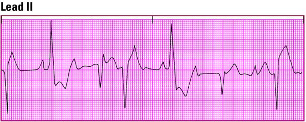
- Records the heart's electrical activity as waveforms that depict depolarization (contraction) and repolarization (relaxation)
- Is used to diagnose and monitor certain disorders, such as myocardial infarction and pericarditis
- Allows identification of rhythm disturbances, conduction abnormalities, and electrolyte imbalances
Leads
- Each lead provides a view of the heart's electrical activity between one positive pole and one negative pole.
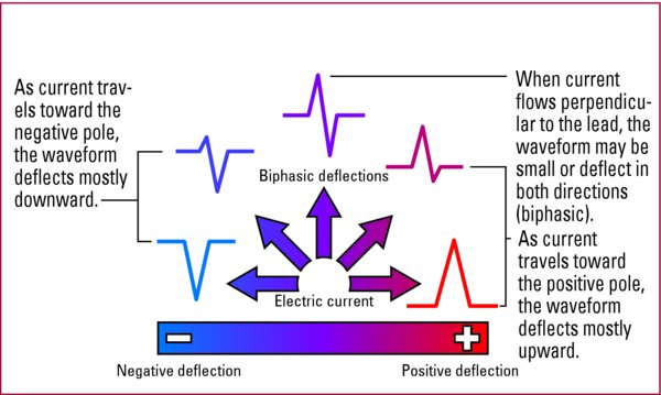
- Each lead generates characteristic waveforms based on the direction electrical current is flowing.
- Each plane is a cross-section of the heart that provides a different view of the heart's electrical activity.
- The six limb leads are viewed from the frontal plane.
- The six precordial leads are viewed from the horizontal plane.
Current direction and waveform deflection The direction of electrical current through the heart determines the upward or downward deflection of an ECG waveform. This illustration shows possible directions of electrical current and their corresponding waveform deflections. Waveform deflection
|
12-lead ECG
- Records information on 12 different views of the heart using a series of electrodes placed on the patient's limbs and chest
- Six limb leads (I, II, III, aVR, aVL, aVF)
- Provide information about the frontal plane of the heart
- May be bipolar and thus require a negative and positive electrode for monitoring (leads I, II, and III)
- May be unipolar and thus need only a positive electrode (augmented leads aVR, aVL, and aVF)
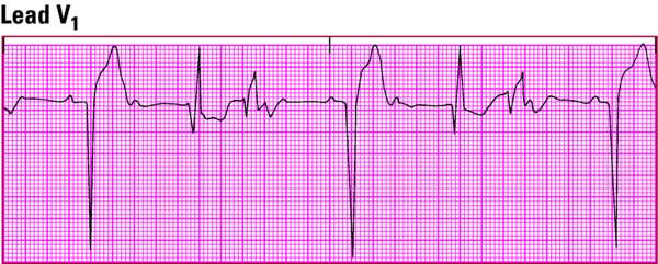
- Six precordial leads (V1, V2, V3, V4, V5, V6)
- Provide information about the horizontal plane of the heart
- Are unipolar (need only a positive electrode)
- Allow calculation of the negative pole of the lead (in the center of the heart) by the ECG monitor
Single-lead or dual-lead ECG
- Monitor up to two different leads
- Display heart rate
- Commonly monitored leads
- I, II, and III
- MCL1 and MCL6 (modified chest leads, similar to unipolar leads V1 and V6 of the 12-lead ECG, which are also used in dual-lead monitoring)
ECG monitoring systems
- May be hardwire or telemetry based on the patient's clinical status
- Hardwire monitoring
- Permits continuous observation of one or more patients from more than one area of the unit
- Connects electrodes directly to cardiac monitor
- Limits mobility (patient is tethered to a monitor)
- Provides a continuous cardiac rate and rhythm display
- Transmits the ECG tracing to a console at the nurses' station
- Has alarms
- Has attachments that can track pulse oximetry, blood pressure, hemodynamic status, and other values
- Telemetry monitoring
- Allows mobility (patient carries a small, battery-powered transmitter that sends electrical signals to another location for display on a monitor)
- Useful for detecting arrhythmias during rest, sleep, exercise, and stressful situations
- Monitors only heart rate and rhythm