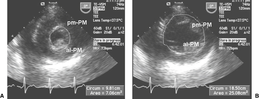
Echocardiographic evaluation of the systolic function of the LV.

The transgastric midpapillary short-axis view of the LV is shown in systole (A) and diastole (B). The endocardial border is traced (without including the endocardium of the two papillary muscles), and the end-systolic area (ESA) and end-diastolic area (EDA) are calculated. The percentage area change (fractional area change [FAC]) is calculated as: FAC = (EDA - ESA) / EDA. The FAC correlates with but does not substitute the percentage ejection fraction. al-PM, anterolateral papillary muscle; pm-PM, posteromedial papillary muscle.