Nejm 2011;364:1046; 2004;351:1860
Cause:Neoplastic B cells
Pathophys:
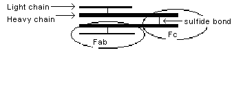
Fig 8.1 Immunoglobulin structure
Natural evolution of MM: from normal plasma cells to "monoclonal gammopathy of unknown significance (MGUS)" by chromosomal translocation or deletion; then, after years, to MM as osteoblastic and angiogenesis capacities of cells increase
of all M-protein disease: 50% IgG (60% with Bence-Jones protein), 24% IgA (70% with BJ), Bence-Jones protein only (21%), IgD (3%), no M protein (1.5%), IgM + Waldenström's (0.5%)
IgG and IgA have specific heavy chains and common light ( and
and  ) chains. M-protein is a homogeneous protein electrophoresis spike. Myelomas in which one can identify a specific antigen to which the M component is directed (eg, ASLO, RA, cold agglutinin anti-I) raise interesting questions of pathogenesis (Nejm 1971;284:831)
10 000 deaths/yr in US; incidence in blacks twice that in whites. Older patients, peak incidence in 50- to 60-yr age group; benzene workers (Nejm 1987;316:1044)
Sx:Recurrent infections; skeletal pain and pathologic fractures; Raynaud's if M component is an IgG cryoprotein; arthritis, 1st sx in 5% (R. Ritchie 1975)
Si:Bone pain (infiltration), hepatosplenomegaly
POEMS syndrome(Nejm 2010;362:929) seen with myeloma and plasmacytomas: Polyneuropathy; Organomegaly, esp adenopathy, splenomegaly, and hepatomegaly; Endocrinopathy, including hypogonadism and hypothyroidism; Monoclonal gammopathy; Skin changes, including hyperpigmentation and thickening
Many die in <2 yr of diagnosis; probably takes 5-10 yr for isolated lesion to spread and kill. Smoldering asx form converts to active disease at 10%/yr for 1st 5 yr (Mayo—Nejm 2007;356:2582)
r/o benign monoclonal gammopathy of undetermined significance (MGUS) (Jama 2010;304:2397; Nejm 2002;346:564; 2006;354:1362; 2006;355:2765) w <3 gm % monoclonal protein, <10% marrow plasma cells; present in 3.2% of the population >50 yr and 5.3% over 70 yr; 1%/yr progress to myeloma or related diseases
- Sepsis esp with H. flu and pneumococcus, at least with IgG type
- Anemia
- Pancytopenia
- Renal failure: ATN often precipitated by IVP, RTA, Fanconi's syndrome, urate deposition, amyloid
- Hypercalcemia
- Amyloidosis (10%)
- Neuropathy due to myelin antibodies (Nejm 1980;303:618)
- Leukemia in 15% at 4 yr after rx (Nejm 1979;301:743)
- Hyperviscosity (9% of IgGs) correlates with Sia water test (Ann IM 1972;77:853)
- Angioneurotic edema due to autoantibody against M component, consumes C'1 inhibitor (Hereditary Angioneurotic Edema)
Lab:
Chem:Anion gap low, cationic proteins increased (Nejm 1977;296:858)
Hem:Anemia with decreased rbc half-life due to IgG coating; peripheral smear shows plasma cells (5%). Marrow shows >10% atypical plasma cells in 90%, usually immature. ESR increased in most, though may be temperature-dependent if cryoprecipitate and hence low at room temperature
Serol:
- SPEP shows >3 gm monoclonal protein; false pos in 0.1% at age 40 yr, 5% at age 80 yr; spurious spikes from aggregated IgG after standing, fibrinogen, or bacterial or Hgb contamination in ß area, hyperlipidemia in
 2 area.
 - Urine protein electrophoresis: monoclonal light-chain ( or ), Bence-Jones protein; either this or SPEP is positive in 99% (r/o amyloid, malignant lymphomas, occasionally benign)
Xray: Skull, spine show osteolytic (punched out) lesions and/or diffuse osteoporosis
Rx:
High-dose chemRx followed by autologous stem cell transplant (over age 65-70 yr, only if anemia, elevated creatinine or calcium) w melphalan + steroids, or VAD (vincristine, doxorubicin, dexamethasone), or perhaps w bortezomib proteasome inhibitor (Nejm 2008;359:906) or thalidomide (Nejm 2006;354:1021, 1076) or analog lenalidomide (Nejm 2007;357:2123, 2133) for refractory cases or relapses
IgG iv prophylactically may prevent infection (Lancet 1994;343:1059) but costs $25 000/yr (Ann IM 1994;121[suppl 2]:32)
of anemia: erythropoietin tiw helps (Nejm 1990;322:1693)
of bone pain and hypercalcemia, and perhaps w initiation of chemoRx: pamidronate iv (Nejm 1996;335:1836) or other biphosphonate (Ann IM 2000;132:734)
of pathologic fx's: Coincident monthly pamidronate infusion w chemo (Nejm 1996;334:488)