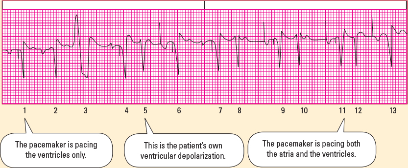
This DDD pacemaker rhythm strip reveals how this pacemaker ensures AV synchrony. In the atrium: The pacemaker paces in the atrium if it does not sense an intrinsic P wave at a certain beats per minute (bpm) rate, as in beats 6, 9, 11, and 13. Therefore, a paced P wave should and does follow the atrial spike. Sensed P waves cause withholding of pacing in the atrium as in beats 1, 2, 3, 4, and 7. When the patient has an intrinsic P wave, the pacemaker serves only to make sure the ventricles respond. In the ventricle: The pacemaker paces in the ventricle if it does not sense an intrinsic QRS complex in the ventricle, as in beats 1, 4, 6, 7, 9, and 11. Sensed QRS complexes cause withholding of pacing in the ventricle as in beats 2, 3, 5, 8, 10, and 12. As you can see, there are several different possible ECG scenarios: AV-paced (e.g., beat 6), AV-sensed (e.g., beat 2), A-paced and V-sensed (e.g., beat 13), and A-sensed and V-paced (e.g., beat 4). This ECG strip has all four possibilities in one strip. However, patients typically have one or two scenarios in one strip, for example, a patient in sinus rhythm with only a very rare paced beat. Also of note, these ventricularly paced beats have a QRS complex that is narrower than right ventricular paced beats. This may be because of pacing lead position (e.g., left ventricular or His bundle pacing) or a QRS complex that is a result of fusion between ventricular pacing and an inherent beat. 
|