Each of the 12 leads views the heart from a different angle. These illustrations show the direction of each lead relative to the wave of depolarization (shown in color) and list the 12 views of the heart. | Views reflected on a 12-lead ECG | Leads | View of the heart |
|---|
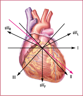
| Stand ard limb leads (bipolar) | | I | Lateral wall | | II | Inferior wall | | III | Inferior wall | | Augmented limb leads (unipolar) | | aVR | No specific view | | aVL | Lateral wall | | aVF | Inferior wall | 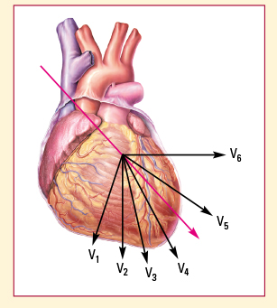
| Precordial, or chest, leads (unipolar) | | V1 | Septal wall | | V2 | Septal wall | | V3 | Anterior wall | | V4 | Anterior wall | | V5 | Lateral wall | | V6 | Lateral wall |
|
|