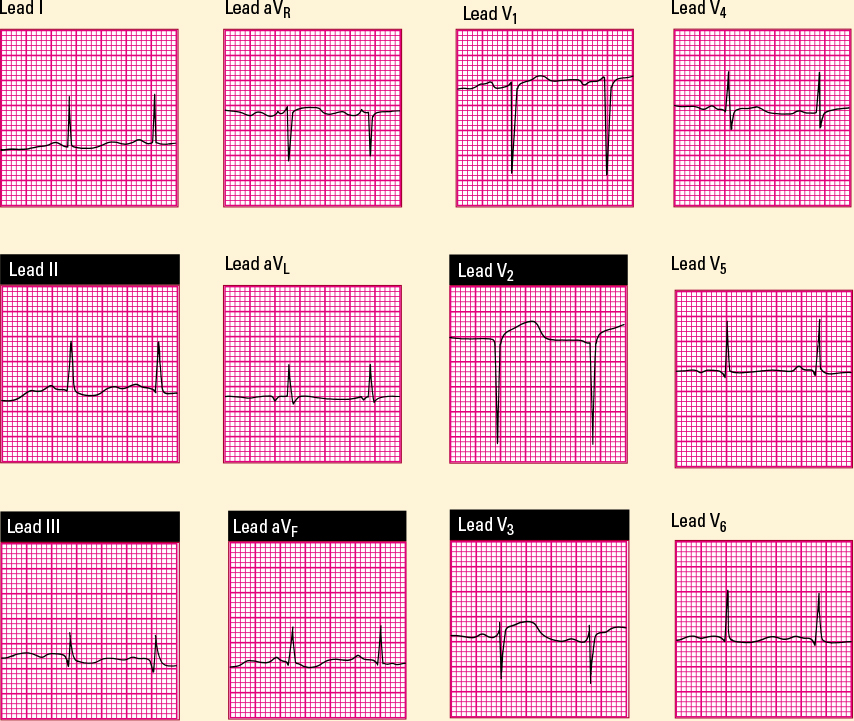
This 12-lead ECG shows typical characteristics of an anterior wall MI. Note that the R waves don’t progress through the precordial leads. Also note the ST-segment elevation in leads V2 and V3. As expected, the reciprocal leads II, III, and aVF show slight ST-segment depression. Axis deviation is normal at +60 degrees. 
|