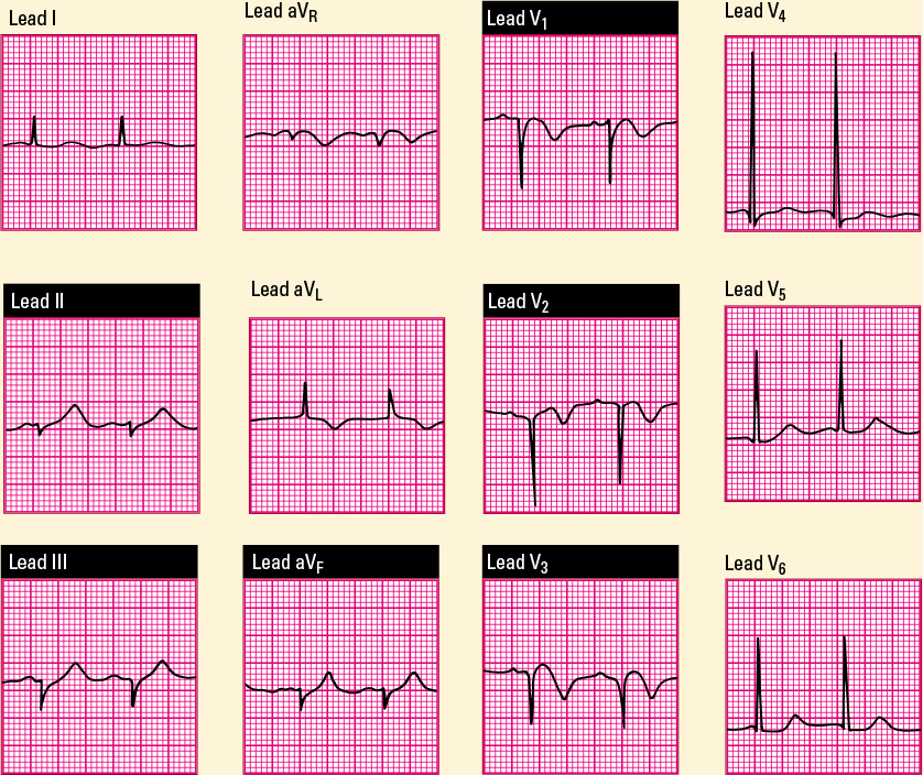
This 12-lead ECG shows typical characteristics of an anteroseptal wall MI. Note the poor R-wave progression, the elevated ST segments, and the inverted T waves in leads V1, V2, and V3. Reciprocal changes are seen in leads II, III, and aVF with depressed ST segments and tall, peaked T waves. 
|