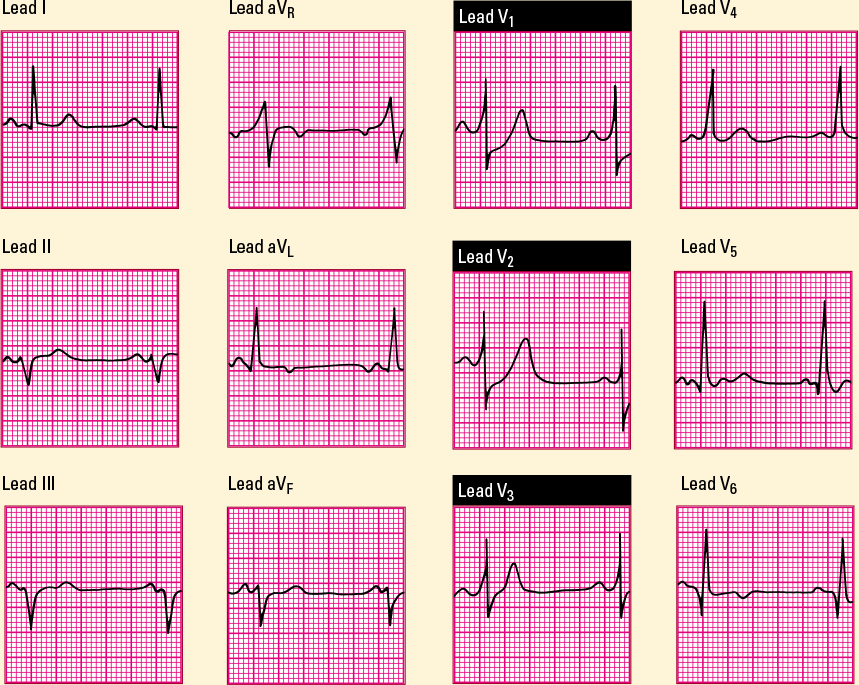
This 12-lead ECG shows typical characteristics of a posterior wall MI. Note the tall R waves, the depressed ST segments, and the upright T waves in leads V1, V2, and V3 that represent reciprocal changes. These are reciprocal changes, because the leads that best monitor a posterior wall MI (V7, V8, and V9) aren’t on a stand ard 12-lead ECG. 
|