A Look at Cardiac Anatomy
Cardiac anatomy includes the location of the heart; the structure of the heart, heart wall, chambers, and valves; and the layout and structure of coronary circulation. A thorough comprehension of the relationship between the various heart structures is critical to the understand ing of current and emerging concepts in cardiac electrophysiology, as well as the management of cardiac arrhythmias.
Outside the Heart
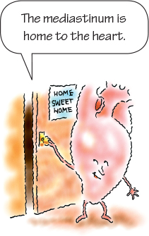
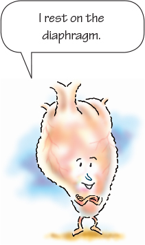
The heart is a cone-shaped, muscular organ. It’s located in the chest, behind the sternum in the mediastinal cavity (or mediastinum), between the lungs, and in front of the spine. The heart lies tilted in this area like an upside-down triangle. The top of the heart, or its base, lies just below the second rib; the bottom of the heart, or its apex, tilts forward and down, toward the left side of the body, and rests on the diaphragm. (See Location of the pediatric heart)
The heart varies in size depending on the person’s body size, but the organ is roughly 5″ (12.5 cm) long and 3½″ (9 cm) wide, or about the size of the person’s fist. The heart’s weight, typically 9 to 12 oz (255 to 340 g), varies depending on the person’s size, age, sex, and athletic conditioning. An athlete’s heart usually weighs more than that of the average person and an elderly person’s heart weighs less. (See The older adult heart.) There are differences in male and female cardiovascular anatomy and physiology. A smaller heart and coronary vessels are seen in females versus males. Varying levels of estrogen may be reasons for gender variants in cardiac disease.
| Ages and stages |
 The older adult heart The older adult heartAs a person ages, his heart usually becomes slightly smaller and loses its contractile strength and efficiency (although exceptions occur in people with hypertension or heart disease). By age 70, cardiac output at rest has diminished by 30% to 35% in many people. Irritable with Age As the myocardium of the aging heart becomes more irritable, extra systoles may occur, along with sinus arrhythmias and sinus bradycardias. In addition, increased fibrous tissue infiltrates the sinoatrial (SA) node and internodal atrial tracts, which may cause atrial fibrillation and flutter. |
Layer Upon Layer
The heart’s wall is made up of three layers: the epicardium, myocardium, and endocardium. (See Layers of the heart wall.) The epicardium, the outer layer (and the visceral layer of the serous pericardium), is made up of squamous epithelial cells overlying connective tissue. The myocardium, the middle layer, makes up the largest portion of the heart’s wall. This layer of muscle tissue contracts with each heartbeat. The endocardium, the heart’s innermost layer, contains endothelial tissue with small blood vessels and bundles of smooth muscle.
A layer of connective tissue called the pericardium surrounds the heart and acts as a tough, protective sac. It consists of the fibrous pericardium and the serous pericardium. The fibrous pericardium, composed of tough, white, fibrous tissue, fits loosely around the heart protecting it. The fibrous pericardium attaches to the great vessels, diaphragm, and sternum. The serous pericardium, the thin, smooth, inner portion, has two layers:
- the parietal layer, which lines the inside of the fibrous pericardium
- the visceral layer, which adheres to the surface of the heart
Between the Layers
The pericardial space separates the visceral and parietal layers and contains 10 to 20 mL of thin, clear pericardial fluid that lubricates the two surfaces and cushions the heart. Excess pericardial fluid, a condition called pericardial effusion, compromises the heart’s ability to pump blood.
Inside the Heart
The heart contains four chambers—two atria and two ventricles. (See Inside a normal heart) The right and left atria serve as volume reservoirs for blood being sent into the ventricles. The right atrium receives deoxygenated blood returning from the body through the inferior and superior vena cava and from the heart through the coronary sinus. The left atrium receives oxygenated blood from the lungs through the four pulmonary veins. The interatrial septum divides the chambers and helps them contract. Contraction of the atria forces blood into the ventricles below. Although much emphasis is placed on left heart function, the right ventricle acts as a key contributor to hemodynamic stability.
Pump Up the Volume
The right and left ventricles serve as the pumping chambers of the heart. The right ventricle receives blood from the right atrium and pumps it through the pulmonary arteries to the lungs, where it picks up oxygen and drops off carbon dioxide. The left ventricle receives oxygenated blood from the left atrium and pumps it through the aorta and then out to the rest of the body. The interventricular septum separates the ventricles and also helps them to pump.
The chamber wall’s thickness depends on the amount of high-pressure work the chamber does. Because the atria collect blood for the ventricles and don’t pump it far, their walls are considerably thinner than the walls of the ventricles. Likewise, the left ventricle has a much thicker wall than the right ventricle because the left ventricle pumps blood against the higher pressures in the body’s arterial circulation, whereas the right ventricle pumps blood against the lower pressures in the lungs.
One-Way Valves
The heart contains four valves—two atrioventricular (AV) valves (tricuspid and mitral) and two semilunar valves (aortic and pulmonic). The valves open and close in response to changes in pressure within the chambers they connect. They serve as one-way doors that keep blood flowing through the heart in a forward direction.
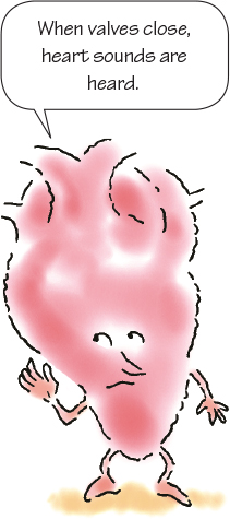
When the valves close, they prevent backflow, or regurgitation, of blood from one chamber to another. The closing of the valves creates the heart sounds that are heard through a stethoscope.
The two AV valves, located between the atria and ventricles, are called the tricuspid and mitral valves. The tricuspid valve is located between the right atrium and the right ventricle. The mitral valve is located between the left atrium and the left ventricle.
Cardiac Cords
The mitral valve has two cusps, or leaflets, and the tricuspid valve has three. The cusps are anchored to the papillary muscles in the heart wall by fibers called chordae tendineae. These cords work together to prevent the cusps from bulging backward into the atria during ventricular contraction. If damage occurs, blood can flow backward into a chamber, resulting in a heart murmur.
Under Pressure
The semilunar valves are the pulmonic valve and the aortic valve. These valves are called semilunar because the cusps resemble three half-moons. Because of the high pressures exerted on the valves, their structure is much simpler than that of the AV valves.
They open because of pressure within the ventricles and close because of the back pressure of blood in the pulmonary arteries and aorta, which pushes the cusps closed. The pulmonic valve, located where the pulmonary artery meets the right ventricle, permits blood to flow from the right ventricle to the pulmonary artery and prevents blood backflow into that ventricle. The aortic valve, located where the left ventricle meets the aorta, allows blood to flow from the left ventricle to the aorta and prevents blood backflow into the left ventricle.
Blood Flow through the Heart
Understand ing how blood flows through the heart is critical to understand ing the heart’s overall functions and how changes in electrical activity affect peripheral blood flow. Deoxygenated blood from the body returns to the heart through the inferior and superior vena cava and empties into the right atrium. From there, blood flows through the tricuspid valve into the right ventricle.
Circuit City
The right ventricle pumps blood through the pulmonic valve into the pulmonary arteries and then into the lungs. From the lungs, blood flows through the pulmonary veins and empties into the left atrium, which completes a circuit called pulmonary circulation.
When pressure rises to a critical point in the left atrium, the mitral valve opens and blood flows into the left ventricle. The left ventricle then contracts and pumps blood through the aortic valve into the aorta, and then throughout the body. Blood returns to the right atrium through the veins, completing a circuit called systemic circulation.
Getting into Circulation
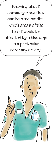
Like the brain and all other organs, the heart needs an adequate supply of blood to survive. The coronary arteries, which lie on the surface of the heart, supply the heart muscle with blood and oxygen. Understand ing coronary blood flow can help you provide better care for a patient with a myocardial infarction (MI) because you’ll be able to predict which areas of the heart would be affected by a blockage in a particular coronary artery. Damaged heart tissue, once thought irreparable, may now be repaired, according to recent research. The “Singheart” RNA molecule may stimulate damaged cells to regenerate or “self-heal.” This could be especially helpful to those with heart failure.
Open that Ostium
The coronary ostium, an opening in the aorta that feeds blood to the coronary arteries, is located near the aortic valve. During systole, when the left ventricle is pumping blood through the aorta and the aortic valve is open, the coronary ostium is partially covered. During diastole, when the left ventricle is filling with blood, the aortic valve is closed and the coronary ostium is open, enabling blood to fill the coronary arteries.
With a shortened diastole, which occurs during periods of tachycardia, less blood flows through the ostium into the coronary arteries. Tachycardia also impedes coronary blood flow because contraction of the ventricles squeezes the arteries and lessens blood flow through them.
That’s Right, Coronary
The right coronary artery as well as the left coronary artery (also known as the left main artery) originate as a single branch off the ascending aorta from the area known as the sinuses of Valsalva. The right coronary artery supplies blood to the right atrium, the right ventricle, and part of the inferior and posterior surfaces of the left ventricle. In about 50% of the population, the artery also supplies blood to the SA node. The bundle of His and the AV node also receive their blood supply from the right coronary artery.
What’s Left, Coronary?
The left coronary artery runs along the surface of the left atrium, where it splits into two major branches: the left anterior descending and the left circumflex arteries. The left anterior descending artery runs down the surface of the left ventricle toward the apex and supplies blood to the anterior wall of the left ventricle, the interventricular septum, the right bundle branch, and the left anterior fasciculus of the left bundle branch. The branches of the left anterior descending artery—the septal perforators and the diagonal arteries—help supply blood to the walls of both ventricles.
Circling Circumflex
The circumflex artery supplies oxygenated blood to the lateral walls of the left ventricle, the left atrium and , in about half of the population, the SA node. In addition, the circumflex artery supplies blood to the left posterior fasciculus of the left bundle branch. This artery circles the left ventricle and provides blood to the ventricle’s posterior portion.
Circulation, Guaranteed
When two or more arteries supply the same region, they usually connect through anastomoses, junctions that provide alternative routes of blood flow. This network of smaller arteries, called collateral circulation, provides blood to capillaries that directly feed the heart muscle. Collateral circulation commonly becomes so strong that even if major coronary arteries become clogged with plaque, collateral circulation can continue to supply blood to the heart.
Veins in the Heart
The heart has veins just like other parts of the body. Cardiac veins collect deoxygenated blood from the capillaries of the myocardium. The cardiac veins join to form an enlarged vessel called the coronary sinus, which returns blood to the right atrium, where it continues through the circulation.
A Look at Cardiac Physiology
This discussion of cardiac physiology includes descriptions of the cardiac cycle, how the cardiac muscle is innervated (distribution/supply of nerves), how the depolarization–repolarization cycle operates, how impulses are conducted, and how abnormal impulses work. (See Events of the cardiac cycle)
Cardiac Cycle Dynamics
During one heartbeat, ventricular diastole (relaxation) and ventricular systole (contraction) occur.
During diastole, the ventricles relax, the atria contract, and blood is forced through the open tricuspid and mitral valves. The aortic and pulmonic valves are closed.
During systole, the atria relax and fill with blood. The mitral and tricuspid valves are closed. Ventricular pressure rises, which forces open the aortic and pulmonic valves. The ventricles then contract, and blood flows through the circulatory system.
The atrial contraction, or atrial kick, contributes about 30% of the cardiac output—the amount of blood pumped by the ventricles in 1 minute. (See Quick facts about circulation.) Certain arrhythmias, such as atrial fibrillation, can cause a loss of atrial kick and a subsequent drop in cardiac output. Tachycardia also affects cardiac output by shortening diastole and allowing less time for the ventricles to fill. Less filling time means less blood will be ejected during ventricular systole and less will be sent through the circulation.
A Balancing Act
The cardiac cycle produces cardiac output, which is the amount of blood the heart pumps in 1 minute. It’s measured by multiplying heart rate times stroke volume. (See Understand ing preload, afterload, and contractility.) The term stroke volume refers to the amount of blood ejected with each ventricular contraction.

Normal cardiac output is 4 to 8 L/min, depending on body size. The heart pumps only as much blood as the body requires. Three factors affect stroke volume—preload, afterload, and myocardial contractility. A balance of these three factors produces optimal cardiac output.
Preload
Preload is the stretching of muscle fibers in the ventricles and is determined by the pressure and amount of blood remaining in the left ventricle at the end of diastole.
Afterload
Afterload is the amount of pressure the left ventricle must work against to pump blood into the circulation. The greater this resistance, the more the heart works to pump out blood.
Contractility
Contractility is the ability of muscle cells to contract after depolarization. This ability depends on how much the muscle fibers are stretched at the end of diastole. Over-stretching or under-stretching these fibers alters contractility and the amount of blood pumped out of the ventricles. To better understand this concept, imagine you are trying to shoot a rubber band across the room. If you don’t stretch the rubber band enough, it won’t go far. If you stretch it too much, it will snap. However, if you stretch it just the right amount, it will go as far as you want it to go.
Nerve Supply to the Heart
The heart is supplied by the two branches of the autonomic nervous system—the sympathetic, or adrenergic, and the parasympathetic, or cholinergic.
The sympathetic nervous system is basically the heart’s accelerator. Two sets of chemicals—norepinephrine and epinephrine—are highly influenced by this system. These chemicals increase heart rate, automaticity, AV conduction, and contractility.
Braking the Heart
The parasympathetic nervous system, on the other hand , serves as the heart’s brakes. One of this system’s nerves, the vagus nerve, carries impulses that slow heart rate and the conduction of impulses through the AV node and ventricles. Stimulating this system releases the chemical acetylcholine, slowing the heart rate.
The vagus nerve is stimulated by baroreceptors, specialized nerve cells in the aorta, and the internal carotid arteries. Conditions that stimulate the baroreceptors also stimulate the vagus nerve.
For example, a stretching of the baroreceptors, which can occur during periods of hypertension or when applying pressure to the carotid artery, stimulates the receptors. In a maneuver called carotid sinus massage, baroreceptors in the carotid arteries are purposely activated in an effort to slow a rapid heart rate.
Transmission of Electrical Impulses
The heart can’t pump unless an electrical stimulus occurs first. Generation and transmission of electrical impulses depend on four characteristics of cardiac cells:
- Automaticity refers to a cell’s ability to spontaneously initiate an impulse. Pacemaker cells possess this ability.
- Excitability results from ion shifts across the cell membrane and indicates how well a cell responds to an electrical stimulus.
- Conductivity is the ability of a cell to transmit an electrical impulse to another cardiac cell.
- Contractility refers to how well the cell contracts after receiving a stimulus.
“de”-Cycle and “re”-Cycle
As impulses are transmitted, cardiac cells undergo cycles of depolarization and repolarization. (See Depolarization–repolarization cycle) Cardiac cells at rest are considered polarized, meaning that no electrical activity takes place. Cell membranes separate different concentrations of ions, such as sodium and potassium, and create a more negative charge inside the cell. This is called the resting potential. After a stimulus occurs, ions cross the cell membrane and cause an action potential, or cell depolarization. Researchers have discovered a “calcium-sensing-gate,” which initiates calcium waves responsible for calcium-triggered arrhythmias. This discovery may aid in the development of new drug treatments. When a cell is fully depolarized, it attempts to return to its resting state in a process called repolarization. Electrical charges in the cell reverse and return to normal.
A cycle of depolarization–repolarization consists of five phases—0 through 4. The action potential is represented by a curve that shows voltage changes during the five phases. (See Action potential curve)
Many Phases of the Curve
During phase 0, the cell receives an impulse from a neighboring cell and is depolarized. Phase 1 is marked by early, rapid repolarization. Phase 2, the plateau phase, is a period of slow repolarization.
During phases 1 and 2 and at the beginning of phase 3, the cardiac cell is said to be in its absolute refractory period. During that period, no stimulus, no matter how strong, can excite the cell.
Phase 3, the rapid repolarization phase, occurs as the cell returns to its original state. During the last half of this phase, when the cell is in its relative refractory period, a very strong stimulus can depolarize it.
Phase 4 is the resting phase of the action potential. By the end of phase 4, the cell is ready for another stimulus.
All that electrical activity is represented on an electrocardiogram (ECG). Keep in mind that the ECG represents electrical activity only, not actual pumping of the heart.
Pathway through the Heart
After depolarization and repolarization occur, the resulting electrical impulse travels through the heart along a pathway called the conduction system. (See The cardiac conduction system)
Impulses travel out from the SA node and through the internodal tracts and Bachmann’s bundle to the AV node. From there, they travel through the bundle of His, the bundle branches, and lastly to the Purkinje fibers.
Setting the Pace
The SA node is located in the upper right corner of the right atrium, where the superior vena cava joins the atrial tissue mass. It’s the heart’s main pacemaker, generating impulses 60 to 100 times per minute. When initiated, the impulses follow a specific path through the heart. They usually can’t flow backward because the cells can’t respond to a stimulus immediately after depolarization.
Bachmann’s Bundle of Nerves
Impulses from the SA node next travel through Bachmann’s bundle, tracts of tissue extending from the SA node to the left atrium. Impulses are thought to be transmitted throughout the right atrium through the anterior, middle, and posterior internodal tracts. Impulse transmission through the right and left atria occurs so rapidly that the atria contract almost simultaneously.
AV: The Slow Node
The AV node, located in the inferior right atrium near the ostium of the coronary sinus, is responsible for delaying the impulses that reach it. Although the nodal tissue itself has no pacemaker cells, the tissue surrounding it (called junctional tissue) contains pacemaker cells that can fire at a rate of 40 to 60 times/min.
The AV node’s main function is to delay impulses by 0.04 seconds to keep the ventricles from contracting too quickly. This delay allows the ventricles to complete their filling phase as the atria contract. It also allows the cardiac muscle to stretch to its fullest for peak cardiac output.

Branch Splitting
The bundle of His, a tract of tissue extending into the ventricles next to the interventricular septum, resumes the rapid conduction of the impulse through the ventricles. The bundle eventually divides into the right and left bundle branches.
The right bundle branch extends down the right side of the interventricular septum and through the right ventricle. The left bundle branch extends down the left side of the interventricular septum and through the left ventricle.
The left bundle branch then splits into two branches, or fasciculi: the left anterior fasciculus, which extends through the anterior portion of the left ventricle, and the left posterior fasciculus, which runs through the lateral and posterior portions of the left ventricle. Impulses travel much faster down the left bundle branch (which feeds the larger, thicker-walled left ventricle) than the right bundle branch (which feeds the smaller, thinner-walled right ventricle).
The difference in the conduction speed allows both ventricles to contract simultaneously. The entire network of specialized nervous tissue that extends through the ventricles is known as the His–Purkinje system.
Those Perky Purkinje Fibers
Purkinje fibers extend from the bundle branches into the endocardium, deep into the myocardial tissue. These fibers conduct impulses rapidly through the muscle to assist in its depolarization and contraction.
Purkinje fibers can also serve as a pacemaker and are able to discharge impulses at a rate of 20 to 40 times/min, sometimes even more slowly. (See Pacemakers of the heart) Purkinje fibers usually aren’t activated as a pacemaker unless conduction through the bundle of His becomes blocked or a higher pacemaker (SA or AV node) doesn’t generate an impulse. (See Pediatric pacemaker rates)
Abnormal Impulses
Now that you understand how the heart generates a normal impulse, let’s look at some causes of abnormal impulse conduction, including automaticity, backward conduction of impulses, reentry abnormalities, and ectopy.
When the Heart Goes on “manual”
Automaticity is a special characteristic of pacemaker cells that generate impulses automatically, without being stimulated to do so. If a cell’s automaticity is increased or decreased, an arrhythmia can occur. Tachycardia, for example, is commonly caused by an increase in the automaticity of pacemaker cells below the SA node. Likewise, a decrease in automaticity of cells in the SA node can cause the development of bradycardia or an escape rhythm (a compensatory beat generated by a lower pacemaker site).
Out of Synch
Impulses that begin below the AV node can be transmitted backward toward the atria. This backward, or retrograde, conduction usually takes longer than normal conduction and can cause the atria and ventricles to beat out of synch.
Coming Back for More
Sometimes impulses cause depolarization twice in a row at a faster-than-normal rate. Such events are referred to as reentry events. In reentry, impulses are delayed long enough that cells have time to repolarize. In these cases, the active impulse reenters the same area and produces another impulse.
| That’s a wrap! |
 Cardiac anatomy and physiology review Cardiac anatomy and physiology reviewThe Heart’s Valves
Blood Flow
Coronary Arteries and Veins
Cardiac Cycle Dynamics
Innervation of the Heart Two branches of the autonomic nervous system supply the heart:
Transmission of Electrical Impulses Generation and transmission of electrical impulses depend on the following cell characteristics:
Depolarization–repolarization Cycle Cardiac cells undergo the following cycles of depolarization and repolarization as impulses are transmitted:
Cardiac Conduction
Intrinsic Firing Rates
Abnormal Impulses
|
Repeating Itself
An injured pacemaker (or nonpacemaker) cell may partially depolarize, rather than fully depolarizing. Partial depolarization can lead to spontaneous or secondary depolarization, which involves repetitive ectopic firings called triggered activity.
The resultant depolarization is called afterdepolarization. Early afterdepolarization occurs before the cell is fully repolarized and can be caused by hypokalemia, slow pacing rates, or drug toxicity. If it occurs after the cell has been fully repolarized, it’s called delayed afterdepolarization. These problems can be caused by digoxin toxicity, hypercalcemia, or increased catecholamine release. Atrial or ventricular tachycardia may result. You’ll learn more about these and other arrhythmias in later chapters.