AUTHORS: Averill Guo, MD and Jeanette G. Smith, MD


DefinitionPrimary biliary cholangitis (PBC), previously known as primary biliary cirrhosis, is a chronic, variably progressive immune-mediated cholestatic liver disease manifested by fatigue and pruritus. Most often diagnosed in middle-aged women, this condition is characterized by autoimmune destruction of small intralobular bile ducts leading to portal inflammation, hepatic cell necrosis, fibrosis, and, if left untreated, cirrhosis, liver failure, and death.1
SynonymsPBC
Primary biliary cirrhosis
Biliary cirrhosis
Nonsuppurative destructive cholangitis
Autoimmune cholangiopathy (AIC)
| ICD-10CM CODE | | K74.3 | Primary biliary cholangitis |
|
Epidemiology & DemographicsIncidence
- Most recent annual incidence rate in European countries is 1.87 new cases per 100,000 patients.1
- Most recent annual incidence rate in the Asia-Pacific region is 0.86 new cases per 100,000 patients.2
- Most recent annual incidence rate in North America is 2.75 new cases per 100,000 patients.3
- Pooled global annual incidence is estimated at 1.76 new cases per 100,000 patients.3
PrevalencePrevalence is greatest in North America and Northern Europe and varies tremendously by time and geographic areas, but most recent data show an incidence of 22.27 cases per 100,000 patients in European countries, 11.88 cases per 100,000 patients in the Asia-Pacific region, and 21.81 per 100,000 patients in North America.1,3 Pooled global prevalence is estimated at 14.60 per 100,000 patients. Disease incidence and prevalence appear to be increasing worldwide, particularly in North America.3,4
Predominant SexWhile a female:male ratio of 9:1 is often described, recent studies suggests that PBC may be more common in men than previously understood with a female:male ratio of 4 or 5:1.5,6 Men with PBC typically present with more advance disease at the time of diagnosis, and those with well-compensated cirrhosis have been reported to have a higher risk of death and liver-related death or transplantation.6
Predominant AgeOnset typically occurs between the ages of 30 and 65 yr and is uncommon before age 25 yr.
Predominant RaceMost often described in Caucasians, but PBC affects all races.
Genetics
- Pathogenesis is unknown, but it is thought to be in the setting of environmental influences and genetic predisposition.7
- There are no clearly identified genetic factors associated with PBC; however, there is a clear familial occurrence.
- Up to 73% of patients with PBC have at least one other extrahepatic autoimmune disorder such as thyroiditis, Sjögren syndrome, rheumatoid arthritis, cutaneous scleroderma (including CREST syndrome), systemic lupus erythematosus, pernicious anemia, celiac disease, inflammatory bowel disease, autoimmune thrombocytopenia purpura, autoimmune diabetes mellitus, and/or other autoimmune diseases.8
- A variant form of PBC exists as an overlap syndrome with autoimmune hepatitis (AIH).
- PBC is closely associated with a greater risk of hepatocellular carcinoma as well as an overall greater risk of cancer.
Physical Findings & Clinical PresentationClinical stages:
- Asymptomatic
- Symptomatic
- Cirrhotic
- Hepatic failure
Symptoms:
- 50% to 65% of patients may be asymptomatic at time of diagnosis; between 35% and 89% become symptomatic within 4.5 to 17.8 yr.9
- Fatigue (50% to 78% of patients) and pruritus (20% to 70% of patients) are the usual presenting symptoms and are independent of disease severity.9
- Fatigue can be chronic and correlate with daytime somnolence and autonomic dysfunction.
- Pruritus is present predominantly on the palm and soles, is worse at night and with constricting garments, and is worse with dry skin and humid weather. The cause is unknown but elevated histamine, bile salt concentration, endogenous opioids, lysophosphatidic acid, and female steroid hormones and their metabolites have been discussed as potential causes. Pruritus may first occur during pregnancy but is distinguished from pruritus of pregnancy because it persists into the postpartum period and beyond.
- Symptoms include jaundice, unexplained right upper quadrant pain, manifestations of portal hypertension, dyslipidemia, xanthomas, and osteoporosis, and may be associated with Sjögren syndrome, rheumatoid arthritis, systemic lupus erythematosus, celiac disease, and thyroid disorders (with the most common being Hashimoto thyroiditis).
- Other symptoms can include steatorrhea, osteopenia, fat-soluble vitamin deficiencies, and anemia.
Physical examination:
- Variable: Findings depend on stage of disease at time of presentation; patients at the early stage may be completely unaffected.
- Excoriations may be present due to extensive scratching from pruritus and can be severe enough to cause bleeding.
- Hepatomegaly and splenomegaly can worsen with disease progression.
- Xanthomas and jaundice generally appear in advanced disease. Kayser-Fleischer rings are rare and result from copper retention. Hyperpigmentation of the skin due to melanin deposition may also occur.
- Late physical findings mirror those of cirrhosis: Spider nevi, caput medusae, temporal and proximal limb wasting, ascites, palmar erythema, digital clubbing, gynecomastia, and edema.
Etiology
- Although the cause of PBC remains unknown, it is believed to require both a genetic susceptibility as well as an environmental trigger ultimately leading to the modification of mitochondrial proteins triggering a persistent T lymphocyte-mediated attack on intrahepatic biliary epithelial cells.
- PBC is associated most strongly with HLA alleles DRA, DRB1, DPB1, DQB1, BTNL2, and c6orf10. PBC is also associated with ORMDL3, CD80, STAT1/STAT4, IL12A, NF-κB, and RPL3/SYNGR1. However, there is variation across ethnic groups.
- Possible environmental triggers include infectious agents, cigarette smoking, environmental pollutants, radiation, urinary tract infections, reproductive hormone replacement, prior pregnancy, toxic waste sites (particularly exposure to halogenated hydrocarbons), electrophilic drugs, and xenobiotics found in food additives and cosmetics.
- The enzyme complex subunit PDC-E2 is an autoantigen that plays a major role in the early pathogenesis of PBC. Patients with PBC have a tenfold increased concentration of PDC-E2-specific cytotoxic CD8+ lymphocytes in their livers compared to their blood, and antimitochondrial antibodies (AMAs), which are the serologic hallmark of this disease, react to the PDC-E2 subunit leading to a strong inflammatory response. In addition, biliary epithelial cells handle PDC-E2 in a unique way that exposes them to immune-mediated attack. Future therapies may be specific immunomodulation directed at these peptides.
- Damage to bile ducts results in bile leaking into liver parenchyma resulting in hepatocyte necrosis, which can lead to fibrosis and eventual cirrhosis.

The diagnosis of PBC can be established when two of the following three criteria are met in the absence of extrahepatic biliary obstruction.
- Positive serum AMA (titer >1:40) or PBC-specific antibodies to sp100 or gp210 in AMA-negative patients9
- Biochemical evidence of cholestasis (mainly alkaline phosphatase elevation [ALP] ≥1.5 times the upper limit of normal [ULN])
- Characteristic liver histology demonstrating nonsuppurative destruction of small to medium-sized interlobular biliary ducts
- Recent guidelines suggest that the above criteria may lead to a delay in diagnosis, so clinical judgment and expert referral should be considered4
Differential Diagnosis
- Drug-induced cholestasis (common medications: Phenothiazines, anabolic steroids, some antibiotics as TMP-SMX, oxacillin, and ampicillin)
- PBC-AIH overlap syndrome; reported in 1% to 14.2% of patients initially diagnosed with PBC. Transition from stable PBC to AIH and vice versa also seen
- Other etiologies of chronic liver disease and cirrhosis, such as alcoholic cirrhosis, chronic viral hepatitis, primary sclerosing cholangitis, AIH, sarcoidosis, hepatic amyloidosis, chemical/toxin-induced cirrhosis, other hereditary or familial disorders (e.g., cystic fibrosis, α-1-antitrypsin deficiency)
- Biliary obstruction
- Secondary biliary cirrhosis or secondary sclerosing cholangitis
WorkupHistory, physical examination, laboratory evaluation, liver biopsy
Laboratory Tests
- AMAs are found in 90% to 95% of patients with PBC and are 98% specific, although ANA-negative PBC is possible.9 ANA is also found in about 30% to 50% of patients.10 Most patients have ANA or AMAs, or both.
- Cholestatic pattern of liver biochemical markers; markedly increased ALP (of hepatic origin). ALP levels below 1.5 times ULN are associated with more favorable prognosis.
- γ-Glutamyl transpeptidase is increased and may be indicative of biliary origin of ALP elevation.
- Serum IgM levels are increased (lower in AMA-negative PBC).
- Bilirubin level is normal early on and increases with disease progression (direct and indirect). Increased serum bilirubin level is generally regarded as a poor prognostic indicator.
- Rising serum hyaluronate levels correlate with the serum bilirubin and histologic worsening of the disease.
- Aminotransferase level may be normal and, if increased, is rarely more than 5× ULN.
- Bile acid levels are strikingly elevated and this accumulation is thought to cause foamy degeneration of hepatocytes due to toxic effects.
- Serum ceruloplasmin may also be elevated.
- Markedly increased serum lipids is largely due to increased lipoprotein X (LpX). Total cholesterol may surpass 1000 mg/dl (with increased xanthomas rather than xanthelasmas). In the early stages of PBC, patients can have relatively higher HDL in comparison to LDL and VLDL. This rise in HDL might explain the lack of increased risk for cardiovascular disease. However, cardiovascular risk may still exist due to other risk factors (e.g., family history and metabolic syndrome).
- Percutaneous liver biopsy is helpful to rule out or confirm PBC in patients with superimposed NASH or AIH overlap syndrome, but is not always necessary for diagnosis.11 Biopsy is also helpful for disease staging.
- Histology is not uniform, so histologic stage is based on the most advanced lesion present.
- Stage I: Nonsuppurative cholangitis indicated by lymphocytic infiltration of small bile ducts with or without epithelioid granulomas or plasma cells, limited to portal areas
- Stage II: Extension of inflammatory cells to periportal parenchyma, ductular proliferation
- Stage III: Bridging necrosis or fibrous septa linking portal triads
- Stage IV: Frank cirrhosis with regenerative nodules
Imaging StudiesIf history, physical examination, blood tests, and liver biopsy are all consistent with PBC, neither imaging nor cholangiography (Fig. E1) is necessary. MRI or transient elastography (TE) is important for determination of degree of cirrhosis if present and may be indicated for monitoring of disease progression. Notably, the cutoffs for transient elastography are different than those established for other diseases, such as hepatitis C.
Figure E1 Primary Biliary Cholangitis Demonstrated by Endoscopic Retrograde Cholangiopancreatography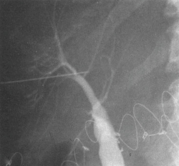
From Berk RN et al: Radiology of the gallbladder and bile ducts: diagnosis and intervention, Philadelphia, 1983, Saunders.
Prognosis
- There is widespread variation in progression between phases of PBC. Some patients advance rapidly to cirrhosis and require transplant whereas others remain asymptomatic for decades.
- Median time to development of extensive fibrosis in untreated patients is 2 yr.9
- Median survival is 7.5 yr in untreated patients and 16 yr in asymptomatic patients; however, this has improved with earlier diagnosis and initiation of treatment.9Table 1 summarizes time course of histologic progression to a higher stage in patients with PBC.
- Neither presence nor total titer level of AMAs predicts survival, disease progression, or response to therapy. As such, AMA should not be serially measured.
- Serum bilirubin is the best predictor of survival and the most heavily weighted factor in prognostic models. Box 1 summarizes independent predictors of survival in patients with PBC.1
- Serum ALP >1.5 ULN has been shown to be a risk factor for a more progressive course of PBC including cirrhosis.12
- Response to ursodeoxycholic acid (UDCA) therapy can be prognostic, with approximately 40% of patients failing to respond. There are multiple biochemical response criteria that, if met after 1 yr of treatment with UDCA, are associated with improved clinical outcomes. Three of these criteria are Barcelona (decrease in ALP level of at least 40% or to the reference range), Paris I (ALP <3 × ULN, AST <2 × ULN and bilirubin within normal limits), and Paris II (ALP <1.5 × ULN, ALT <1.5 × ULN and bilirubin within normal limits).13-15
- Nonresponse to UDCA is also associated with increased HCC risk compared to UDCA-responsive PBC.
- Similarly, the Mayo Risk score, a predictor of short-term survival probability in nontransplanted patients (www.mayoclinic.org/medical-professionals/transplant-medicine/the-updated-natural-histo/the-updated-natural-history-model-for-primary-biliary-cirrhosis/itt-20434724), can also reliably predict life expectancy when calculated after 6 mo of UDCA therapy.16
- Several predictive models based upon laboratory and clinical data have been proposed, and two such models (GLOBE score and UK-PBC score) are based on multicenter studies including large cohorts of patients with PBC.
- GLOBE score: Includes the following five variables: Serum bilirubin, albumin, serum ALP, platelet count after 1 yr of UDCA treatment, and age at start of therapy. It estimates the duration of transplant-free survival.17
- UK-PBC score: Includes serum ALP, aminotransferases, and bilirubin after 12 mo of UDCA therapy, in addition to baseline albumin and platelet count. This model estimates the risk of liver transplantation or liver-related death.18
- Transient elastography is a valuable noninvasive means of determining prognosis and treatment response. It has been reported to be significantly superior to biochemical markers and risk scores (APRI, FIB-4, Mayo risk score) in predicting fibrosis and cirrhosis in PBC. In a prospective study of PBC patients treated with UDCA, noncirrhotic PBC patients (F0 to F3 stages) were found to have either limited or no significant progression of liver stiffness whereas cirrhotic PBC patients developed significant increases in liver stiffness. Increasing liver stiffness (>2.1 kPa/yr) has been associated with an 8.4-fold increased risk of decompensation, liver transplantation, or death.19
- Poorer prognosis is associated with jaundice, advanced histologic stage, elevated bilirubin or ALP, low albumin, hepatocellular carcinoma, nonresponse to UDCA, and esophageal varices.
BOX 1 Independent Predictors of Survival in Patients With PBC in Various Clinical Studies
Clinical
- Age
- Ascites
- Edema
- Hepatomegaly
- Variceal bleeding
Laboratory
- Serum albumin level
- Serum alkaline phosphatase level
- Serum bilirubin level
- Prothrombin time
Liver Histology
- Cholestasis
- Cirrhosis
- Fibrosis
- Mallory hyaline
|
From Feldman M et al: Sleisenger and Fordtran’s gastrointestinal and liver disease, ed 10, Philadelphia, 2016, Elsevier.
TABLE 1 Time Course of Histologic Progression to a Higher Stage in Patients With PBC
| Histologic Progression∗ | Initial Histologic Stage |
|---|
| 1 | 2 | 3 |
|---|
| 1 yr | 41 | 43 | 35 |
| 2 yr | 62 | 62 | 50 |
From Feldman M et al: Sleisenger and Fordtran’s gastrointestinal and liver disease, ed 10, Philadelphia, 2016, Elsevier.
