section name header

Basic Information

AUTHOR: Patricia Cristofaro, MD

Definition

Sporotrichosis is a granulomatous/pyogenic disease commonly caused by pathogenic strains of the dimorphic fungus Sporothrix schenckii, although at least six other pathogenic Sporothrix species exist in various geographic regions, such as Sporothrix brasiliensis.

Synonyms

Lymphocutaneous sporotrichosis

Cutaneous sporotrichosis

Pulmonary sporotrichosis

ICD-10CM CODES
B42.0Pulmonary sporotrichosis
B42.1Lymphocutaneous sporotrichosis
B42.7Disseminated sporotrichosis
B42.8Other forms of sporotrichosis
B42.9Sporotrichosis, unspecified
Epidemiology & Demographics
Predominant Sex

The most common form, lymphocutaneous sporotrichosis, occurs equally in both sexes. Males predominate in both pulmonary and osteoarticular sporotrichosis.

Predominant Age

Generally, lymphocutaneous sporotrichosis occurs in people 35 yr of age or younger, usually through occupation or hobbies such as landscaping or gardening. Pulmonary sporotrichosis occurs in people between the ages of 30 and 60 yr, often alcoholic men with chronic obstructive pulmonary disease and diabetics. Those with HIV are at risk for disseminated infection.

Genetics

Neonatal infection: At least one case of transmission from the cheek lesions of the mother to the skin of the infant has been reported.

Physical Findings & Clinical Presentation

  • Cutaneous disease:
    1. Arises at the site of inoculation, often the site of soil exposure (Fig. E1)
    2. Initial lesion usually located on the distal part of an extremity (Fig. E2), although any area may be affected, including the face
    3. Variable incubation period of approximately 3 wk once introduced into the skin
    4. Granulomatous reaction provoked
    5. Lesion becomes papulonodular, erythematous, elastic, variable in size
    6. Subsequently, nodule becomes fluctuant, undergoes central necrosis, breaks down, discharges mucoid material from which fungus may be isolated
    7. Indolent ulcer with raised erythematous or violaceous borders
    8. Secondary lesions:
      1. Develop along superficial lymphatic channels
      2. Evolve in the same manner as the primary lesion, with subsequent inflammation, induration, and suppuration
  • Fixed, or plaque form:
    1. Erythematous verrucous, ulcerated, or crusted lesions
    2. Does not spread locally
    3. Does not involve lymphatic vessels
    4. Rarely undergoes spontaneous resolution
    5. More often persists for years without systemic symptoms and within a setting of normal laboratory examinations
  • Osteoarticular involvement:
    1. Most common extracutaneous form
    2. Usually presents as monoarticular arthritis
    3. Left untreated, may progress to:
      1. Synovitis
      2. Osteitis
      3. Periostitis
      4. All involving elbows, knees, wrists, and ankles
  • Joint inflamed:
    1. Associated with an effusion
    2. Painful on motion
  • Early pulmonary disease:
    1. Usually associated with a paucity of clinical findings:
      1. Low-grade fever
      2. Cough
      3. Fatigue
      4. Malaise
      5. Weight loss
    2. Untreated:
      1. Cavitary pulmonary disease
      2. Frank pulmonary dysfunction
      3. Hemoptysis
      4. Mimics tuberculosis
    3. Meningitis:
      1. Uncommon except perhaps in the immunocompromised patient
      2. Presents with few signs or symptoms of neurologic involvement, usually headache
    4. Few reported cases:
      1. Infection of the ocular adnexa
      2. Endophthalmitis without antecedent trauma
      3. Infection of the testes and epididymis
      4. Certain strains favor cooler body areas such as the scrotum, lower extremities

Figure E1 Sporotrichosis.

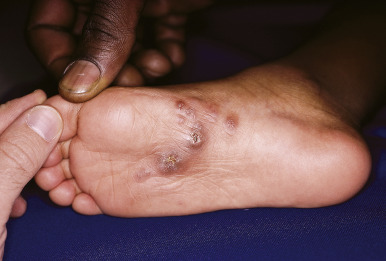
Erythematous papules and nodules on the plantar surface with early lymphangitic (sporotrichoid) spread.

From Paller AS, Mancini AJ: Hurwitz clinical pediatric dermatology, a textbook of skin disorders of childhood and adolescence, ed 5, Philadelphia, 2016, Elsevier.

Figure E2 Sporotrichosis of the fifth finger in a gardener.

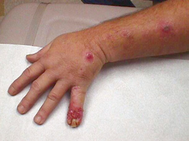
Three nodular lesions are visible on the hand and arm.

From Mandell GL et al: Principles and practice of infectious diseases, ed 7, Philadelphia, 2010, Saunders.

Etiology

  • Sporothrix schenckii:
  • Sporothrix brasiliensis emerging in South America; increased virulence
    1. Global in distribution
    2. Often isolated from soil, plants, and plant products
    3. Majority of case reports from tropical and subtropical regions of the Americas
  • Occupational or recreational exposure:
    1. Hay
    2. Straw
    3. Sphagnum moss
    4. Timber
    5. Thorny plants (e.g., roses and barberry bushes)
  • Animal contact:
    1. Armadillos
    2. Cats
    3. Squirrels
  • Tattooing

Diagnosis

Differential Diagnosis

  • Fixed, or plaque, sporotrichosis:
    1. Bacterial pyoderma
    2. Foreign body granuloma
    3. Tularemia
    4. Anthrax
    5. Other mycoses: Blastomycosis, chromoblastomycosis
  • Lymphocutaneous sporotrichosis:
    1. Nocardia brasiliensis
    2. Leishmania braziliensis
    3. Atypical mycobacterial disease: Mycobacterium marinum, Mycobacterium kansasii
  • Pulmonary sporotrichosis:
    1. Pulmonary tuberculosis (TB)
    2. Histoplasmosis
    3. Coccidioidomycosis
  • Osteoarticular sporotrichosis:
    1. Pigmented villonodular synovitis
    2. Gout
    3. Rheumatoid arthritis
    4. Infection with Mycobacterium tuberculosis
    5. Atypical mycobacteria: Mycobacterium marinum, Mycobacterium kansasii, Mycobacterium avium-intracellulare
  • Meningitis:
    1. Histoplasmosis
    2. Cryptococcosis
    3. TB
Workup

  • The diagnosis should be considered in individuals who are occupationally exposed to soil, decaying plant matter, and thorny plants (gardeners, horticulturists, farmers) who present with chronic nonhealing ulcers or lesions with or without associated arthritis or pulmonary symptoms.
  • Diagnosis is made by culture:
    1. Pus
    2. Joint fluid
    3. Sputum
    4. Blood
    5. Skin biopsy
  • Isolation of the fungus from any site is considered diagnostic of infection.
  • Saprophytic colonization of the respiratory tract has been described.
  • A positive blood culture may indicate infection in an immunocompromised host.
  • Increasingly sensitive laboratory culturing systems may detect the fungus in the normal host.
  • Biopsy specimens are diagnostic if characteristic cigar-shaped, round, oval, or budding yeast forms are seen.
  • Despite special staining, the yeast may remain difficult to detect unless multiple sections are examined, as organisms are rare.
  • No standard method of serologic testing is available.
Laboratory Tests

  • CBCs and serum chemistries are generally normal.
  • Elevated erythrocyte sedimentation rate is seen with extracutaneous disease.
  • Cerebrospinal fluid analysis in meningeal disease reveals:
    1. Lymphocytic pleocytosis
    2. Elevated protein
    3. Hypoglycorrhachia
  • Nested polymerase chain reaction assay represents a future clinical modality to rapidly detect Sporothrix schenckii.
  • Matrix-assisted laser desorption ionization-time of flight has been used to identify Sporothrix grown in culture to the species level.
Imaging Studies

  • Chest x-ray examination: Unilateral or bilateral upper lobe cavitary or noncavitary lesions
  • Radiographic findings of affected joints:
    1. Loss of articular cartilage
    2. Periosteal reaction
    3. Periarticular osteopenia
    4. Cystic changes

Treatment

Nonpharmacologic Therapy

Local heat and prevention of bacterial superinfection in cutaneous or plaque form. This treatment is important for pregnant women as azoles are teratogenic.

Acute General Rx
Cutaneous and Lymphocutaneous Sporotrichosis

  • Itraconazole at doses of 200 mg/day is the drug of choice and should be given for 2 to 4 wk after the lesions have resolved.
  • Alternative treatment: Use saturated solution of potassium iodide (SSKI) 5 to 10 drops orally (PO) tid or 1.5 ml PO tid, gradually increasing to 40 to 50 drops PO tid or 3 ml PO tid after meals.
  • Maximum tolerated dose should be continued until cutaneous lesions have resolved, approximately 6 to 12 wk.
  • Adjunctive therapy with heat is useful and occasionally curative.
  • Side effects of SSKI:
    1. Nausea
    2. Anorexia
    3. Diarrhea
    4. Parotid or lacrimal gland hypertrophy
    5. Acneiform rash
Deep-Seated Mycoses (Osteoarticular, Noncavitary Pulmonary Disease)

  • Itraconazole:
    1. Appropriate initial chemotherapy
    2. Less toxic than amphotericin B
    3. 200 mg PO bid for 1 to 2 yr with continued lifelong suppressive therapy in selected patients
    4. Absence of relapses from 40 to 68 mo has been documented when at least 200 mg/day administered for 24 mo
    5. Insufficient data for use in disseminated disease (e.g., fungemia and meningitis)
    6. Drug levels should be monitored to assess absorption
  • Parenteral amphotericin B, total course of 2 to 2.5 g or more, results in cure in approximately two thirds of cases. Lipid amphotericin B 3 to 5 mg/kg/day is also recommended and less toxic.
    1. Relapses are common.
    2. Amphotericin B-resistant isolates of Sporothrix schenckii have been reported.
    3. Remains the drug of choice for severely ill patients with disseminated disease.
    4. In cavitary pulmonary disease, given perioperatively as an adjunct to surgical resection.
    5. When patient is improved, can continue treatment with itraconazole 200 mg PO bid.
    6. Treat for at least 12 mo.
    7. The latest Infectious Diseases Society of America guidelines recommend fluconazole only as an alternative treatment for lymphocutaneous disease.
    8. Requires daily doses of 400 mg/day for lymphocutaneous disease and 800 mg/day for visceral or osteoarticular disease.
    9. Fluconazole much less effective than itraconazole.
    10. All azoles are teratogenic in pregnancy.
Chronic Rx

  • For lymphocutaneous and visceral disease, therapy with itraconazole 200 mg/day for periods of 24 mo or greater. Sporothrix is resistant to voriconazole. It is sensitive in vitro to posaconazole, but existing data are very limited at present.
  • Sporothrix appears to be resistant to isavuconazole.
Disposition

  • Prognosis for cutaneous disease is good.
  • Prognosis is less satisfactory for extracutaneous disease, especially if associated with abnormal immunologic states or other underlying systemic diseases.
Referral

  • To surgeon; with an established diagnosis of pulmonary sporotrichosis, cavitary lesions require resection of involved tissue and possible pretreatment with amphotericin
  • To infectious disease physician

Pearls & Considerations

Comments

  • In patients with underlying immunosuppression (e.g., hematologic malignancy or infection with HIV), progression of the initial infection may develop into multifocal extracutaneous sporotrichosis.
  • In this subset of patients, dissemination of cutaneous lesions is accompanied by hematogenous spread to lungs, bone, mucous membranes, central nervous system.
  • Osteoarticular and pulmonary manifestations predominate with the development of polyarticular arthritis and osteolytic bone lesions.
  • In the absence of therapy, the infection is ultimately fatal.
  • Patients with underlying immunosuppressive states should be carefully evaluated even when presenting with single cutaneous lesions.
  • Diagnostic modalities should include:
    1. Radiographic examination of chest
    2. Technetium pyrophosphate bone scan
    3. Culture of synovial fluid, blood, skin lesion(s)
  • In patients with acquired immunodeficiency syndrome (AIDS), itraconazole appears to be the drug of choice, although meningitis and pulmonary disease may warrant the use of amphotericin B.
  • In patients with AIDS, lifetime suppressive therapy with itraconazole should follow initial therapy because of the potential for relapse and dissemination. Treatment may be terminated when CD4 counts exceed 200 for more than 1 yr.
  • Disseminated cases have been reported with the use of tumor necrosis factor alpha inhibitors.

Suggested Readings

  1. Barros M.B. : Sporothrix schenckii and sporotrichosisClin Microbiol Rev. ;24, 2011.
  2. Bernardos-Engemann A.R. : Validation of a serodiagnostic test for sporotrichosis: a follow-up study of patients related to the Rio de Janeiro zoonotic outbreakMed Mycol. ;53, 2015.
  3. Bonifaz A. : Cutaneous and extracutaneous sporotrichosis: current status of a complex diseaseJ Fungi (Basel). ;3(1), 2017.
  4. Espinel-Ingroff A. : Multicenter international study of MIC/MEC distributions for definition of epidemiological cutoff values for Sporothrix species identified by molecular methodsAntimicrob Agents Chemother. ;61(1), 2017.
  5. Hessler C. : The upside of bias: a case of chronic meningitis due to Sporothrix schenckii in an immunocompetent hostNeurohospitalist. ;7(1), 2017.
  6. Kauffman C.A. : Central nervous infection with other endemic mycoses: rare manifestations of blastomycosis, paracoccidiomycosis, talaromycosis, and sporotrichosisJ Fungi (Basel). ;5(3), 2019.
  7. Li J. : Prevalence and antifungal susceptibility of Sporothrix species in Jiangxi, central ChinaMed Mycol. ;57, 2019.
  8. Oliveira M.M. : Development and optimization of a new MALDI-TOF protocol for the identification of the Sporothrix species complexRes Microbiol. ;166, 2015.
  9. Queiroz-Telles : Sporotrichosis in immunocompromised hostsJ Fungi (Basel). ;5(1), 2019.
  10. Rangel-Gamboa : Update of phylogenetic and genetic diversity of Sporothrix schenckii sensu latoMed Mycol. ;54, 2016.
  11. Rodrigues A.M. : Genetic diversity and antifungal susceptibility profiles in causative agents of sporotrichosisBMC Infect Dis. ;14, 2014.
  12. Rodrigues A.M. : Molecular diagnosis of pathogenic Sporothrix speciesPLoS Negl Trop Dis. ;9, 2015.
  13. Rodrigues A.M. : The threat of emerging and re-emerging Sporothrix speciesMycopathologica. ;185, 2020.
  14. Rudramurthy S.M. : Sporotrichosis: update on diagnostic techniquesCurr Fungal Infect Rep. ;11, 2017.
  15. Soto R. : Sporotrichosis among children of a hyperendemic area in Peru: an 8-year retrospective studyInt J Dermatol. ;56, 2017.
  16. Tirado-Sanchez : Sporotrichosis in children: an updateCurr Fungal Infections Rep. ;10, 2016.
  17. Tirado-Sanchez : Nodular lymphangitis (sporothrichid lymphocutaneous infections): clues to differential diagnosisJ Fungi (Basel). ;4, 2018.