section name header

Basic Information

AUTHOR: Shiva Kumar R. Mukkamalla, MD, MPH, FACP

Definition

Pernicious anemia (PA) is an autoimmune disease resulting from antibodies against gastric intrinsic factor and gastric parietal cells that results in vitamin B12 deficiency leading to megaloblastic anemia.

Synonyms

Megaloblastic anemia resulting from vitamin B12 deficiency

Addison-Biermer anemia

Anemia, pernicious

ICD-10CM CODES
D51.0Vitamin B12 deficiency anemia due to intrinsic factor deficiency
D51.8Other vitamin B12 deficiency anemias
D51.9Vitamin B12 deficiency anemia, unspecified
D51.1Vitamin B12 deficiency anemia due to selective vitamin B12 malabsorption with proteinuria
Epidemiology & Demographics

  • Increased incidence in females and older adults (40 to 70 yr)
  • More frequent in patients of northern European ancestry
  • Overall prevalence of undiagnosed PA after age 60 yr is 1.9%
  • Prevalence is higher in women (2.7%), particularly in black women (4.3%)
  • Associated with other autoimmune diseases (e.g., type 1 diabetes mellitus, Graves disease, Addison disease), along with possible Helicobacter pylori association
Physical Findings & Clinical Presentation12

  • Mucosal pallor and/or glossitis (“beefy red tongue”)
  • Angular cheilosis
  • Mild jaundice (representative of intramedullary hemolysis of megaloblastic cells); “lemon yellow” skin due to pallor and jaundice
  • Peripheral sensory neuropathy with paresthesias initially and absent reflexes in advanced disease
  • Delirium or dementia
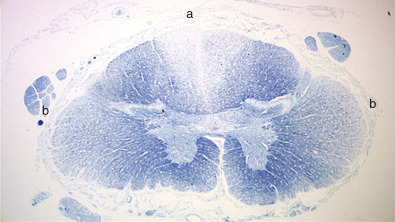
  • Worsening weakness and possible subacute combined degeneration of spinal cord (Fig. E1)
  • Loss of proprioception and an unsteady gait
  • Gastrointestinal symptoms including anorexia, pyrosis, nausea, and vomiting
  • Possible splenomegaly and mild hepatomegaly

Figure E1 Spinal cord in cobalamin deficiency.

From Hoffman R et al: Hematology, basic principles and practice, ed 7, Philadelphia, 2018, Elsevier.

Etiology34

  • Parietal cell antibodies are present in >70% of patients, while intrinsic factor antibodies are noted in >50% of patients.
  • Atrophic gastric mucosa (Fig. E2) with achlorhydria.
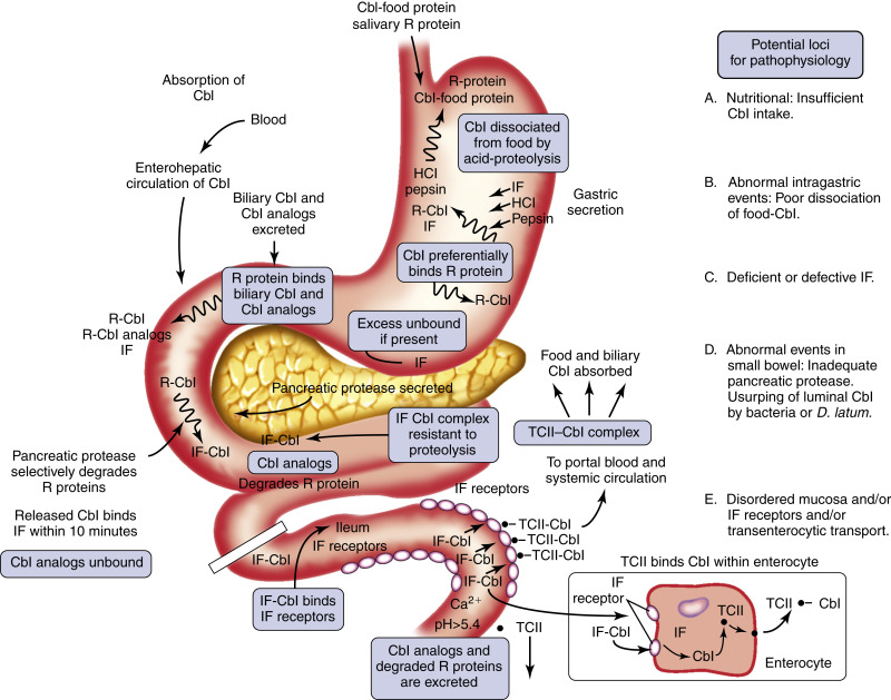
  • Inborn errors of cobalamin-cofactor synthesis are rare. Fig. E3 illustrates the components and mechanism of cobalamin absorption. An etiopathophysiologic classification of cobalamin deficiency is described in Section II.

Figure E2 Histologic features of stomach in pernicious anemia compared with normal.

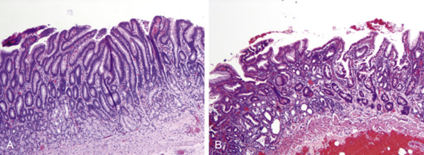
The normal gastric mucosa (A) is contrasted with that seen in pernicious anemia (B), in which there is atrophy of gastric glands, intestinal metaplasia with goblet cells, and loss of parietal cells (not visible at this magnification).

From Hoffman R et al: Hematology, basic principles and practice, ed 7, Philadelphia, 2018, Elsevier.

Figure E3 Components and mechanism of cobalamin absorption.

Cbl, Cobalamin; HCl, hydrochloric acid; IF, intrinsic factor; R, protein ligand; TCII, transcobalamin II.

From Hoffman R et al: Hematology, basic principles and practice, ed 6, Philadelphia, 2013, Elsevier.

TREATMENT5-7

Diagnosis

Differential Diagnosis

  • Nutritional vitamin B12 deficiency
  • Malabsorption (e.g., celiac disease)
  • Chronic alcoholism (multifactorial)
  • Chronic gastritis related to H. pylori infection
  • Folic acid deficiency
  • Myelodysplasia
  • Thyroid abnormalities
  • Atrophic gastritis
  • Paraproteinemias
  • Gastrectomy or use of H2 blockers
  • Insufficient pancreatic enzymes (Zollinger-Ellison syndrome, chronic pancreatitis, post-Whipple procedure)
Workup

  • The clinical presentation of PA varies with the stage. Initially, patients may be asymptomatic. In advanced stages, patients may have impaired memory, depression, gait disturbances, paresthesias, and generalized weakness.
  • Investigation consists primarily of laboratory evaluation. Table 1 describes a step-wise approach to the diagnosis of cobalamin and folate deficiency.
  • Endoscopy and biopsy for atrophic gastritis may be performed in selected cases.
  • Diagnosis is crucial because failure to treat may result in irreversible neurologic deficits.

TABLE 1 Stepwise Approach to the Diagnosis of Cobalamin and Folate Deficiency

Megaloblastic Anemia or Neurologic-Psychiatric Manifestations Consistent With Cobalamin Deficiency Plus Test Results on Serum Cobalamin and Serum Folate
Cobalamina (pg/ml)Folateb (ng/ml)Provisional DiagnosisProceed with Metabolites?c
>300>4Cobalamin or folate deficiency is unlikelyNo
<200>4Consistent with cobalamin deficiencyNo
200-300>4Rule out cobalamin deficiencyYes
>300<2Consistent with folate deficiencyNo
<200<2Consistent with (1) combined cobalamin plus folate deficiency or (2) isolated folate deficiencyYes
>3002-4Consistent with (1) folate deficiency or (2) an anemia unrelated to vitamin deficiencyYes
Test Results on Metabolites: Serum Methylmalonic Acid and Total Homocysteine
Methylmalonic Acid (Normal, 70-270 nM)Total Homocysteine (Normal, 5-14 μM)Diagnosis
IncreasedIncreasedCobalamin deficiency confirmed; folate deficiency still possible (i.e., combined cobalamin plus folate deficiency possible)
NormalIncreasedFolate deficiency is likely
NormalNormalCobalamin and folate deficiency is excluded

a Serum cobalamin levels: Abnormally low, <200 pg/ml; clinically relevant low-normal range, 200-300 pg/ml.

b Serum folate levels: Abnormally low, <2 ng/ml; clinically relevant low-normal range, 2-4 ng/ml.

c Any frozen-over sample from serum folate/cobalamin determination can be subjected to metabolite tests.

From Hoffman R et al: Hematology, basic principles and practice, ed 7, Philadelphia, 2018, Saunders.

Laboratory Tests

  • Complete blood count generally reveals macrocytic anemia, thrombocytopenia, and mild leukopenia with hypersegmented neutrophils (Fig. E4).
  • Mean corpuscular volume (MCV) is significantly elevated in advanced stages.
  • Reticulocyte count is low to normal.
  • False low serum cobalamin levels can occur in patients who are pregnant or taking oral contraceptives, have multiple myeloma, have transcobalamin I (TCI) deficiency, have severe folic acid deficiency, or are taking large doses of ascorbic acid. False high normal levels in patients with cobalamin deficiency can occur in several conditions including hepatomas, severe liver disease, or monoblastic leukemias (Table 2).
  • The absence of anemia or macrocytosis does not exclude the diagnosis of cobalamin deficiency. Anemia is absent in 20% of patients with cobalamin deficiency, and macrocytosis is absent in >30% of patients at the time of diagnosis. Macrocytosis can be masked by concurrent iron deficiency, anemia of chronic disease, or thalassemia trait.
  • Laboratory tests used for detecting cobalamin deficiency in patients with normal vitamin B12 levels include serum and urinary methylmalonic acid (MMA) level (elevated), total homocysteine level (elevated), and intrinsic factor antibody (positive). Cobalamin is a cofactor for the enzymes L-methylmalonyl coenzyme A mutase and methionine synthase. Inadequate levels of cobalamin will thus result in increased MMA and homocysteine levels. Plasma MMA levels can also be used to differentiate cobalamin deficiency from folate deficiency because patients with folate deficiency have normal or mild elevations of MMA levels.
  • An increased concentration of plasma MMA does not predict clinical manifestations of vitamin B12 deficiency and should not be used as the only marker for diagnosis of B12 deficiency.
  • Additional laboratory abnormalities can include elevated lactate dehydrogenase, direct hyperbilirubinemia, and decreased haptoglobin, due to rapid destruction of red blood cells.
  • Bone marrow aspirate is not necessary to diagnose cobalamin deficiency. It may show giant C-shaped neutrophil bands and megaloblastic normoblasts (Fig. E5).
  • Schilling test: No longer used. It was historically used to identify the locus of cobalamin malabsorption and the cause of cobalamin deficiency.

TABLE 2 Serum Cobalamin: False-Positive and False-Negative Test Results

Falsely Low Serum Cobalamin in the Absence of True Cobalamin Deficiency
  • Folate deficiency (one third of patients)
  • Multiple myeloma
  • TCI deficiency
  • Megadose vitamin C therapy
  • Pregnancy
  • Oral contraceptives
Falsely Raised Cobalamin Levels in the Presence of a True Deficiencya
  • Cobalamin binders (TCI and II) increased (e.g., myeloproliferative states, hepatomas, and fibrolamellar hepatic tumors)
  • TCII-producing macrophages are activated (e.g., autoimmune diseases, monoblastic leukemias and lymphomas)
  • Release of cobalamin from hepatocytes (e.g., active liver disease)
  • High serum anti-IF antibody titer

IF, Intrinsic factor; TC, transcobalamin.

a Although a low serum cobalamin level is not synonymous with cobalamin deficiency, 5% of patients with true cobalamin deficiency have low-normal cobalamin levels, a potentially serious problem because the patient’s underlying cobalamin deficiency will progress if uncorrected.

From Hoffman R et al: Hematology, basic principles and practice, ed 6, Philadelphia, 2013, Saunders.

Figure E4 Macroovalocytes (A) and Hypersegmented Neutrophils (B) are Typical Features of Megaloblastic Anemia

From Jaffe ES et al: Hematopathology, Philadelphia, 2011, Saunders.

Figure E5 Bone Marrow Aspirate from a Patient with Cobalamin Deficiency Illustrates a Giant C-Shaped Neutrophil Band and Megaloblastic Normoblasts

From Jaffe ES et al: Hematopathology, Philadelphia, 2011, Saunders.

Pearls & Considerations

Comments

  • Early manifestations of negative cobalamin balance are increased serum methylmalonic acid and total homocysteine levels. This occurs when the total cobalamin in serum is still in the low-normal range.
  • Vitamin B12 deficiency that is allowed to progress for longer than 3 mo may produce permanent degenerative lesions of the spinal cord (e.g., subacute combined degeneration of spinal cord).
  • Vitamin B12 deficiency may suppress signs of polycythemia vera; treatment of B12 deficiency may unmask this disorder.
  • Blunted or impeded therapeutic response to vitamin B12 may be due to concurrent iron or folic acid deficiency, uremia, infections, or use of drugs with bone marrow suppressant properties. Causes of megaloblastosis not responding to therapy with cobalamin or folate are summarized in Table 3.
  • Drugs that interfere with B12 absorption include metformin, colchicine, neomycin, and aminosalicylic acid.
  • Patients must understand that cobalamin replacement therapy is lifelong.
  • Self-injection of vitamin B12 may be taught in selected patients. Cost of monthly injections is less than $10.
  • Patients who have had bariatric surgery should receive 1 mg of oral vitamin B12 per day indefinitely.

TABLE 3 Causes of Megaloblastosis Not Responding to Therapy With Cobalamin or Folate

Wrong Diagnosis
Combined folate and cobalamin deficiencies being treated with only one vitamin
Associated iron deficiency
Associated hemoglobinopathy (e.g., sickle cell disease, thalassemia)
Associated anemia of chronic disease
Associated hypothyroidism

From Hoffman R et al: Hematology, basic principles and practice, ed 7, Philadelphia, 2018, Elsevier.

Related Content

Pernicious Anemia (Patient Information)

NONPHARMACOLOGIC THERAPY

Avoid folic acid supplementation without proper vitamin B12 supplementation. Folic acid supplementation alone may result in hematologic remission in patients with vitamin B12 deficiency but will not treat or prevent neurologic manifestations.

ACUTE GENERAL Rx

Traditional therapy of cobalamin deficiency consists of intramuscular (IM) or deep subcutaneous (SC) injections of vitamin B12 1000 mcg/day for 1 wk, followed by 1000 mcg/mo, indefinitely. Monitor response and increase dosing if serum B12 levels decline.

CHRONIC Rx

DISPOSITION

Anemia generally resolves with appropriate cobalamin replacement therapy. Neurologic deficits, on the other hand, may be corrected only if treated early on.

REFERRAL

Gastroenterology referral for endoscopy on diagnosis of PA followed by periodic surveillance endoscopies to rule out gastric adenocarcinoma or carcinoid tumors.

Related Content

    1. Bizzaro N., Antico A. : Diagnosis and classification of pernicious anemiaAutoimmun Rev. ;13:565-568, 2014.
    2. Fernando M.M.A. : Defining the role of the MHC in autoimmunity: a review and pooled analysisPLoS Genet. , 2008.doi:10.1371/journal.pgen.1000024
    3. Green R., Datta Mitra A. : Megaloblastic anemias: nutritional and other causesMed Clin North Am. ;101(2):297-317, 2017.
    4. Hesdorffer C.S., Longo D.L. : Drug-induced megaloblastic anemiaN Engl J Med. ;373:1649-1658, 2015.
    5. Langan R.C., Goobred A.J. : Vitamin B12 deficiency: recognition and managementAm Fam Physician. ;96(6):384-389, 2017.
    6. Stabler S. : Vitamin B12 deficiencyN Engl J Med. ;368(21):2041-2042, 2013.
    7. Yousaf F. : Pernicious anemia associated cobalamin deficiency and thrombotic microangiopathy: case report and review of the literatureCase Rep Med. , 2017.doi:10.1155/2017/9410727