|
Figure 25 Workup of older adult presenting with anemia.

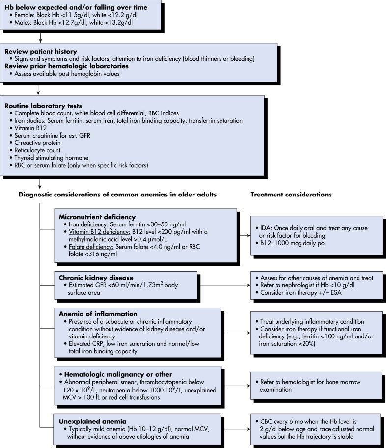
From Warshaw G et al: Ham’s primary care geriatrics, ed 7, Philadelphia, 2022, Elsevier.
| Parameters | Interpretation | |
|---|---|---|
| History | Diet (meat and green vegetables) | Risk of malnutrition if lacking: Suggests susceptibility to iron and folic acid deficiency |
| Blood loss | Obvious gastrointestinal bleeding Chronic subclinical blood loss (e.g., NSAIDs, colon polyps, hereditary telangiectasias, menorrhagia) | |
| Gastrointestinal surgery | Gastrectomy or ileal resection: Vitamin B12 deficiency Small bowel resections: Iron, folate deficiency | |
| Comorbidity | Chronic infections and inflammatory disorders suggest risk of anemia of chronic disease | |
| Examination | Pallor | Severity of anemia |
| Icterus | May suggest hemolysis | |
| Lymphadenopathy, hepatomegaly, splenomegaly, bone tenderness | Coexistence of another primary hematologic disorder | |
| Evidence of portal hypertension | Hypersplenism and potential for variceal bleed | |
| Red cell size and hemoglobinization | Microcytic hypochromic | Iron deficiency Thalassemia Anemia of chronic disease Sideroblastic anemia |
| Normocytic | Acute bleeding Bone marrow infiltration Aplastic anemia Renal failure | |
| Macrocytic | Vitamin B12/folate deficiencies Myelodysplasia Medication effects Other medical conditions: Hypothyroidism, liver impairment |
NSAIDs, Nonsteroidal antiinflammatory drugs.
From Talley NJ et al: Essentials of internal medicine, ed 4, Chatswood, NSW, 2021, Elsevier Australia.
Table 9 Differentiating Features of Microcytic Anemias∗
| Test | Iron-Deficiency Anemia | Thalassemia Minor† | Anemia of Inflammation‡ |
|---|---|---|---|
| Serum iron | Low | Normal | Low |
| Serum iron-binding capacity | High | Normal | Low or normal |
| Serum ferritin | Low | Normal or high | Normal or high |
| Marrow iron stores | Low or absent | Normal or high | Normal or high |
| Marrow sideroblasts | Decreased or absent | Normal or increased | Normal or increased |
| Free erythrocyte protoporphyrin | High | Normal or slightly increased | High |
| Hemoglobin A2 or F | Normal | High beta-thalassemia; normal alpha-thalassemia | Normal |
| Red blood cell distribution width§ | High | Normal | Normal/↑ |
∗See Table 11 for definition of microcytosis.
† Alpha-thalassemia minor can be diagnosed by the presence of Bart hemoglobin on newborn screening.
‡ Usually normochromic; 25% of cases are microcytic.
§ Red blood cell distribution width quantitates the degree of anisocytosis (different sizes) of red blood cells.
From Marcdante KJ et al: Nelson essentials of pediatrics, ed 9, Philadelphia, 2023, Elsevier.