section name header

Basic Information

AUTHOR: Glenn G. Fort, MD, MPH

Definition

Sinusitis is inflammation of the mucous membranes lining one or more of the paranasal sinuses. The various presentations are:

  • Acute sinusitis: Infection lasting <4 wk, with complete resolution of symptoms.
  • Subacute infection: Lasts from 4 to 12 wk, with complete resolution of symptoms.
  • Recurrent acute infection: Episodes of acute infection lasting <30 days, with resolution of symptoms, which recur at intervals at least 10 days apart.
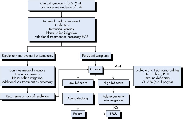
  • Chronic sinusitis: Inflammation of the paranasal sinuses and nasal cavities lasting >12 wk, with persistent upper respiratory symptoms. It accounts for 1% to 2% of total physician encounters.
  • Acute bacterial sinusitis superimposed on chronic sinusitis: New symptoms that occur in patients with residual symptoms from prior infection(s). With treatment, the new symptoms resolve, but the residual ones do not.
Synonym

Rhinosinusitis: Sinusitis is almost always accompanied by inflammation of the nasal mucosa; thus it is now the preferred term.

ICD-10CM CODES
J32.9Chronic sinusitis, unspecified
J01.90Acute sinusitis, unspecified
J01.00Acute maxillary sinusitis, unspecified
J01.01Acute recurrent maxillary sinusitis
J01.10Acute frontal sinusitis, unspecified
J01.11Acute recurrent frontal sinusitis
J01.20Acute ethmoidal sinusitis, unspecified
J01.21Acute recurrent ethmoidal sinusitis
J01.30Acute sphenoidal sinusitis, unspecified
J01.31Acute recurrent sphenoidal sinusitis
J01.80Other acute sinusitis
J01.81Other acute recurrent sinusitis
J01.91Acute recurrent sinusitis, unspecified
J32.0Chronic maxillary sinusitis
J32.1Chronic frontal sinusitis
J32.2Chronic ethmoidal sinusitis
J32.3Chronic sphenoidal sinusitis
J32.8Other chronic sinusitis
Epidemiology & Demographics
Incidence (In U.S.)

Seems to correlate with the incidence of upper respiratory tract infections and higher in women than men; 30 million cases per yr in the U.S.

Peak Incidence

  • Fall, winter, spring: September through March
  • In adults: Greatest incidence between 45 and 74 yr of age
  • Approximately 6% to 7% of children presenting with respiratory symptoms have acute sinusitis
Physical Findings & Clinical Presentation

  • Patients often give a history of a recent upper respiratory illness with some improvement, then a relapse.
  • Mucopurulent secretions in the nasal passage:
    1. Purulent nasal and postnasal discharge lasting 7 to 10 days
    2. Facial tightness, pressure, or pain
    3. Nasal obstruction
    4. Headache
    5. Decreased sense of smell
    6. Purulent pharyngeal secretions, brought up with cough, often worse at night
  • Erythema, swelling, and tenderness over the infected sinus in a small proportion of patients:
    1. Diagnosis cannot be excluded by the absence of such findings.
    2. These findings are not common, and do not correlate with number of positive sinus aspirates.
  • Intermittent low-grade fever in about half of adults with acute bacterial sinusitis.
  • Toothache is a common complaint when the maxillary sinus is involved.
  • Periorbital cellulitis and excessive tearing with ethmoid sinusitis:
    1. Orbital extension of infection: Chemosis, proptosis, impaired extraocular movements
  • Characteristics of acute sinusitis in children with upper respiratory tract infections:
    1. Persistence of symptoms
    2. Cough
    3. Bad breath
  • Symptoms of chronic sinusitis (may or may not be present):
    1. Nasal or postnasal discharge
    2. Fever
    3. Facial pain or pressure
    4. Headache
  • Nosocomial sinusitis is typically seen in patients with nasogastric tubes or nasotracheal intubation.
Etiology

  • Each of the four paranasal sinuses is connected to the nasal cavity by narrow tubes (ostia), 1 to 3 mm in diameter; these drain directly into the nose through the turbinates. The sinuses are lined with a ciliated mucous membrane (mucoperiosteum).
  • Acute viral infection:
    1. Infection with the common cold or influenza
    2. Mucosal edema and sinus inflammation
    3. Decreased drainage of thick secretions/obstruction of the sinus ostia
    4. Subsequent entrapment of bacteria
      1. Multiplication of bacteria
      2. Secondary bacterial infection
  • Other predisposing factors:
    1. Tumors
    2. Polyps
    3. Foreign bodies
    4. Congenital choanal atresia
    5. Other entities that cause obstruction of sinus drainage
    6. Allergies
    7. Asthma
  • Dental infections lead to maxillary sinusitis.
  • Viruses recovered alone or in combination with bacteria (in 16% of cases):
    1. Rhinovirus
    2. Coronavirus
    3. Adenovirus
    4. Parainfluenza virus
    5. Respiratory syncytial virus
  • The principal bacterial pathogens in sinusitis are Streptococcus pneumoniae, nontypable Haemophilus influenzae, and Moraxella catarrhalis (Table 1).
  • In the remainder of cases Streptococcus pyogenes, Staphylococcus aureus, beta-hemolytic streptococci, and mixed anaerobic infections (Peptostreptococcus, Fusobacterium, Bacteroides, and Prevotella spp.) are found.
  • Infection is polymicrobial in about one third of cases.
  • Anaerobic infections are seen more often in cases of chronic sinusitis and in cases associated with dental infection; anaerobes are unlikely pathogens in sinusitis in children.
  • Fungal pathogens are isolated with increasing frequency in immunocompromised patients but remain uncommon pathogens in the paranasal sinuses. Fungal pathogens include Phaeohyphomycosis, Aspergillus, Pseudallescheria, Sporothrix, and Zygomycetes spp.
  • Nosocomial infections: Occur in patients with nasogastric tubes, nasotracheal intubation, cystic fibrosis, and immunocompromised state.
    1. S. aureus (including MRSA)
    2. Pseudomonas aeruginosa
    3. Klebsiella pneumoniae
    4. Enterobacter spp.
    5. Proteus mirabilis
  • Organisms typically isolated in chronic sinusitis:
    1. S. aureus
    2. S. pneumoniae
    3. H. influenzae
    4. P. aeruginosa
    5. Anaerobes

TABLE 1 Microbiology of Acute Bacterial Rhinosinusitis in Adults

OrganismRange of Prevalence (%)
Streptococcus pneumoniae20-43
Haemophilus influenzae22-35
Streptococcus spp3-9
Anaerobes0-9
Moraxella catarrhalis2-10
Staphylococcus aureus0-8
Other4

From Broaddus VC et al: Murray & Nadel’s textbook of respiratory medicine, ed 7, Philadelphia, 2022, Elsevier.

Diagnosis

Differential Diagnosis

  • Temporomandibular joint disease
  • Migraine headache
  • Cluster headache
  • Dental infection
  • Trigeminal neuralgia
  • Allergic rhinitis
  • Drugs (cocaine, decongestant overuse)
  • Gastroesophageal reflux disease
  • Wegener granulomatosis
  • Cystic fibrosis
Workup

  • The diagnosis is generally based on clinical signs and symptoms (purulent rhinorrhea and facial pain). Radiologic tests and cultures are not recommended initially and should be considered only when treatment is ineffective, and sinusitis persists.
  • In the normal healthy host, the paranasal sinuses should be sterile. Although the contiguous structures are colonized with bacteria and likely contaminate the sinuses, the mucociliary lining functions to remove these bacteria.
  • Gold standard for diagnosis: Recovery of bacteria in high-density 104 colony-forming units/ml from a paranasal sinus, in the setting of a patient with history of upper respiratory infection and symptoms persisting for 7 to 10 days. Sinus aspiration is the best method for obtaining cultures; however, it must be performed by an otorhinolaryngologist and is not practical for the primary care practitioner. Therefore, most diagnoses are based on the clinical history and presentation, possibly supported by radiologic evaluations.
    1. Overall, standard radiographs are of limited use in diagnosis, although negative films are strong evidence against the diagnosis
    2. Computed tomography (CT) scans (Figs. E1 and E2):
      1. Much more sensitive than plain x-rays in detecting acute changes and disease in the sinuses
      2. Recommended for patients requiring surgical intervention, including sinus aspiration; it is a useful adjunct to guide therapy
    3. Transillumination:
      1. Used for diagnosis of frontal and maxillary sinusitis
      2. Absence of light transmission indicates that sinus is filled with fluid
      3. Dullness (decreased light transmission) is less helpful in diagnosing infection
    4. Endoscopy:
      1. Used to visualize secretions coming from the ostia of infected sinuses
      2. Culture collection via endoscopy often contaminated by nasal flora; not nearly as good as sinus puncture
  • Sinus puncture:
    1. Gold standard for collecting sinus cultures
    2. Generally reserved for treatment failures, suspected intracranial extension, and nosocomial sinusitis

Figure E1 Acute and chronic sinusitis.

Postgadolinium fat-saturation T1-weighted image demonstrates an air-fluid level (arrow) in the right maxillary sinus and is diagnostic of acute sinusitis (superimposed on chronic sinusitis). Left maxillary sinus is filled with low-intensity secretions and has a peripheral ring of enhancing inflamed mucosa (arrowheads) typical of chronic sinusitis. Mastoid air cells and the left middle ear cavity (asterisk), which normally appear black, are filled with enhancing inflammatory tissue.

From Flint PW et al: Cummings otolaryngology, head and neck surgery, ed 7, Philadelphia, 2021, Elsevier.

Figure E2 Coronal computed tomography (CT) imaging of the paranasal sinuses.

(A) Normal anatomy, including well-aerated maxillary (M) and ethmoid (E) sinuses bilaterally, patency of the osteomeatal complex (arrow), and normal appearance of the inferior turbinates (IT). Incidentally noted is a left concha bullosa (CB), a normal variant involving aeration of the middle turbinate, which arises in approximately 30% of patients. (B) CT findings consistent with acute sinusitis. There is unilateral opacification of the ethmoid sinuses (asterisk) and a fluid level within the right maxillary sinus (arrows). Acute sinusitis may present with unilateral or bilateral disease, and routine CT imaging is not recommended. (C) Bilateral chronic sinusitis with nasal polyposis. There is complete opacification of the maxillary (M) and ethmoid (E) sinuses bilaterally. Arrowheads demonstrate bilateral nasal polyps, a soft tissue density within the nasal cavity and adjacent to the inferior turbinates (IT).

From Broaddus VC et al: Murray & Nadel’s textbook of respiratory medicine, ed 7, Philadelphia, 2022, Elsevier.

Treatment

Nonpharmacologic Therapy

To help promote sinus drainage:

  • Air humidification with vaporizers (for steam) or humidifiers (for a cool mist)
  • Application of hot, wet towel over the face
  • Sipping hot beverages
  • Hydration
Acute General Rx

  • Sinus drainage:
    1. Nasal vasoconstrictors, such as phenylephrine nose drops, 0.25% or 0.5%.
    2. Topical decongestants should not be used for more than a few days because of the risk of rebound congestion.
    3. Systemic decongestants.
    4. Corticosteroids: Nasal or systemic corticosteroids, such as nasal beclomethasone. Oral corticosteroids combined with antibiotics may be associated with modest benefit for short-term relief of symptoms in adults with severe symptoms of acute sinusitis compared with antibiotics alone. Oral corticosteroids as monotherapy are not associated with improved clinical outcomes in adults with clinically diagnosed acute sinusitis.
    5. Nasal irrigation, with hypertonic or normal saline (saline may act as a mild vasoconstrictor of nasal blood flow).
    6. Use of antihistamines has no proven benefit, and the drying effect on the mucous membranes may cause crusting, which blocks the ostia, thus interfering with sinus drainage.
  • Analgesics, antipyretics

Antimicrobial therapy:

  • Most cases of acute sinusitis have a viral cause and will resolve within 2 wk without antibiotics.
  • Current treatment recommendations favor symptomatic treatment for those with mild symptoms. 85% of persons have a reduction or resolution of symptoms within 7 to 15 days without antibiotic therapy. Physicians grossly overprescribe antibiotics for presumed bacterial sinusitis despite a much higher prevalence of viral infections.
  • Antibiotics should not be prescribed for mild to moderate sinusitis within the first wk of illness. They should be reserved for those with persistent symptoms for more than 10 days, high fever and purulent nasal discharge or facial pain lasting for at least 3 consecutive days, or worsening symptoms after a typical viral illness lasting >5 days that had initially improved (“double sickening”).
  • Antibiotic therapy is usually empiric, targeting the common pathogens:
    1. First-line antibiotics in children include amoxicillin or amoxicillin/clavulanate. For adults, amoxicillin/clavulanate or doxycycline is first-line agent, with quinolones (levofloxacin or moxifloxacin) reserved as second-line agents unless patient is penicillin allergic.
    2. Second-line antibiotics include the newer macrolides: Clarithromycin and oral cephalosporins: Cefuroxime axetil, cefprozil, cefaclor, loracarbef, but high rate of resistance of S. pneumoniae is a concern with these agents as is H. influenzae resistance with TMP-SMX and azithromycin such that they should no longer be used as first-line agents.
    3. For patients with uncomplicated acute sinusitis, the less expensive first-line agents appear to be as effective as the costlier second-line agents.
  • Hospitalization and intravenous (IV) antibiotics may be required for more severe infection and those with suspected intracranial complications. Broader-spectrum antibiotic coverage may be indicated in severe cases, to cover for MRSA, Pseudomonas, and fungal pathogens.
  • Duration of therapy generally 5 to 7 days in adults rather than 10 to 14 days as recommended in the past.
    1. Optimal duration of treatment in children varies from 10 to 28 days.

Surgery:

  • Surgical drainage indicated
    1. If intracranial or orbital complications suspected
    2. Many cases of frontal and sphenoid sinusitis
    3. Chronic sinusitis recalcitrant to medical therapy
  • Surgical debridement imperative in the treatment of fungal sinusitis

Complications:

  • Untreated, sinusitis may lead to a number of serious, life-threatening complications.
  • Intracranial complications include meningitis, brain abscess, and epidural and subdural empyema.
  • Intracranial sequelae are more common with frontal and ethmoid infections.
  • Extracranial complications include orbital cellulitis, blindness, orbital abscess, osteomyelitis.
  • Extracranial sequelae are more commonly seen with ethmoid sinusitis.
Chronic Rx

  • Chronic sinusitis: Evidence supports daily high-volume saline irrigation with topical corticosteroid therapy as a first-line therapy for chronic sinusitis. A short course of systemic corticosteroids (1 to 3 wk), short course of doxycycline (3 wk), or a leukotriene antagonist may be considered in patients with nasal polyps. A prolonged course (3 mo) of macrolide antibiotic may be considered for patients without polyps. A clinical algorithm for the management of chronic rhinosinusitis in children is illustrated in Fig. 3.
  • Dupilumab (Dupixent), a monoclonal antibody that targets interleukin -4 and -13 was recently FDA approved for chronic rhinosinusitis with nasal polyps (CRSwNP). Cost is a limiting factor.
  • Surgical intervention may be necessary in nonresponders.
Figure 3 General Clinical Algorithm for the Management of Chronic Rhinosinusitis (Crs) in Children

!!flowchart!!

If symptoms improve or resolve after maximal medical therapy, one can continue conservative medical management with intranasal steroids and nasal saline irrigation as necessary. If a child has persistent symptoms after maximal medical management, then one should obtain a computed tomography (CT) scan at the end of the course of treatment to evaluate for extent of disease. One would also initiate investigations into possible coexisting comorbidities such as allergic rhinitis, immune deficiencies, and primary ciliary dyskinesia. Surgical management would be informed by the severity of disease on CT scan. If the severity is low, as evidenced by a Lund MacKay score <5, then one can move forward with an adenoidectomy. If the score is greater than 5, then most would choose adenoidectomy with maxillary sinus irrigation. Clearly, factors such as age, the presence of asthma and the severity of disease on the CT scan might mandate concomitant adenoidectomy and limited FESS. Finally, in patients with significant disease such as nasal polyps, mucoceles associated with cystic fibrosis, or expansile disease such as with allergic fungal sinusitis, one would go directly to FESS. AFS, Allergic fungal sinusitis; AR, allergic rhinitis; CF, cystic fibrosis; FESS, functional endoscopic sinus surgery; LM, Lund MacKay; PCD, primary ciliary dyskinesia.

From Flint PW et al: Cummings otolaryngology, head and neck surgery, ed 7, Philadelphia, 2021, Elsevier.

Referral

  • To infectious disease specialist if failure to respond to initial therapy
  • To otorhinolaryngologist for:
    1. Failure to respond to therapy
    2. Suspected fungal infection
    3. Suspected intracranial or orbital complications
Related Content

Sinusitis (Patient Information)

Suggested Readings

    1. Arora H.S. : Sinusitis in childrenPediatr Ann. ;47:e396-e401, 2018.
    2. Demuri G.P., Wald E.R. : Acute bacterial sinusitis in childrenN Engl J Med. ;367:1128-1134, 2012.
    3. LibMan H. : Should we prescribe antibiotics to this patient with persistent upper respiratory symptomsAnn Int Med. ;166:201-208, 2017.
    4. McCoul E.D., Tabaee A. : A practical approach to refractory chronic rhinosinusitisOtolaryngol Clinic North Am. ;50:183-198, 2017.
    5. Rosenfeld R.M. : Acute sinusitis in adultsN Engl J Med. ;375:962-970, 2016.
    6. Stevens W.W. : A new treatment for chronic rhinosinusitis with nasal polypsLancet. ;394:1595-1597, 2019.
    7. Venekamp R.P. : Systemic corticosteroid therapy for acute sinusitisJAMA. ;313(12):1258-1259, 2015.
    8. Wyler B., Mallon W.K. : Sinusitis updateEmerg Med Clin North Am. ;37:41-54, 2019.