section name header

Basic Information

AUTHOR: Glenn G. Fort, MD, MPH

Definition

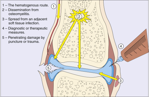
Septic arthritis is a highly destructive form of joint disease most often caused by hematogenous spread of organisms from a distant site of infection. Direct penetration of the joint as a result of trauma or surgery and spread from adjacent osteomyelitis may also cause bacterial arthritis. Any joint in the body may be affected.

Synonyms

Infectious arthritis

Bacterial arthritis

Pyogenic arthritis

ICD-10CM CODE
M00.9Pyogenic arthritis, unspecified
Epidemiology & Demographics
Incidence (In U.S.)

Between 2 and 6 cases per 100,000 people per year

Prevalence (In U.S.)

Unknown

Predominant Sex

Gonococcal arthritis in females

Predominant Age

Gonococcal arthritis in sexually active adults

Peak Incidence

  • Gonococcal arthritis: Young adults
  • Other bacterial causes: All ages
Physical Findings & Clinical Presentation

  • Hallmark: Acute onset of monoarticular joint pain, erythema, heat, and immobility
  • Limited range of motion of the joint
  • Effusion, with varying degrees of erythema and increased warmth around the joint
  • Single joint affected in 80% to 90% of cases of nongonococcal arthritis
  • Gonococcal dermatitis-arthritis syndrome:
    1. Typical pattern is a migratory polyarthritis or tenosynovitis
    2. Small pustules on the trunk or extremities
  • Febrile patient at presentation
  • Most commonly affected joints in adult: Knee and hip, but any joint may be involved; in children-hip
Etiology

  • Bacteria spread from another locus of infection.
    1. Highly vascular synovium is invaded by hematogenously spread bacteria.
    2. White blood cell (WBC) enzymes cause necrosis of synovium, cartilage, and bone.
    3. Extensive joint destruction is rapid if infection is not treated with appropriate intravenous (IV) antibiotics and drainage of necrotic material.
  • Predisposing factors: Rheumatoid arthritis, prosthetic joints, advanced age, immunodeficiency (HIV, diabetes mellitus [DM], immunosuppressive drugs), gout, sexual activity (gonococcal arthritis), skin infections, cutaneous ulcers (contiguous spread), recent joint surgery, recent intraarticular infection. Risk factors for development of septic arthritis are summarized in Table 1. Fig. E1 illustrates routes by which bacteria can reach the joint.
  • The most common nongonococcal organisms are staphylococci (40%), streptococci (28%), and gram-negative bacilli (19%). Less common are mycobacteria (8%), gram-negative cocci (3%), anaerobes (1%), and gram-positive bacilli (1%).
  • Staphylococci (S. aureus and coagulase-negative staphylococcal species) account for >50% of prosthetic-hip and prosthetic-knee infections. S. aureus is very common in patients with rheumatoid arthritis.

TABLE 1 Risk Factors for Development of Septic Arthritis

Age >80 yr
Diabetes mellitus
Presence of a prosthetic joint in the knee or the hip
Recent joint surgery
Skin infection
Previous septic arthritis
Recent intra-articular injection
HIV or AIDS
Intravenous drug abuse
End-stage renal disease on hemodialysis
Advanced hepatic disease
Hemophilia with or without AIDS
Sickle cell disease
Underlying malignancy
Hypogammaglobulinemia (susceptible to Mycoplasma infections)
Late complement-component deficiency (susceptible to Neisseria infections)
Low socioeconomic status with high rate of comorbidities

AIDS, Acquired immunodeficiency virus; HIV, human immunodeficiency virus.

From Firestein GS et al: Firestein & Kelley’s textbook of rheumatology, ed 11, Philadelphia, 2021, Elsevier.

Figure E1 Routes by which bacteria can reach the joint.

From Hochberg MC et al: Rheumatology, ed 5, St. Louis, 2011, Mosby.

Diagnosis

Differential Diagnosis

  • Gout
  • Pseudogout
  • Trauma
  • Hemarthrosis
  • Rheumatic fever
  • Adult or juvenile rheumatoid arthritis
  • Spondyloarthropathies such as reactive arthritis (Reiter syndrome)
  • Osteomyelitis
  • Viral arthritides
  • Septic bursitis
  • Lyme disease caused by Borrelia burgdorferi
Workup

  • Joint aspiration, Gram stain, and culture of the synovial fluid. Fig. 2 describes an algorithm for synovial fluid analysis in septic arthritis.
  • Immediate arthrocentesis before other studies are undertaken or antibiotics instituted. Synovial fluid should be evaluated at bedside and then sent for lab evaluation.
  • Criteria for diagnosis of prosthetic joint infections are summarized in Table 2.

TABLE 2 Criteria for Diagnosis of Prosthetic Joint Infection

Major Criteria (One or More of the Following)Decision
Two positive cultures of same organism or sinus tract with communication to the joint/prosthesisInfected
Minor CriteriaScorea
Elevated serum CRP or D-dimer2
Elevated ESR1
Elevated synovial WBC or leukocyte esterase3
Positive synovial alpha-defensin3
Elevated synovial PMN (%)2
Elevated synovial CRP1

CRP, C-reactive protein; ESR, erythrocyte sedimentation rate; PMN, polymorphonuclear leukocyte; WBC, white blood count.

a Score of 6 or more, infected; score of 2-5, possibly infected; score of 0-1, not infected.

From Firestein GS et al: Firestein & Kelley’s textbook of rheumatology, ed 11, Philadelphia, 2021, Elsevier.

Figure 2 Algorithm for synovial fluid analysis in septic arthritis.

!!flowchart!!

OA, Osteoarthritis; RA, rheumatoid arthritis; WBC, white blood cell count.

From Harris ED et al: Kelley’s textbook of rheumatology, ed 7, Philadelphia, 2005, Saunders.

Laboratory Tests

  • Joint fluid analysis:
    1. Synovial fluid leukocyte count is usually elevated >50,000 cells/mm3 with >80% polymorphonuclear cells.
    2. Counts are highly variable, with similar findings in gout, pseudogout, or rheumatoid arthritis. Lower WBC counts can occur in joint replacement, disseminated gonococcal disease, and peripheral leukopenia.
    3. Synovial fluid glucose or protein is not helpful because results are not specific for septic arthritis. The differential diagnosis of synovial fluid abnormalities is described in Section IV.
    4. Polymerase chain reaction (PCR) testing: Useful for detection of uncommon organisms (e.g., Lyme disease).
    5. Crystal analysis: Septic arthritis can coexist with crystal arthropathy; therefore the presence of crystals does not preclude a diagnosis of septic arthritis.
  • Blood cultures: Positive in 25% to 50% of patients with septic arthritis.
  • Culture of possible extraarticular sources of infection.
  • Elevated peripheral WBC count, erythrocyte sedimentation rate (ESR) (nonspecific), C-reactive protein (CRP) (nonspecific). When elevated, ESR and CRP may be useful to monitor therapeutic response.
  • If gonococcus is suspected, perform nucleic acid amplification tests (NAATs) on synovial fluid.
Imaging Studies

  • Radiograph of the affected joint (Fig. 3): Useful to rule out osteomyelitis, fractures, chondrocalcinosis, or inflammatory arthritis
  • MRI: Findings that suggest an acute intraarticular infection include the combination of bony erosions with marrow edema
  • Computed tomography (CT) scan: Useful for early diagnosis of infections of the spine, hips, and sternoclavicular and sacroiliac joints
  • Ultrasound: Can be useful for detecting effusions in joints that are more difficult to examine (e.g., hip)

Treatment

Nonpharmacologic Therapy

  • Affected joints aspirated daily to remove necrotic material and to follow serial WBC counts and cultures
  • If no resolution with IV antibiotics and closed drainage: Open debridement and lavage, particularly in nongonococcal infections
  • Prevention of contractures:
    1. After acute stage of inflammation, range-of-motion exercises of the affected joint
    2. Physical therapy helpful
Acute General Rx

  • IV antibiotics immediately after joint aspiration and Gram stain of the synovial fluid. Empiric antibiotic therapy (Table 3) is based on organism found on Gram stain of synovial fluid:
    1. Gram-positive cocci: Vancomycin: 15 to 20 mg/kg IV q8 to 12h. Keep trough levels at 15 to 20 mcg/ml. Alternatives include daptomycin and linezolid
    2. Gram-negative cocci: Ceftriaxone: 1 to 2 g IV daily in adults (children: 50-100 mg/kg IV daily). Alternative includes cefotaxime
    3. Gram-negative rods: Ceftriaxone, cefepime: 1 to 2 g IV q8 to 12h in adults (children: 100-150 mg/kg/day divided in q8h dosing), piperacillin-tazobactam: 3.375 g IV to 4.5 g IV q6h. Aztreonam or fluoroquinolones can be used in patients with allergy to penicillin or cephalosporins
    4. Negative Gram stain: Vancomycin plus either cefepime or a carbapenem such as meropenem: 1 g IV q8h in adults (children: 60 mg/kg/day divided in q8h dosing) or ertapenem
  • The optimal duration of antibiotic use in septic arthritis has traditionally been 3 to 6 wk usually following surgical drainage. Recent trials (Gjika et al, Ann Rheum Dis Aug 2019, 78: 1114) support a 2-wk course in conjunction with surgery. The duration of antimicrobial therapy in patients with prosthetic joint infections remains unclear however trials have shown that outcomes are better with a 12-wk antibiotic course than with a 6-wk course.1

TABLE 3 Antibiotic Agents Used in Adults

Synovial Fluid Gram StainOrganismAntibioticDose
Gram-positive cocci (clusters)Staphylococcus aureus (methicillin-sensitive)Nafcillin/oxacillin2 g IV q4h or 12 g daily continuous infusion
Or
Cefazolin1-2 g IV q8h
S. aureus (methicillin-resistant)Vancomycin or15 mg/kg IV q12ha (serum trough level of 15-20 μg/ml)
Daptomycin6-8 mg/kg q24h
Or
Linezolid600 mg IV or po q12h
Gram-positive cocci (chains)StreptococcusPenicillin or2-4 million units IV q4h or 18-24 million units daily continuous infusion
Cefazolin1-2 g IV q8h
Gram-negative diplococciNeisseria gonorrhoeaeCeftriaxone2 g IV q24h
Or
Cefotaxime1 g IV q8h
Or
Ciprofloxacin400 mg IV q12h
Gram-negative bacilliEnterobacteriaceae(Escherichia coli, Proteus, Serratia)Ceftriaxone or2 g IV daily
Ciprofloxacin750 mg po q12h
Ertapenem1 g IV daily
PseudomonasCefepime2 g IV q8h
Or
Piperacillin-tazobactam or3.375 g IV q6h or 3.375 g IV q8h infused over 4 h or 14 g daily continuous infusion
Ciprofloxacin
Meropenem
750 mg po q12h or 400 mg IV q 12h
1-2 g IV q8h
Plus
Gentamicin/tobramycin7 mg/kg IV daily
Polymicrobial infectionS. aureus, Streptococcus, gram-negative bacilliNafcillin/oxacillina2 g IV q4h or 12 g daily continuous infusion
Plus
Ceftriaxone2 g IV q24h
Or
Cefotaxime2 g IV q8h
Or
Ciprofloxacin400 mg IV q12h or 750 mg po q12h

IV, Intravenous; q4h, every 4 hours; q6h, every 6 hours; q8h, every 8 hours; q12h, every 12 hours; q24h, every 24 hours.

a If patient is penicillin allergic, use vancomycin plus third-generation cephalosporin or ciprofloxacin.

From Firestein GS et al: Firestein & Kelley’s textbook of rheumatology, ed 11, Philadelphia, 2021, Elsevier.

Related Content

Septic Arthritis (Patient Information)

Related Content

    1. Bernard L. : Antibiotic therapy for 6 or 12 weeks for prosthetic joint infectionN Engl J Med. ;384:1991-2001, 2021.
    2. Costales C., Butler-Wu S.M. : A real pain: diagnostic quandaries and septic arthritisJ Clin Microbiol. ;56(2), 2018.
    3. Gjika E. : Two weeks versus four weeks of antibiotic therapy after surgical drainage for native joint bacterial arthritis: a prospective, randomized, non-inferiority trialAnn Rheum Dis. ;78(8):1114-1121, 2019.
    4. Hassan A.S. : Peripheral bacterial septic arthritis: review of diagnosis and managementJ Clin Rheumatol. ;23:435-442, 2017.
    5. Lim S.Y. : Septic arthritis in gout patients: a population-based cohort studyRheumatology. ;54:2095-2099, 2015.
    6. Ross J.J. : Septic arthritis of native jointsInfect Dis Clin North Am. ;31:203-218, 2017.