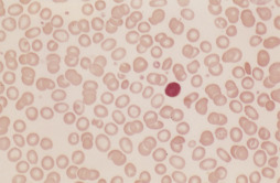
AUTHOR: Fred F. Ferri, MD


Definition
Anemia is defined as a hemoglobin level 2 standard deviations below normal for age and sex. Iron deficiency anemia is anemia resulting from inadequate iron supplementation or excessive blood loss.
Synonyms
| ||||||||||||||||||||
Epidemiology & Demographics
- Dietary iron deficiency occurs often in infants as a result of unsupplemented milk diets. It is also commonly seen in women during their reproductive yr, as a result of heavy menstrual periods, and during pregnancy (increased demand).
- Iron deficiency is the most common nutritional deficiency worldwide.
- The prevalence of iron deficiency is greatest among toddlers ages 1 to 2 yr (7%) from inadequate intake and female individuals ages 12 to 49 yr (9%-16%) from menstrual losses.
- The prevalence of iron deficiency is 2% in adult men, 9% to 12% in non-Hispanic White women, and 20% in Black and Mexican American women.
- GI cancer is diagnosed in 10% of elderly patients with iron deficiency anemia.
Physical Findings & Clinical Presentation
- Most patients have normal examination results.
- Skin pallor and conjunctival pallor may be present.
- Signs and symptoms specific for iron deficiency are koilonychias, pica, pagophagia, blue sclera, glossitis, and angular stomatitis (Fig. E1).
- Patients with severe anemia can have palpitations, headache, weakness, dizziness, and easy fatigability.
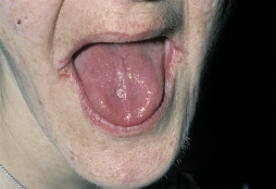
Etiology
- Blood loss from GI or menstrual bleeding (genitourinary blood loss less often the cause)
- Dietary iron deficiency (rare in adults)
- Poor iron absorption in patients with gastric or small-bowel surgery
- Repeated phlebotomy
- Increased requirements (e.g., during pregnancy)
- Other: Traumatic hemolysis (abnormally functioning cardiac valves), idiopathic pulmonary hemosiderosis (iron sequestration in pulmonary macrophages), paroxysmal nocturnal hemoglobinuria (intravascular hemolysis)
- The most common cause worldwide is hookworm infection