section name header

Basic Information

AUTHOR: Fred F. Ferri, MD

Definition

Impetigo is a highly contagious superficial skin infection generally caused by Staphylococcus aureus and/or Streptococcus spp.

Common presentations are bullous impetigo (generally caused by staphylococcal disease) and nonbullous impetigo (from streptococcal infection and possible staphylococcal infection); the bullous form is caused by an epidermolytic toxin produced at the site of infection.

Synonyms

Impetigo vulgaris

Pyoderma

Impetigo contagiosa

Bullous impetigo

ICD-10CM CODES
L01.00Impetigo, unspecified
L01.01Nonbullous impetigo
L01.02Bockhart impetigo
L01.03Bullous impetigo
L01.09Other impetigo
Epidemiology & Demographics

  • Impetigo is the most common bacterial skin infection in children 2 to 5 yr of age. Bullous impetigo accounts for 30% of cases and nonbullous for 70% of cases. Impetigo is most common in temperate zones, mostly during the summer in hot, humid weather. Common sources for children are dirty fingers, pets, and other children in school or day care centers. Impetigo often complicates insect bites, pediculosis, scabies, eczema, and poison ivy.
  • Bullous impetigo is most common in infants and children. The nonbullous form is most common in children ages 2 to 5 yr with poor hygiene in warm climates.
  • The overall incidence of acute nephritis with impetigo varies between 2% and 5%.
Physical Findings & Clinical Presentation

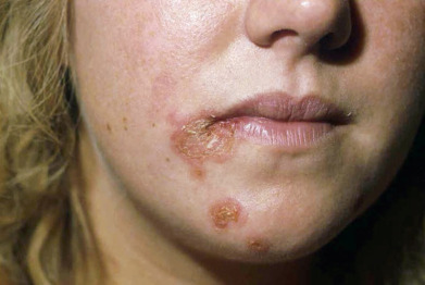
  • Nonbullous impetigo begins as a single red macule or papule that quickly becomes a vesicle. Rupture of the vesicle produces an erosion of which the contents dry to form honey-colored crusts. Multiple lesions with golden yellow crusts (Fig. E1) and weeping areas are often found on the skin around the nose, mouth, and limbs.
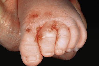
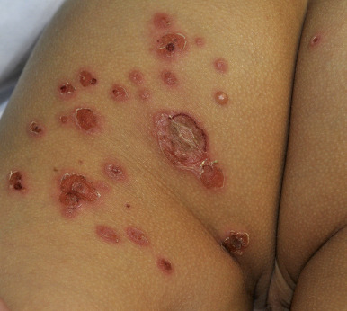
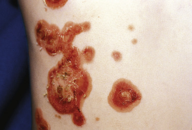
  • Bullous impetigo is manifested by the presence of vesicles that enlarge rapidly to form bullae with contents that vary from clear to cloudy. There is subsequent collapse of the center of the bullae (Fig. E2); the peripheral areas may retain fluid, and a honey-colored crust may appear in the center (Figs. E3 and E4). As the lesions enlarge and become contiguous with the others, a scaling border replaces the fluid-filled rim; there is minimal erythema surrounding the lesions.
  • Regional lymphadenopathy is most common with nonbullous impetigo.
  • Constitutional symptoms are generally absent.
Etiology

  • S. aureus coagulase positive is the dominant microorganism (50% to 70% of cases).
  • S. pyogenes (group A β-hemolytic streptococci): M-T serotypes of this organism associated with acute nephritis are 2, 49, 55, 57, and 60. Group B streptococci are associated with newborn impetigo.

Diagnosis

Differential Diagnosis

  • Atopic dermatitis
  • Herpes simplex infection
  • Ecthyma
  • Folliculitis
  • Dermatitis herpetiformis
  • Insect bites
  • Scabies, pediculosis
  • Tinea corporis, cutaneous candidiasis
  • Pemphigus vulgaris and bullous pemphigoid
  • Chickenpox
  • Thermal burns
  • Contact dermatitis
  • Stevens-Johnson syndrome, Sweet syndrome
Workup

Diagnosis is clinical.

Laboratory Tests

  • Generally not necessary
  • Skin swabs for microbiologic assessment (Gram stain), culture, and sensitivity
  • Nasal swabs from patients and immediate relatives in recalcitrant cases

Figure E1 Nonbullous (crusted) impetigo.

Erythematous papules with honey yellow-colored crusting.

From Paller AS, Mancini AJ: Hurwitz clinical pediatric dermatology: a textbook of skin disorders of childhood and adolescence, ed 5, Philadelphia, 2016, Elsevier.

Figure E2 Bullous impetigo.

Thin-walled vesicles and shallow erosions with peripheral collarettes and mild crusting on the buttock and posterior thigh.

From Paller AS, Mancini AJ: Hurwitz clinical pediatric dermatology: a textbook of skin disorders of childhood and adolescence, ed 5, Philadelphia, 2016, Elsevier.

Figure E3 Bullous impetigo.

Multiple tender, erythematous patches with a peripheral collarette, representing remnants of the blister roof.

From Paller AS, Mancini AJ: Hurwitz clinical pediatric dermatology: a textbook of skin disorders of childhood and adolescence, ed 5, Philadelphia, 2016, Elsevier.

Figure E4 Impetigo.

From Micheletti RG et al: Andrews’ diseases of the skin: clinical atlas, ed 2, Philadelphia, 2023, Elsevier.

Treatment

Nonpharmacologic Therapy

Remove crusts by soaking with wet cloth compresses (crusts block the penetration of antibacterial creams).

General Rx

  • Treatment consists of topical or oral antibiotics that are active against both S. aureus and β-hemolytic streptococci.
  • Application of 2% mupirocin ointment tid for 10 days or retapamulin 1% applied bid for 5 days to the affected area or until all lesions have cleared.
  • Ozenoxacin is a topical nonfluorinated quinolone antibiotic recently FDA-approved for treatment of impetigo in patients 2 mo old. Cost is a limiting factor.
  • Oral antibiotics are used in severe cases: Commonly used agents are dicloxacillin 250 mg qid for 7 to 10 days, cephalexin 250 mg qid for 7 to 10 days, azithromycin 500 mg on day 1, 250 mg on days 2 through 5, amoxicillin/clavulanate 500 mg q8h.
  • Impetigo can be prevented by prompt application of mupirocin or triple-antibiotic ointment (bacitracin, Polysporin, and neomycin) to sites of skin trauma.
  • Patients who are carriers of S. aureus in their nares should be treated with mupirocin ointment applied to their nares bid for 5 days or a 10-day course of rifampin, 600 mg/day, combined with dicloxacillin (for methicillin-sensitive Staphylococcus aureus [MSSA]) or trimethoprim-sulfamethoxazole [TMP-SMX] (for methicillin-resistant Staphylococcus aureus [MRSA]).
  • Fingernails should be kept short, and patients should be advised not to scratch any lesions to avoid spread of infection.
Disposition

Most cases of impetigo resolve promptly with appropriate treatment. Both bullous and nonbullous forms of impetigo heal without scarring.

Referral

Nephrology referral in patients with acute nephritis

Pearls & Considerations

Comments

  • Patients should be instructed on use of antibacterial soaps and avoidance of sharing of towels and washcloths because impetigo is extremely contagious.
  • Children attending day care should be removed until 48 to 72 h after initiation of antibiotic treatment.
  • Bullous impetigo may be an early manifestation of HIV infection.
Related Content

Impetigo (Patient Information)