section name header

Basic Information

AUTHOR: Fred F. Ferri, MD

Definition

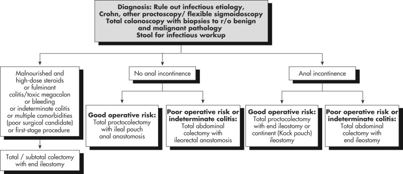
Ulcerative colitis (UC) is an idiopathic, remitting and relapsing, chronic inflammatory bowel disease (IBD) that starts in the rectum and extends proximally.

Synonyms

UC

Inflammatory bowel disease (IBD)

Idiopathic proctocolitis

Pancolitis

ICD-10CM CODES
K51.0Ulcerative pancolitis
K51.2Ulcerative proctitis
K51.3Ulcerative rectosigmoiditis
K51.5Left-sided colitis
K51.90Ulcerative colitis, unspecified, without complications
K51.80Other ulcerative colitis without complications
K51.811Other ulcerative colitis with rectal bleeding
K51.812Other ulcerative colitis with intestinal obstruction
K51.813Other ulcerative colitis with fistula
K51.814Other ulcerative colitis with abscess
K51.818Other ulcerative colitis with other complication
K51.819Other ulcerative colitis with unspecified complications
K51.911Ulcerative colitis, unspecified with rectal bleeding
K51.912Ulcerative colitis, unspecified with intestinal obstruction
K51.913Ulcerative colitis, unspecified with fistula
K51.914Ulcerative colitis, unspecified with abscess
K51.918Ulcerative colitis, unspecified with other complication
K51.919Ulcerative colitis, unspecified with unspecified complications
Epidemiology & Demographics
Incidence

The incidence of UC is 9 to 12 cases/100,000 persons per yr in the U.S.; worldwide, the estimated incidence ranges from 1.2 to 20.3 cases/100,000 person-yr, and its prevalence ranges from 7.6 to 246.0/100,000 persons. It is most common between ages 15 and 40 yr, with a second peak between 50 and 80 yr. The disease affects men and women at similar rates. Infection with nontyphoid Salmonella or Campylobacter is associated with an 8 to 10 times higher risk of developing UC in the following year. Worldwide, UC is more common than Crohn disease.

Prevalence

The prevalence of UC is 7.6 to 246.0 cases/100,000 per yr. Higher prevalence in Ashkenazi Jewish descendants.

Genetics

  • Both specific and nonspecific gene variants are associated with UC.
  • There are 47 loci associated with UC, of which 19 are specific for UC and 28 are shared with Crohn disease.
  • Abnormalities in humoral and cellular adaptive immunity are also found in UC.
Geographic Distribution

The highest incidence and prevalence of IBD are seen in northern Europe and North America, and the lowest in continental Asia.

Physical Findings & Clinical Presentation

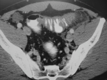
  • Patients with UC often present with acute onset of bloody diarrhea accompanied by tenesmus, fever, and dehydration. At presentation 40% of adults have proctitis, 40% have left-sided colitis, and 20% have pancolitis. Diarrhea is not always present in UC patients with proctosigmoiditis and proctitis, and patients may have constipation.
  • Abdominal pain is not usually a prominent symptom. Abdominal distention and tenderness may indicate the presence of complications such as toxic megacolon.
  • The onset of symptoms is typically acute and is generally followed by periods of spontaneous remission and frequent relapses.
  • Fever, evidence of dehydration may be present during the acute flare-up.
  • Evidence of extraintestinal manifestations may be present in nearly 25% of patients: Liver disease, sclerosing cholangitis, iritis, uveitis, episcleritis, arthritis, erythema nodosum, pyoderma gangrenosum, aphthous stomatitis. Box 1 summarizes common extraintestinal manifestations of UC.

BOX 1 Common Extraintestinal Manifestations of Ulcerative Colitis

Cutaneous/Oral

Angular stomatitis

Aphthous stomatitis

Erythema nodosum

Oral ulcerations

Psoriasis

Pyoderma gangrenosum

Pyostomatitis vegetans

Sweet syndrome (acute febrile neutrophilic dermatosis)

Ophthalmologic

Conjunctivitis

Episcleritis

Retinal vascular disease

Scleritis

Uveitis, iritis

Musculoskeletal

Ankylosing spondylitis

Osteomalacia

Osteonecrosis

Osteopenia

Osteoporosis

Peripheral arthropathy

Sacroiliitis

Hepatobiliary

Autoimmune hepatitis

Cholangiocarcinoma

Pericholangitis

Primary sclerosing cholangitis

Hepatic steatosis

Hematologic

Anemia of chronic disease

Autoimmune hemolytic anemia

Hypercoagulable state

Iron deficiency anemia

Leukocytosis or thrombocytosis

Leukopenia or thrombocytopenia

From Feldman M et al: Sleisenger and Fordtran’s gastrointestinal and liver disease, ed 10, Philadelphia, 2016, Elsevier.

Etiology & Pathogenesis

Accumulating evidence suggests that it may result from an inappropriate inflammatory response to environmental triggers and immune dysregulation involving CD4+ T-cell Th2 response in a genetically susceptible host.

Diagnosis

Differential Diagnosis (Table 1Box 2

  • Crohn disease
  • Bacterial infections:
    1. Acute: Campylobacter, Yersinia, Salmonella, Shigella, Chlamydia, Escherichia coli, Clostridioides difficile, gonococcal proctitis
    2. Chronic: Whipple disease, tuberculosis, enterocolitis
  • Irritable bowel syndrome
  • Protozoal and parasitic infections (amebiasis, giardiasis, cryptosporidiosis)
  • Neoplasm (intestinal lymphoma, carcinoma of colon)
  • Ischemic bowel disease
  • Diverticulitis
  • Celiac sprue, lymphocytic or collagenous colitis, radiation enteritis, endometriosis
  • Solitary rectal ulcer
  • Acute self-limited colitis
  • Medication (NSAIDs, chemotherapy)

BOX 2 Differential Diagnosis of Ulcerative Colitis

Infectious Causes

Aeromonas hydrophila

Campylobacter jejuni

Chlamydia spp.

Clostridioides difficile

Cytomegalovirus

Entamoeba histolytica

Escherichia coli O157:H7, other EHEC

Herpes simplex virus (HSV)

Listeria monocytogenes

Neisseria gonorrhoeae

Salmonella spp.

Schistosomiasis

Shigella spp.

Yersinia enterocolitica

Noninfectious Causes

Acute self-limited colitis

Behçet disease

Crohn disease

Diversion colitis

Diverticulitis

Drugs and toxins

  • Chemotherapy
  • Gold
  • Penicillamine

Eosinophilic colitis

Graft-versus-host disease

Ischemic colitis

Microscopic colitis

  • Collagenous
  • Lymphocytic

Neutropenic colitis (typhlitis)

NSAIDs

Radiation colitis

Segmental colitis associated with diverticulosis

Solitary rectal ulcer syndrome

From Feldman M et al: Sleisenger and Fordtran’s gastrointestinal and liver disease, ed 10, Philadelphia, 2016, Elsevier.

TABLE 1 Features That Distinguish Ulcerative Colitis From Other Diagnoses

DiagnosisClinical FeaturesRadiologic and Colonoscopic FeaturesHistologic Features
UCBloody diarrheaExtends proximally from rectum; fine mucosal ulcerationDistortion of crypts; acute and chronic diffuse inflammatory cell infiltrate; goblet cell depletion; crypt abscesses; lymphoid aggregates
Crohn colitisPerianal lesions are common; may be associated with ileitis; frank bleeding is less common than in UCSegmental disease; rectal sparing; strictures, fissures, ulcers, fistulas; small bowel involvementFocal inflammation; submucosal involvement; granulomas; goblet cell preservation; transmural inflammation; fissuring
Ischemic colitisOccurs in older adults; sudden onset, often painful; usually resolves spontaneously in several daysSegmental splenic flexure and sigmoid involvement are most common, with thumbprinting early and ulceration after 24-72 hr; rectal involvement is rareMucosal necrosis with ghost cells; congestion with red blood cells; hemosiderin-laden macrophages and fibrosis (when disease is chronic)
Microscopic colitisWatery diarrhea; normal-appearing mucosa at colonoscopyUsually normalChronic inflammatory infiltrate; increased intraepithelial lymphocytes (lymphocytic colitis) and/or subepithelial collagen band (collagenous colitis)
Infectious colitisSudden onset; identifiable source in some cases (e.g., Salmonella spp.); pain may predominate (e.g., Campylobacter spp.); pathogens are present in stoolNonspecific findingsCrypt architecture is usually normal; edema, superficial neutrophilic infiltrate, crypt abscesses
Amebic colitisHistory of travel to endemic area; amebae may be detected in a fresh stool specimen, but ELISA for amebic lectin antigen is the preferable diagnostic testDiscrete ulcers; ameboma or stricturesSimilar to UC; amebae present in lamina propria or in flask-shaped ulcers, identified by periodic acid-Schiff stain
Gonococcal proctitisRectal pain; pusGranular changes in rectumIntense polymorphonuclear neutrophil infiltration; purulent exudate; gram-negative diplococci
Pseudomembranous colitisOften a history of antibiotic use; characteristic pseudomembranes may be seen on sigmoidoscopy; Clostridioides difficile toxin is detectable in stoolsEdematous; shaggy outline of colon; pseudomembranes may be identified radiologically or seen at colonoscopyMay resemble acute ischemic colitis; summit lesions of fibrinopurulent exudate

ELISA, Enzyme-linked immunosorbent assay; UC, ulcerative colitis.

From Feldman M et al: Sleisenger and Fordtran’s gastrointestinal and liver disease, ed 10, Philadelphia, 2016, Elsevier.

Workup

An accurate diagnosis of UC should define the extent and severity of inflammation. Diagnostic workup includes:

  • Comprehensive history, physical examination
  • Laboratory tests (see “Laboratory Tests”)
  • Colonoscopy to establish the presence of mucosal inflammation; typical endoscopic findings in UC are areas of continuous friable mucosa; diffuse, uniform erythema replacing the usual mucosal vascular pattern (Table 2); and pseudopolyps. The transition from abnormal to normal tissue tends to be abrupt. Rectal involvement is invariably present if the disease is active. Pathologic findings suggestive of UC include crypt abscesses and atrophy, mucin depletion, basal plasmacytosis, basal lymphoid aggregates, increased lamina propria cellularity, and Paneth cell metaplasia

TABLE 2 Endoscopic Differentiation of Ulcerative Colitis and Crohn Disease

FeatureUlcerative ColitisCrohn Disease
DistributionDiffuse inflammation that extends proximally from the anorectal junctionRectal sparing, frequent skip lesions
InflammationDiffuse erythema, early loss of vascular markings with mucosal granularity or friabilityFocal and asymmetric, cobblestoning; granularity and friability less commonly seen
UlcerationSmall ulcers in a diffusely inflamed mucosa; deep, ragged ulcers in severe diseaseAphthoid ulcers, linear or serpiginous ulceration; intervening mucosa is often normal
Colonic lumenOften narrowed in long-standing chronic disease; tubular colon; strictures are rareStrictures are common

From Feldman M et al: Sleisenger and Fordtran’s gastrointestinal and liver disease, ed 10, Philadelphia, 2016, Elsevier.

Laboratory Tests

  • Anemia and high erythrocyte sedimentation rate (in severe colitis) are common, but normal levels do not rule out the disorder.
  • Potassium, magnesium, calcium, and albumin may be decreased.
  • Antineutrophil cytoplasmic antibodies (ANCA) with a perinuclear staining pattern (pANCA) can be found in >45% of patients; there is an increased frequency in treatment-resistant left-sided colitis, suggesting a possible association between these antibodies and a relative resistance to medical therapy in patients with UC.
  • Calprotectin is a protein that is measured in feces as a marker of intestinal mucosa leukocyte activity that may be useful for screening of patients with suspected IBD. Trials have shown that based on a pretest probability of IBD of 32% in adults, an abnormal fecal calprotectin test would increase the posttest probability to 91% and a normal result would reduce the probability to 3%.
  • Fecal lactoferrin is also a sensitive marker of intestinal inflammation.
  • Stool examinations for ova and parasites, stool culture, and testing for C. difficile toxin and E. coli O157:H7 may be useful to eliminate other causes of diarrhea in selected patients with risk factors.
Imaging Studies

Image studies (plain radiography, CT scan [Fig. E1]) are generally reserved for suspected complications such as perforation of bowel or toxic megacolon.

Figure E1 CT scan showing colonic wall thickening in a patient with ulcerative colitis.

From Adams JG et al: Emergency medicine: clinical essentials, ed 2, Philadelphia, 2013, Elsevier.

Treatment

Nonpharmacologic Therapy

  • Correct nutritional deficiencies; total parenteral nutrition with bowel rest may be necessary in severe cases. Folate supplementation may reduce the incidence of dysplasia and cancer in chronic UC.
  • Avoid oral feedings during acute exacerbation to decrease colonic activity; a low-roughage diet may be helpful in early relapse.
  • Psychotherapy is useful in most patients. Referral to self-help groups is also important because of the chronicity of the disease and the young age of the patients.
Acute General Rx

The therapeutic options (Table 3) vary with the degree of disease (mild, severe, fulminant) and areas of involvement (distal, extensive).

  • Mild disease can be treated with 5-aminosalicylate agents (mesalamine, olsalazine, balsalazide, sulfasalazine). It can be administered as an enema (40 mg once daily at bedtime for 3 to 6 wk) or suppository (500 mg bid) for patients with distal colonic disease. Oral forms in which the 5-acetyl salicylic acid is in a slow-release or pH-dependent matrix (Pentasa 1 g qid, Asacol 800 mg PO tid) can deliver therapeutic concentrations to the more proximal small bowel or distal ileum. Olsalazine can be useful for maintenance of remission of UC in patients intolerant to sulfasalazine. Usual dose is 500 mg bid taken with food. Balsalazide is indicated for mild to moderately active UC. Usual dose is three 750-mg capsules tid. Probiotics may also be helpful in inducing remission in mild-to-moderate UC.
  • Mild-to-moderate UC is often treated with a combination of rectal and oral 5-aminosalicylate. Refractory patients are candidates for oral glucocorticoids or immunosuppressive agents (e.g., cyclosporine).
  • Severe disease usually responds to oral corticosteroids (e.g., prednisone 40 to 60 mg/day); the FDA has recently approved an extended-release formulation of the corticosteroid budesonide for induction of remission in mild to moderate UC. Corticosteroid suppositories or enemas are also useful for distal colitis. The immunosuppressant azathioprine or mercaptopurine also provides effective long-term treatment for Crohn disease. In patients with moderately to severely active disease, a tumor necrosis factor (TNF) inhibitor (infliximab, adalimumab, golimumab), an integrin receptor antagonist (vedolizumab), or an interleukin (IL) 12-23 antagonist (ustekinumab) are useful for both induction and maintenance of remission. In patients with moderately to severely active ulcerative colitis who cannot tolerate or respond poorly to TNF inhibitors oral Janus kinase inhibitors (tofacitinib or upadacitinib) may be effective. The FDA has also approved ozanimod, an oral sphingosine 1-phosphate (S1P) receptor modulator for treatment of adults with moderately to severe active ulcerative colitis who had an inadequate response to or would not tolerate other drugs.
  • Fulminant disease generally requires hospital admission and parenteral corticosteroids (e.g., IV hydrocortisone 100 mg q6h). When bowel movements have returned to normal and the patient is able to eat normally, PO prednisone is resumed. IV cyclosporine can also be used in severe refractory cases; renal toxicity is a potential complication.
  • Surgery is indicated in patients who do not respond to intensive medical therapy. Fig. 2 illustrates the surgical management of chronic UC.
  • Proctocolectomy is usually curative in these patients and also eliminates the high risk of developing adenocarcinoma of the colon (10% to 20% of patients develop it after 10 yr with the disease). Total proctocolectomy with ileal pouch-anal anastomosis (IPAA) is the procedure of choice for most patients who require elective surgery, since it preserves anal sphincter function. Continent ileostomy is an alternative procedure.

Figure 2 Surgical management of chronic ulcerative colitis.

!!flowchart!!

From Cameron JL, Cameron AM: Current surgical therapy, ed 12, Philadelphia, 2017, Elsevier.

TABLE 3 Medical Therapy Used in Ulcerative Colitis

DrugRelease SiteTreatment of UCSide Effects
Oral 5-aminosalicylates
SulfasalazineColonInduce and maintain remission in mild-to-moderate UC
Use in combination with topical mesalamine for distal colitis
Sulfasalazine: Idiosyncratic (rash, hepatitis, aplastic anemia), dose-related (nausea, hemolytic anemia, inhibits folic acid transport), oligospermia (reversible)
Mesalamine (mesalazine)Distal ileum, colon
Mesalamine (mesalazine) (controlled-release)Duodenum, jejunum, ileum, colon
OlsalazineColon
BalsalazideColon
Topical 5-aminosalicylates
Mesalamine (mesalazine) enemaRectum, sigmoidInduce and maintain remission in mild-to-moderate distal disease
Combination therapy with oral aminosalicylates are superior to monotherapy
Mesalamine (mesalazine) suppositoryRectum
Antibiotics
Ciprofloxacin
Metronidazole
Amoxicillin/clavulanic acid
Rifaximin
SystemicAntibiotics are not used for the treatment of UC but are used for pouchitisCiprofloxacin: Tendonitis and rupture
Metronidazole: Neuropathy
Amoxicillin/clavulanic acid: Hepatitis
Corticosteroids
Budesonide extended-releaseColonInduce remission in mild-to-moderate UC
Should not be used for maintenance
High first-pass metabolism
More favorable side-effect profile than prednisone
PrednisoneSystemicInduce remission in mild-to-moderate and some moderate-to-severe UC flares
Not used for maintenance therapy
Infection, diabetes mellitus, osteoporosis, osteonecrosis, cataracts, glaucoma, and myopathy. Increase risk of mortality
MethylprednisoloneSystemicInduce remission in severe UCAs for prednisone
Immunomodulators
6-MercaptopurineSystemicMaintain remission in steroid-dependent, steroid-refractory, or steroid-induced-remission UC
Not used to induce remission
Allergic reactions, pancreatitis, myelosuppression, nausea, infections, hepatotoxicity, and malignancy, in particular lymphoma
AzathioprineSystemic
CyclosporineSystemicRescue therapy to induce remission in severe UC not responding to IV methylprednisoloneInfections, hypertension, renal insufficiency, tremor, headache, hepatotoxicity
Biologicals
InfliximabSystemicCan be used to induce and maintain moderate-to-severe UC and are steroid-sparing
Infliximab is also used as a rescue therapy to induce remission in severe UC not responding to IV methylprednisolone
Infections (tuberculosis, fungal infections), autoantibody formation, psoriasis, drug-induced lupus
Infusion reactions (infliximab), injection-site reaction (adalimumab and golimumab), delayed hypersensitivity reaction (infliximab)
Lymphoma (higher in combination therapy)
AdalimumabSystemic
GolimumabSystemic
VedolizumabSystemicEffective in the induction and maintenance of remission in moderate-to-severe UCHeadache, infections, abdominal pain, infusion reactions
Tofacitinib (small molecules)SystemicEffective in induction and maintenanceInfections (especially herpes zoster), lymphoma

UC, Ulcerative colitis.

From Talley NJ et al: Essentials of internal medicine, ed 4, Chatswood, NSW, 2021, Elsevier Australia.

Chronic Rx

  • Colonoscopic surveillance and multiple biopsies should be instituted approximately 10 yr after diagnosis because of the increased risk of colon carcinoma.
  • Erythropoietin is useful in patients with anemia refractory to treatment with iron and vitamins.
  • In patients on long-term steroid therapy, periodic bone density scans are recommended to screen for glucocorticoid-induced osteoporosis.
Disposition

  • The natural history of the disease is one of remission and episodic flares.
  • The clinical course is variable. 66% of patients will achieve clinical remission with medical therapy, and nearly 80% of treatment-compliant patients maintain remission. 15% to 20% of patients eventually require colectomy. Pouchitis is the most common long-term complication of IPAA (up to 40% of patients). >75% of patients treated medically will experience relapse.
Referral

  • GI consultation for initial diagnostic sigmoidoscopy/colonoscopy in suspected cases.
  • Surgical referral for patients with severe disease unresponsive to medical therapy. Indications for surgery in patients with UC are summarized in Box 3.

BOX 3 Indications for Surgery in Patients With Ulcerative Colitis

Colonic dysplasia or carcinoma

Colonic perforation

Growth retardation

Intolerable or unacceptable side effects of medical therapy

Medically refractory disease

Systemic complications that are recurrent or unmanageable

Toxic megacolon

Uncontrollable colonic hemorrhage

From Feldman M et al: Sleisenger and Fordtran’s gastrointestinal and liver disease, ed 10, Philadelphia, 2016, Elsevier.

Related Content

Ulcerative Colitis (Patient Information)

Suggested Readings

    1. Feuerstein J.D., Cheifetz A.S. : Ulcerative colitis: epidemiology, diagnosis, and managementMayo Clin Proc. ;89(11):1553-1563, 2014.
    2. Sands B.E. : Vedolizumab versus adalimumab for moderate-to-severe ulcerative colitisN Engl J Med. ;381:1215-1226, 2019.