section name header

Basic Information

AUTHORS: Javad Najjar Mojarrab, MD, MBA and James E. Novak, MD, PhD

Definition

Hepatorenal syndrome (HRS) is a functional cause of acute kidney injury (AKI) that occurs in the setting of severe acute or chronic liver disease, most commonly with cirrhosis and portal hypertension. HRS develops because of reduced renal perfusion caused by hemodynamic alterations in the arterial circulation and overactivity of endogenous vasoactive systems.

In HRS, liver dysfunction leads to increased production or activity of vasodilators such as nitric oxide. Vasodilation is most pronounced in the splanchnic circulation and, combined with cardiac dysfunction, results in systemic hypotension. Disturbed vasoconstrictor-vasodilator balance in the kidney favors vasoconstriction. Intense renal arteriolar vasoconstriction, renal cortical hypoperfusion, and AKI ensue (Fig. E1). Fig. E2 illustrates the proposed pathophysiology of HRS and HRS precipitants. HRS involves not only circulatory dysfunction but also systemic inflammation triggered by hepatic injury and bacterial translocation from the gut lumen.

There are two types of HRS (Table E1). The International Club of Ascites (ICA) revised the nomenclature and diagnosis of type 1 HRS (HRS-1), now termed HRS-acute kidney injury (HRS-AKI), and type 2 HRS (HRS-2), also now termed HRS-nonacute kidney injury (HRS-NAKI).

  • HRS-AKI: Acute and rapid deterioration in kidney function (Table E2)
  • HRS-NAKI: Moderate stable chronic kidney disease (CKD) (average serum creatinine [SCr] 1.5 mg/dl [133 mmol/L])

Figure E1 Circulatory dysfunction in hepatorenal syndrome (HRS).

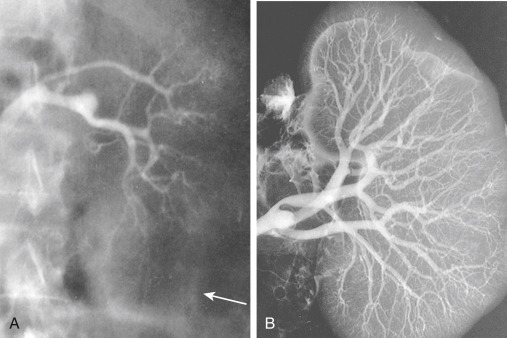
A, Renal angiogram (the arrow marks edge of the kidney). B, The angiogram carried out in the same kidney at autopsy. Note complete filling of the renal arterial system throughout the vascular bed to the periphery of the cortex. The vascular attenuation and tortuosity seen previously (A) are no longer present. The vessels are also histologically normal. This finding indicates the functional nature of the vascular abnormality in HRS.

From Floege J et al: Comprehensive clinical nephrology, ed 4, St Louis, 2010, Saunders.

Figure E2 Proposed pathophysiology and triggers of hepatorenal syndrome.

!!flowchart!!

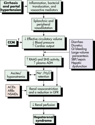
ACEI, Angiotensin-converting enzyme inhibitor; ADH, antidiuretic hormone; ARB, angiotensin receptor blocker; CCM, cirrhotic cardiomyopathy; GFR, glomerular filtration rate; GI, gastrointestinal; H2O, water; NA, sodium; NSAID, nonsteroidal antiinflammatory drug; RAAS, renin-angiotensin-aldosterone system; SBP, spontaneous bacterial peritonitis; SNS, sympathetic nervous system.

From Feldman M et al: Sleisenger and Fordtran’s gastrointestinal and liver disease, ed 10, Philadelphia, 2016, Elsevier.

TABLE E1 Definition of Hepatorenal Syndrome

HRS-AKI (Type 1)
Acute and rapid deterioration in kidney function:
An acute increase in serum creatinine of 0.3 mg/dl within 48 hr and/or urinary output <0.5 ml/kg for >6 hr or increase in serum creatinine of 50% from a stable baseline serum creatinine within 3 mo (presumed to have developed within the past 7 days when no prior readings are available)
Occurs in parallel with the cirrhosis-associated failure of other organs or systems (e.g., coagulopathy, hepatic encephalopathy)
In cirrhosis, HRS is a form of acute-on-chronic liver failure
Frequently follows a precipitating event, mainly bacterial infection; rapidly fatal without treatment
Renal replacement therapy, i.e., hemodialysis, does not prolong life without liver transplantation
HRS-NAKI (Type 2)
HRS-AKDHepatorenal syndrome-acute kidney disease: (a) eGFR <60 ml/min per 1.73 m2 for <3 mo in the absence of other (structural) causes; or (b) SCr increase <50% using the last available outpatient value within 3 mo
HRS-CKDHepatorenal syndrome-chronic kidney disease: eGFR <60 ml/min per 1.73 m2 for 3 mo in the absence of other (structural) causes
Moderate stable CKD
Mean survival of 6 mo

AKI, Acute kidney injury; CKD, chronic kidney disease; eGFR, estimated glomerular filtration rate; HRS, hepatorenal syndrome; NAKI, nonacute kidney injury; SCr, serum creatinine.

From Fernández J, Arroyo V: Hepatorenal syndrome. In Johnson RJ et al (eds): Comprehensive clinical nephrology, ed 5, Philadelphia, 2015, Saunders.

TABLE E2 Hepatorenal Syndrome-AKI Classified by Acute Kidney Injury (AKI) Stage According to the International Club of Ascites and Acute Kidney Injury Network Criteria

Risk/Stage 1Increase in SCr >1.5- to 2-fold from baseline; or by >0.3 mg/dl (26.5 mmol/L)
Increase in SCr of 50% or more to a final value of >1.5 mg/dl (133 mmol/L)
Injury/Stage 2Increase in SCr >2- to 3-fold from baseline
Failure/Stage 3Increase in SCr >3-fold from baseline; or SCr >4 mg/dl (353.6 mmol/L) with an acute increase of 0.5 mg/dl (44 mmol/L); or
initiation of renal replacement therapy

SCr, Serum creatinine.

From Angeli P et al: Diagnosis and management of acute kidney injury in patients with cirrhosis: revised consensus recommendations of the International Club of Ascites, J Hepatol 62:968, 2015.

Synonyms

Hepatorenal failure

Hepatic nephropathy

HRS

Oliguric renal failure of cirrhosis

ICD-10CM CODES
K76.7Hepatorenal syndrome, hepatorenal failure
Z87.19History of hepatorenal syndrome
Z84.1Family history of hepatorenal syndrome
Epidemiology & Demographics

The probability of HRS in patients with cirrhosis is 18% at 1 yr and 39% at 5 yr. HRS is associated with poor prognosis. HRS-AKI is associated with >90% mortality in 3 mo, with higher mortality conferred by a Model for End-stage Liver Disease (MELD) score >30 and by higher stages of AKI. HRS-NAKI is associated with 30% mortality at 3 mo and 60% at 1 yr. Although the cause of liver failure or MELD score does not predict the development of HRS, MELD score predicts mortality when HRS has occurred. Hyponatremia is an independent risk factor for HRS and is associated with elevated renin levels, which are thought to reflect the overactivated neurohumoral response.

Physical Findings & Clinical Presentation

There are no specific physical findings associated with HRS, although it is usually associated with signs and symptoms of acute or chronic decompensated liver failure, such as hypotension, jaundice, spider angiomata, splenomegaly, ascites, fetor hepaticus, edema, asterixis, encephalopathy, or coma. Usually, HRS is accompanied by oliguria and bland urine sediment. Urine chemistries tend to show a low rate of urinary sodium excretion (usually <10 mEq/L). However, a nonoliguric state or an active urine sediment (blood, casts, and/or protein) does not exclude the diagnosis of HRS.

Precipitants

Precipitating events are identified in most cases of HRS. Contributory factors include bacterial infection, alcoholic hepatitis, excessive laxative or NSAID use, large-volume paracentesis without albumin administration, Gl bleeding, or a major surgical procedure. Unlike HRS, prerenal azotemia improves with cessation of diuretic therapy and/or volume resuscitation. Nevertheless, HRS may present in the absence of a clear precipitating factor. Recent studies suggest that, among those not listed for liver transplant, mortality rates are extremely high regardless of whether the etiology of AKI is HRS or acute tubular necrosis (ATN).

Diagnosis

HRS is a diagnosis of exclusion. The revised diagnostic criteria for HRS are summarized in Table E3.

TABLE E3 Revised Diagnostic Criteria for Hepatorenal Syndrome Type 1 or Acute Kidney Injury (HRS-AKI) in Patients With Cirrhosis

  1. Cirrhosis; acute liver failure; acute or chronic liver failure
  2. Diagnosis of AKI according to International Club of Ascites criteria
  3. No improvement in kidney function after at least 48 consecutive hours of diuretic withdrawal and plasma volume expansion with albumin 1 g/kg/day (maximum dose, 100 g/day)
  4. Absence of shock
  5. No current or recent use of nephrotoxic drugs (e.g., nonsteroidal antiinflammatory drugs, aminoglycosides, iodinated contrast media)
  6. No macroscopic signs of structural kidney injury, defined as:
    • Absence of proteinuria (>500 mg/day)
    • Absence of microhematuria (>50 red blood cells per high-power field)
    • Normal findings on renal ultrasonography

This criterion would not be included in cases of known preexisting structural chronic kidney disease (e.g., diabetic or hypertensive nephropathy).

From Angeli P et al: Diagnosis and management of acute kidney injury in patients with cirrhosis: revised consensus recommendations of the International Club of Ascites, J Hepatol 62:968, 2015.

Differential Diagnosis

  • Prerenal azotemia: Must be excluded or treated appropriately before establishing a diagnosis of HRS. Prerenal azotemia typically responds to volume expansion and cessation of diuretic therapy. Both prerenal azotemia and HRS are often associated with fractional excretion of sodium (FENa) <1%.
  • ATN: Urine sodium >30 mEq/L, FENa >1.5%, urine-to-plasma creatinine ratio <30, urine-to-plasma osmolality ratio of 1; urine sediment reveals muddy brown casts and cellular debris. There is no significant response to sustained plasma expansion.
  • Other: Renal artery or vein thrombosis; cardiorenal syndrome; urinary tract obstruction; glomerulonephritis; and toxicity from drugs, organic solvents, heavy metals, heme pigments, and intravenous contrast medium.
Workup

Acute azotemia and oliguria in the setting of liver disease require laboratory evaluation to distinguish HRS from ATN. A volume challenge may be required to differentiate HRS from prerenal azotemia. Diagnostic paracentesis should be done to rule out spontaneous bacterial peritonitis (SBP). SCr and blood urea nitrogen (BUN) are poorly sensitive markers of kidney function in cirrhosis. SCr as a predictor of kidney function in patients with cirrhosis is also obscured by the following factors:

  • Decreased formation of creatinine from creatine in muscles due to muscle wasting
  • Increased renal tubular secretion of creatinine
  • Increased volume of distribution causing dilution of SCr
  • Interference with assays for SCr by elevated bilirubin
Laboratory Tests

  • Serum electrolytes (serum sodium <135 mEq/L), BUN, creatinine, and osmolality
  • Urinalysis, urine sodium, urine creatinine, and urine osmolality
  • Urine sodium <10 mEq/L, FENa <1%, urine-to-plasma creatinine ratio >30, urine plasma-to-urine osmolality ratio >1.5, bland urine sediment. FENa does not adequately differentiate HRS from other common etiologies of AKI in liver dysfunction; however, only a minority of patients with HRS have high urine sodium concentrations. (Note: FENa can be <1% in advanced cirrhotic patients with high sodium avidity; prior literature suggests that FENa <0.2% favors the diagnosis of HRS over ATN in these patients.)
  • Investigational urinary biomarkers such as neutrophil gelatinase-associated lipocalin (NGAL) could be a useful biomarker in differentiating between HRS and ATN. NGAL urine levels are much higher in patients with ATN compared to patients with other causes of AKI.1-3
  • Additional diagnostic tests include cystatin C, which, when combined with SCr, can help calculate glomerular filtration rate more accurately compared to SCr alone.4
Imaging Studies

Kidney, bladder, and ureter ultrasound are indicated if obstructive uropathy is suspected. Duplex Doppler ultrasonography may show increased renal resistive indices (RI) in hilar, medullary, and cortical areas and disappearance of the RI gap (difference between interlobar and cortical RI), indicating reduction of renal blood flow. Computed tomography (CT) may be required to diagnose occult infection(s), and prophylaxis directed at preventing contrast-associated nephropathy is recommended.

Treatment

Pharmacologic Therapy

Most therapy is centered on the use of vasoconstrictor therapy and albumin resuscitation targeted at the reversal of splanchnic arterial vasodilation.

Nonpharmacologic Therapy

  • Liver transplantation alone for candidates with HRS-AKI for <4 wk, and simultaneous liver-kidney transplantation for those at risk of nonrecovery of kidney function.
  • Transjugular intrahepatic portosystemic shunt for patients with HRS-NAKI and refractory ascites.
  • Renal replacement therapy as a bridge to liver transplantation.
Acute General Rx

  • Box E1 summarizes the management of HRS. Avoidance of precipitating factors (Table E4) and increasing blood pressure and volume are the cornerstones of HRS management. A recent guideline from the American Association for the Study of Liver Disease lists terlipressin as the preferred first-line agent; however, this agent has been associated with worse clinical outcomes and is not available in the United States. Norepinephrine is an alternative. Midodrine plus octreotide can be used, but evidence for efficacy is poor. If creatinine does not decline after 4 days on maximal doses of vasopressors, further improvement is unlikely, and treatment can be stopped.5
  • Patients who have survived an episode of SBP should receive long-term prophylaxis with daily norfloxacin or trimethoprim/sulfamethoxazole.
  • Improvement in liver function due to partial resolution of the primary disorder or successful liver transplantation is the optimal treatment. Liver transplantation leads to resolution of HRS-AKI in 76% of cases.
  • Splanchnic and systemic vasoconstrictor agents (e.g., terlipressin, norepinephrine, and midodrine combined with octreotide). Terlipressin plus albumin improves creatinine but not other clinical outcomes, such as mortality, and is associated with increased risk of adverse events such as respiratory failure; this agent was not approved by the U.S. Food and Drug Administration (FDA) in 2021.6
  • Dopamine and prostaglandins are generally ineffective in treating patients with HRS.

TABLE E4 Prevention of Acute Kidney Injury in Patients With Cirrhosis

Risk FactorsPreventive Approaches
Hepatorenal syndrome developmentJudicious use of diuretics and lactulose to avoid hypovolemia
Antibiotic prophylaxis following GI bleeding
Albumin administration during large volume paracentesis (6 to 8 g/L of ascitic fluid removed)
Spontaneous bacterial peritonitis (SBP) prophylaxis in patients with low-protein ascites
Early recognition and treatment of SBP with antibiotics and albumin at the dose of 1.5 g per kg of body weight at the time of diagnosis of SBP and 1 g per kg of body weight at the third day of treatment
Nephrotoxic medication exposureAvoid nephrotoxic medications if possible
Appropriate drug dosing based on pharmacokinetics and close monitoring of drug toxicity, early recognition of drug-induced AKI, and discontinuation of offending agent if possible
Use of lipid formulations of amphotericin B rather than conventional formulations of amphotericin B
Use of azole antifungal agents and/or the echinocandins rather than conventional amphotericin B, if equal therapeutic efficacy can be assumed
Radiocontrast exposureConsider alternative imaging methods or avoidance of IV contrast if possible
Use of low or iso-osmolar agents with lowest volume possible
IV volume replacement prior to contrast exposure
Intraabdominal hypertensionParacentesis, with coadministration of albumin, in patients with tense ascites

AKI, Acute kidney injury; GI, gastrointestinal; IV, intravenous.

From Ronco C et al: Critical care nephrology, ed 3, Philadelphia, 2019, Elsevier.

BOX E1 Management of Hepatorenal Syndrome (HRS)

  • Measures to prevent variceal bleeding (e.g., beta blockers, band ligation)
  • Treatment of severe alcoholic hepatitis
  • Prevention of HRS
    • Avoid intravascular volume depletion (diuretics, excessive lactulose, gastrointestinal bleeding, large-volume paracentesis without adequate volume repletion)
    • Angiotensin-converting enzyme inhibitors (ACEIs) and angiotensin receptor blockers (ARBs) should be avoided or used with caution in patients with cirrhosis and ascites. Judicious management of nephrotoxins (nonsteroidal antiinflammatory drugs [NSAIDs], antibiotics). Prompt diagnosis and treatment of infections (spontaneous bacterial peritonitis [SBP], sepsis)
    • SBP prophylaxis: Long-term use of norfloxacin (or trimethoprim/sulfamethoxazole) can be justified if the ascitic fluid protein <1.5 g/dl along with chronic kidney disease (serum creatinine >1.2 mg/dl, BUN >25 mg/dl, or serum Na <130 mmol/L) or liver failure (Child score >9 and bilirubin >3 mg/dl) (Class I, Level A)
    • Avoid use of proton pump inhibitors without indications as can precipitate SBP
  • Treatment of HRS
    • Stop all nephrotoxic or volume-depleting agents (ACEIs, ARBs, NSAIDs, beta-blockers, diuretics)
    • Antibiotics for infections
    • Intravenous (IV) albumin: bolus of 1 g/kg/day on presentation (maximum dose, 100 g daily). Continue albumin dose at 20-60 g daily as needed to maintain central venous pressure at 10-15 cm H2O
    • Vasopressor therapy (in addition to albumin):
      • Terlipressin: start at 1 mg IV every 4 h and increase up to 2 mg IV every 4 h if baseline serum creatinine level does not improve by 25% at day 3 of therapy OR
      • Midodrine and octreotide: begin midodrine at 2.5-5 mg orally 3 times daily and increase to a maximum dose of 15 mg 3 times daily. Titrate to a mean arterial pressure (MAP) increase of at least 15 mm Hg; begin octreotide at 100 mg subcutaneously 3 times daily and increase to a maximum dose of 200 mg subcutaneously 3 times daily, or begin octreotide as a 25 mg IV bolus and continue at a rate of 25 mg/h OR
      • Norepinephrine: 0.1-0.7 μg/kg/min as an IV infusion. Increase by 0.05 mg/kg/min every 4 h and titrate to an MAP increase of at least 10 mm Hg
      • Duration of vasopressor treatment is generally a maximum of 2 wk until reversal of hepatorenal syndrome or liver transplantation
    • Evaluate patient for liver transplantation

From Feldman M et al: Sleisenger and Fortran’s gastrointestinal and liver disease, ed 10, Philadelphia, 2016, Elsevier.

Referral

Referral for hepatology, nephrology, and liver transplantation as indicated

Not available in the U.S. or Canada.BUN, Blood urea nitrogen.

Pearls & Considerations

Related Content

Hepatorenal Syndrome (Patient Information)

Related Content

  1. Acevedo J.G., Cramp M.E. : Hepatorenal syndrome: update on diagnosis and therapyWorld J Hepatol. ;9, 2017.
  2. Fagundes C. : Urinary neutrophil gelatinase-associated lipocalin as biomarker in the differential diagnosis of impairment of kidney function in cirrhosisJ Hepatol. ;57, 2012.
  3. Yap D.Y.H. : Serum and urinary biomarkers that predict hepatorenal syndrome in patients with advanced cirrhosisDig Liver Dis. ;49, 2017.
  4. Singapura P. : Estimating glomerular filtration rate in cirrhosis using creatinine-based and cystatin C-based equations: systematic review and meta-analysisLiver Transpl. ;27, 2021.
  5. Biggins S.W. : Diagnosis, evaluation, and management of ascites and hepatorenal syndrome: 2021 practice guidance by the American Association for the Study of Liver DiseasesHepatol. ;74, 2021.
  6. Wong F. : Terlipressin plus albumin for the treatment of type 1 hepatorenal syndromeNew Engl J Med. ;384, 2021.
  7. Bera C, Wong F: Management of hepatorenal syndrome in liver cirrhosis: a recent update, Therap Adv Gastroenterol 15:17562848221102679, 2022.
  8. Allegretti A.S. : Prognosis of patients with cirrhosis and AKI who initiate RRTClin J Am Soc Nephrol. ;13, 2018.