section name header

Basic Information

AUTHORS: Helen B. Gomez Slagle, MD and Anthony Sciscione, DO

Definition

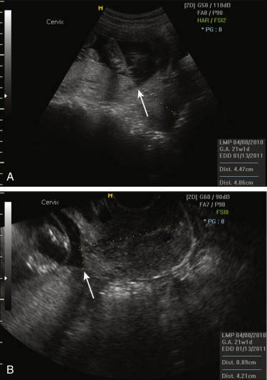
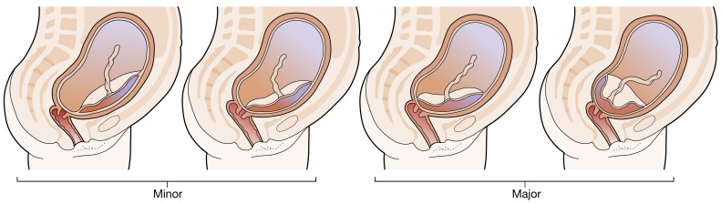
The diagnosis of placenta previa is based on sonography and requires the identification of echogenic homogeneous placental tissue over the internal cervical os. When the placental edge is <2 cm from the internal os, the placenta is called “low-lying.” The historic terms “marginal” and “partial” for characterizing a placenta previa are no longer used. A classification of placenta previa in “major” or “minor” is described in Table 1 and illustrated in Fig. 1.

TABLE 1 Classification of Placenta Previa

MinorIEncroaches the lower uterine segment
IIReaches internal os of the cervix (marginal)
MajorIIICovers part of internal os (partial)
IVCompletely covers the internal os (complete)

From Magowan BA: Clinical obstetrics and gynecology, ed 4, London, 2019, Elsevier.

Figure 1 Classification into “major” and “minor” Placenta Previa Depends on the Distance of the Placenta from the Internal Os of the Cervix

In the presence of a cesarean section scar, an anterior placenta previa may result in abnormal invasion (morbidly adherent placenta, placenta accreta).

From Magowan BA: Clinical obstetrics and gynecology, ed 4, London, 2019, Elsevier.

One hypothesis is that the lower uterine cavity contains more vascularized decidua, which promotes implantation of trophoblast toward the cervical os. Another hypothesis is that a particularly large placental surface area increases the probability that the placenta will implant over the cervical os.

ICD-10CM CODES
O44Placenta previa
O44.0Complete placenta previa NOS or without hemorrhage
O44.1Complete placenta previa with hemorrhage
O44.2Partial placenta previa without hemorrhage
O44.3Partial placenta previa with hemorrhage
O44.4Low-lying placenta NOS or without hemorrhage
O44.5Low-lying placenta with hemorrhage
Epidemiology & Demographics
Incidence

The pooled prevalence of major placenta previa was 4.3 cases/1000 pregnancies. Prevalence was highest among Asian studies (12.2/1000) and lower among studies from Europe (3.6/1000), North America (2.9/1000), and sub-Saharan Africa (2.7/1000).

Risk Factors

  • Previous placenta previa (recurs in 4% to 8% of subsequent pregnancies)
  • Previous cesarean delivery (increases incidence by 47% to 60%)
  • Multiparity
  • Multiple gestation (increases prevalence by 40%)
  • Smoking and cocaine use
  • Previous intrauterine surgical procedure or Asherman syndrome
  • Abnormal or large placenta
Physical Findings & Clinical Presentation

The classic presentation of placenta previa is painless vaginal bleeding, usually in the second or third trimester. 10% to 20% of women present with uterine contractions, pain, and bleeding. On physical examination, the uterus is soft and pain free. The fetus is often in breech, transverse lie, or high. Fetal distress is usually not present.

Diagnosis

Differential Diagnosis

  • Morbidly adherent placenta (accreta, increta, percreta)
  • Vasa previa
  • Abruptio placentae
  • Vaginal or cervical trauma
  • Labor
  • Local malignancy
Workup

  • Do not perform a digital vaginal examination.
  • Perform a speculum examination in a hospital setting to exclude any local bleeding.
  • Perform a transvaginal ultrasonography to assess for placental location.
  • Exclude the presence of placenta previa-accreta in patients with a history of cesarean sections. In a prospective study, the frequency of placenta accreta increased with an increasing number of cesarean deliveries: 3% for first cesarean, 11% for second cesarean, 40% for third cesarean, 61% for fourth cesarean, and up to 67% for fifth or more cesarean sections.
Laboratory Tests

  • A CBC can be used to monitor hemoglobin and hematocrit.
  • A Kleihauer-Betke preparation of maternal blood in all Rh-negative women and Rh-immune globulin when indicated.
Imaging Studies

  • The simplest and safest method of placental localization is transabdominal sonography with confirmatory imaging by transvaginal ultrasonography (TVUS). Transabdominal ultrasonography alone is inaccurate in the diagnosis of placenta previa and should be used only as a screening tool. TVUS (Fig. 2) has become the gold standard for the diagnosis of placenta previa. It is safe even in the presence of active bleeding.
  • MRI has also been effective in detecting placenta previa, although sonography remains the preferred method due to lower cost, widespread availability, and well-established accuracy.
Figure 2 Transabdominal Ultrasonography and Transvaginal Ultrasonography of Marginal Placenta Previa

Arrows identify placental edge.

Courtesy K. Francois. From Gabbe SG: Obstetrics, ed 6, Philadelphia, 2012, Saunders.

Treatment

Management of Asymptomatic Placenta Previa

  • Avoid sexual activity, digital exams, and extraneous exercise.
  • Review bleeding precautions and anticipatory guidance, including the need for cesarean section and the risk for hysterectomy early in pregnancy.

Sequential assessment of placental location:

  • At 32 wk, follow-up is indicated:
    1. If the placental edge is 2 cm from the internal os, placenta previa has resolved, and no further assessment is required.
    2. If the placental edge is over or <2 cm from the internal os, placenta previa persists, and repeat at 36 wk is indicated.
  • At 36 wk:
    1. If the placental edge is 2 cm, the patient is consented for vaginal delivery.
    2. If the placental edge is over the internal os, cesarean delivery is scheduled.

Timing of delivery:

  • Cesarean delivery at 36+0 to 37+6 wk in pregnancies with uncomplicated placenta previa
Management of Symptomatic Placenta Previa

  • Initial assessment for signs of maternal hemodynamic compromise or hemorrhagic shock; large-bore IV access with crystalloid fluid resuscitation.
  • Assess fetal status and gestational age by sonogram and continuous fetal heart rate monitoring.
  • Cross-matched blood should be made available during bleeding episodes.
  • Tocolytic therapy should not be administered in an actively bleeding patient.
  • Magnesium sulfate therapy for fetal neuroprotection should be considered in those with symptomatic preterm (<32 wk) placenta previa if the decision has been made to likely deliver the patient within 24 hr. Emergent delivery should not be delayed to administer magnesium.
  • Cesarean delivery is indicated for active labor, nonreassuring fetal heart rate tracing, active bleeding with hemodynamic instability, and significant bleeding after 34 wks’ gestation.

Expectant management after a resolved bleed:

  • Antenatal corticosteroid should be administered to symptomatic women between 23+0 and 36+6 wks’ gestation to enhance fetal pulmonary maturity.
  • Correct anemia.
  • Administer anti-D immune globulin to D-negative women.
Disposition

Inpatient versus outpatient: Consider discharge for women whose bleeding has stopped for 24 h and live in close proximity to the hospital, demonstrate compliance with medical management, can maintain bed rest, understand bleeding precautions, and have an adult companion available for transport 24 h a day.

Pearls & Considerations

Comments

  • Placenta previa is diagnosed by TVUS, with repeat assessment at 32 and 36 wk when indicated.
  • Asymptomatic placenta previa can be managed expectantly with a planned cesarean delivery at 36+0 to 37+6 wk.
  • Symptomatic placenta previa is managed by assessing hemodynamic stability, considering steroids for fetal lung maturity, magnesium sulfate for fetal neuroprotection when indicated, and cesarean delivery.
Related Content

Placenta Previa (Patient Information)

Vaginal Bleeding During Pregnancy (Related Key Topic)

Suggested Readings

    1. Fan D. : Prevalence of antepartum hemorrhage in women with placenta previa: a systematic review and meta-analysisSci Rep. ;7, 2017.
    2. Gyamfi-Bannerman C: Society for Maternal-Fetal Medicine (SMFM) Consult Series #44: management of bleeding in the late preterm period, Am J Obstet Gynecol 218(1):B2-B8, 2018.
    3. Weiner E. : The effect of placenta previa on fetal growth and pregnancy outcome, in correlation with placental pathologyJ Perinatol. ;36, 2016.