section name header

Basic Information ⬇

AUTHORS: Angad Jolly, MD, PhD, and Joseph S. Kass, MD, JD, FAAN

Definition

Dementia with Lewy bodies (DLB) is a neurodegenerative dementia occurring concurrently with or within 1 yr (either before or after) of the onset of parkinsonism. DLB also has other core features, including fluctuations in attention and alertness and recurrent vivid visual hallucinations. Diagnostic criteria for dementia syndrome associated with Lewy body pathology are described in Table E1. Patients generally respond to cholinesterase inhibitors, are very sensitive to the adverse effects of neuroleptics, and are less responsive to levodopa compared with Parkinson disease (PD) patients.

TABLE E1 Revised Criteria for the Clinical Diagnosis of Probable and Possible Dementia With Lewy Bodies (DLB)

Essential for a diagnosis of DLB is dementia, defined as a progressive cognitive decline of sufficient magnitude to interfere with normal social or occupational functions, or with usual daily activities. Prominent or persistent memory impairment may not necessarily occur in the early stages but is usually evident with progression. Deficits on tests of attention, executive function, and visuoperceptual ability may be especially prominent and occur early.
Core clinical features (The first three typically occur early and may persist throughout the course.)
  • Fluctuating cognition with pronounced variations in attention and alertness.
  • Recurrent visual hallucinations that are typically well formed and detailed.
  • REM sleep behavior disorder, which may precede cognitive decline.
  • One or more spontaneous cardinal features of parkinsonism: These are bradykinesia (defined as slowness of movement and decrement in amplitude or speed), rest tremor, or rigidity.
Supportive clinical features
  • Severe sensitivity to antipsychotic agents; postural instability, repeated falls, syncope or other transient episodes of unresponsiveness; severe autonomic dysfunction, such as constipation, orthostatic hypotension, urinary incontinence; hypersomnia; hyposmia; hallucinations in other modalities; systematized delusions; apathy, anxiety, and depression.
Indicative biomarkers
  • Relative preservation of medial temporal lobe structures on CT/MRI scan.
  • Generalized low uptake on SPECT/PET perfusion/metabolism scan with reduced occipital activity ± the cingulate island sign on FDG-PET imaging.
  • Prominent posterior slow-wave activity on EEG with periodic fluctuations in the pre-alpha/theta range.
Probable DLB can be diagnosed if:
  1. Two or more core clinical features of DLB are present, with or without the presence of indicative biomarkers, or
  1. Only one core clinical feature is present but with one or more indicative biomarkers.
Probable DLB should not be diagnosed on the basis of biomarkers alone.
Probable DLB can be diagnosed if:
  1. a. Only one core clinical feature of DLB is present, with no indicative biomarkers evidence, or
  1. b. One or more indicative biomarkers is present but there are no clinical features.
DLB is less likely:
  1. a. In the presence of any other physical illness or brain disorder, including cerebrovascular disease, sufficient to account in part or in total for the clinical picture, although these do not exclude a DLB diagnosis and may serve to indicate mixed or multiple pathologies contributing to the clinical presentation, or
  1. b. If parkinsonian features are the only core clinical feature and appear for the first time at a state of severe dementia.
DLB should be diagnosed when dementia occurs before or concurrently with parkinsonism. The term Parkinson disease dementia (PDD) should be used to describe dementia that occurs in the context of well-established Parkinson disease. In a practice setting, the term that is most appropriate to the clinical situation should be used, and generic terms such as Lewy body disease are often helpful. In research studies in which distinction needs to be made between DLB and PDD, the existing 1-yr rule between the onset of dementia and parkinsonism continues to be recommended.

CT, Computed tomography; EEG, electroencephalogram; FDG-PET, fluorodeoxyglucose-positron emission tomography; MRI, magnetic resonance imaging; PET, positron emission tomography; REM, rapid eye movements; SPECT, single-photon emission computerized tomography.

From McKeith IG et al: Diagnosis and management of dementia with Lewy bodies: fourth consensus report of the DLB Consortium, Neurology 89(1):88-100, 2017.

Synonyms

DLB

Lewy body dementia

Diffuse Lewy body disease

Lewy body type senile dementia

Cortical Lewy body disease

ICD-10CM CODE
G31.83Dementia with Lewy bodies
Epidemiology & Demographics
Incidence

Accounts for 10% to 15% of all dementias. DLB is the second most common neurodegenerative cause of dementia after Alzheimer disease (AD) and is the third most common cause of dementia when vascular dementia is included in the tally.

Prevalence

  • Estimated 0.7% of individuals >65 yr.
Predominant Sex & Age

  • Sex: Male predominance
  • Mean age of onset: 75 yr. On average, 10 yr greater for dementia with Lewy bodies (DLB) than PD
Peak Incidence

Affects individuals in their sixth decade or older.

Risk Factors

  • Male sex
  • Advanced age
Genetics

  • Most cases are sporadic with a discordance among monozygotic twins, suggesting that either environmental or other epigenetic factors may play a major role in the incidence of DLB.
  • Copy number variation of the alpha-synuclein gene (SNCA) has been reported in families with DLB. Rare autosomal dominant variants in LRRK2 have also been reported. These genes are associated with PD and PD dementia in addition to DLB, suggesting a common molecular etiology with a spectrum of clinical phenotypes.
  • The APOEΕ4 allele has a higher prevalence in DLB than in control individuals, suggesting heightened disease risk conferred by the allele. Conversely, the APOEΕ2 allele is enriched in control individuals, suggesting a neuroprotective role, or at least the lack of a deleterious effect, for the allele.
  • Other factors include glucocerebrosidase genetic mutations, high prevalence of Lewy bodies with presenilin-1 mutations, and polymorphisms of the coding region for the synuclein genes.
Physical Findings & Clinical Presentation

  • Importance of recognizing DLB relates to its pharmacologic management, including responsiveness to cholinesterase inhibitors, sensitivity to the adverse effects of neuroleptics, and relative unresponsiveness to levodopa.
  • The diagnostic criteria for DLB have been revised several times. The latest revision, the Fourth Consensus Report of the DLB Consortium, was published in 2017 and is the basis for the diagnostic information outlined later. 1
  • The presence of dementia is absolute requirement for the diagnosis. The dementia is insidious in onset with a neuropsychologic profile of early impairments in visuoperceptual, attentional, and executive functions, with relative sparing early on of episodic memory (in contradistinction to ADs, in which impairment in episodic memory is a hallmark early finding). Memory impairment typically ensues as the disease progresses.
  • In addition to dementia, DLB has four core clinical features. For the diagnosis of probable DLB, the patient should have dementia plus either two core clinical features or one core clinical feature and one indicative biomarker. Possible DLB can be diagnosed in a patient with dementia who manifests only one core clinical feature or has no core clinical features but has one or more indicative biomarkers of the disease.
  • Core clinical features
    1. Fluctuations in cognition with marked variations in attention and alertness level
    2. Recurrent well formed, vivid visual hallucinations
    3. Rapid eye movement (REM) sleep behavior disorder (RBD), which may precede the cognitive decline by several years
    4. Parkinsonian motor features, which include at least one of the following: bradykinesia, rest tremor, or rigidity
      1. a. NB: The dementia either precedes the onset of parkinsonism or begins at the same time. For research purposes, 1 yr is used as the time in which dementia and parkinsonism should be appear. Because the first three core features often precede the onset of parkinsonism, at the time of diagnosis, parkinsonian may not have yet manifested.
  • Supportive clinical features include:
    1. Severe neuroleptic sensitivity
    2. Repeated falls
    3. Unexplained syncope or other episodes of nonresponsiveness
    4. Severe autonomic dysfunction
    5. Hyposmia
    6. Hallucinations other than visual
    7. Systematized delusions
    8. Apathy, anxiety, and depression
  • Biomarkers
    1. Indicative biomarkers
      1. a. Reduced dopamine transporter uptake in the basal ganglia demonstrated by single-photon emission computerized tomography (SPECT) or PET.
      2. b. Low uptake on 123iodine-MIBG myocardial scintigraphy
      3. c. Polysomnographic confirmation of REM sleep without atonia
    2. Suggestive biomarkers
      1. a. Relative preservation of medial temporal lobe structures on computed tomography/MRI scan.
      2. b. Generalized low uptake on SPECT/PET perfusion/metabolism scan with reduced occipital activity ± the cingulate island sign on fluorodeoxyglucose (FDG)-PET imaging.
      3. c. Prominent posterior slow-wave activity on electroencephalogram (EEG) with periodic fluctuations in the pre-alpha/theta range.
Etiology

  • SNCA encodes for a protein normally found at the synapse with a role in vesicle production. In its insoluble form, SNCA aggregates into Lewy bodies found at the cortical and subcortical levels.
  • Lewy bodies (Fig. E1) are round, eosinophilic, intracytoplasmic inclusions in the nuclei of neurons.
  • Cortical Lewy bodies are found in deep cortical layers of the anterior frontal and temporal lobes, the cingulate gyrus, and insula.
  • As in PD, Lewy bodies aggregate in the following structures: Substantia nigra, locus ceruleus, raphe nuclei, nucleus basalis of Meynert, and brain stem nuclei.
  • Fig. E2 shows the relationships among the subtypes of dementia.
  • Many patients with DLB also have evidence of amyloidosis typical of AD. These patients often have a worse cognitive profile than pure DLB, with a cognitive phenotype typical of both AD (episodic memory loss) and DLB (inattention, executive dysfunction, and visuospatial dysfunction).
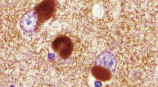
Figure E1 Cortical Lewy Bodies Present in Cerebral Cortex, as Opposed to Parkinson Disease Without Dementia, in Which Lewy Bodies are Found in the Substantia Nigra

Immunostain for alpha synuclein is characteristic for Lewy body immunohistologic profile.

From MacDonald AB: Spirochetal cyst forms in neurodegenerative disorders, hiding in plain sight, Med Hypotheses 67[4]:819-832, 2006.
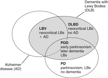
Figure E2 Relationships Among Alzheimer Disease (AD), the Three Subtypes of Dementia with Lewy Bodies (DLB), and Parkinson Disease (PD)

Parkinsonism refers to the clinical symptoms of PD (hypokinesia, tremor, and muscular rigidity). DLBD, Diffuse Lewy body disease; LBs, Lewy bodies; LBV, Lewy body variant of Alzheimer disease; PD, Parkinson disease; PDD, Parkinson disease dementia.

From Lewis KA et al: Abnormal neurites containing C-terminally truncated α-synuclein are present in Alzheimer’s disease without conventional Lewy body pathology, Am J Pathol 177[6]:3037-3050, 2010.

Diagnosis ⬆ ⬇

Differential Diagnosis

  • Diagnosis of DLB when dementia occurs before or concurrently with extrapyramidal features-arbitrarily set as the “1-yr rule” vs. PD with dementia, which occurs in the setting of well-established PD.
  • Dementia: AD, vascular dementia, frontotemporal dementia. However, some patients have pathologic overlap of AD and DLB or DLB and vascular dementia.
  • Parkinsonian features: Parkinson disease dementia, progressive supranuclear palsy (PSP), corticobasal syndrome (CBS), and multisystem atrophy (MSA).
  • Rapidly progressive form: Creutzfeldt-Jakob disease (CJD). Lack of cerebellar signs and lack of typical CJD MRI may help distinguish DLB from classic CJD (but not variant form of CJD).
  • Psychiatric features: Late-onset psychosis or depression with psychotic features.
  • Hallucinations with fluctuations in consciousness: Temporal lobe epilepsy (TLE) or delirium due to toxic/metabolic or autoimmune derangement.
Workup

  • MRI of the brain to evaluate for structural causes of dementia and exclude MRI features of vascular dementia or CJD.
  • Lumbar puncture to rule out underlying chronic infections, only if there are atypical features.
  • EEG if there is concern about TLE. However, both DLB and TLE may show nonspecific slowing or periodic complexes (see supportive biomarkers above).
  • DaTscan [Ioflupane (123I)] imaging is a SPECT scan preceded by injection of a radiopharmaceutical that detects the loss of functional dopaminergic neuron terminals in the striatum, reflecting underlying PD or DLB.
Laboratory Tests

Rule out other potential reversible causes for dementia, including CBC, complete metabolic panel, thyroid-stimulating hormone, and B12. Consider RPR and HIV testing as well.

Imaging Studies

  • MRI typically shows a relative preservation of the hippocampi and medial temporal lobe volumes (in contrast to AD) but generalized atrophy and white matter changes may be present.
  • Generalized low uptake on SPECT/PET perfusion/metabolism scan with reduced occipital activity ± the cingulate island sign on FDG-PET imaging.

Treatment ⬆ ⬇

Patient and caregiver education about benefits, side effects, and limitations of treatment is very important. Caregivers may be encouraged to avoid neuroleptics unless the psychotic features either trouble or endanger the patient. If neuroleptics must be used, typical neuroleptics must be avoided.

Nonpharmacologic Therapy

  • Social interaction and environmental novelty may improve cognitive dysfunction and psychiatric features often exacerbated by low levels of arousal and attention
  • Behavioral methods, such as avoiding previously exposed environmental triggers known to cause anxiety, agitation, or aggression
  • Physical therapy, mobility aids, and daily exercise
Acute General Rx

Atypical neuroleptic for disabling, persistent, bothersome (to the patient) psychotic features despite initiation of a cholinesterase inhibitor. A very low dose of an atypical antipsychotic (quetiapine 12.5 mg/day) may be started after patient/caregiver education regarding the sensitivity to neuroleptics. However, all neuroleptics do carry a boxed warning from the FDA about the increased mortality risk associated with neuroleptic use in patients with dementia. Patients and/or caregivers should be informed of this risk and allowed to balance the risk against the perceived benefit.

Chronic Rx

  • Cholinesterase inhibitors for cognitive and behavioral symptoms. Rivastigmine (6 to 12 mg/day PO or 9.5 mg/day by transdermal patch) has shown in a randomized controlled trial (RCT) to significantly reduce anxiety, delusions, and hallucinations, as well as significantly improved performance on neuropsychologic testing.
  • PD medications for disabling parkinsonian features. Carbidopa/levodopa is reported to be more effective with fewer side effects than dopamine agonists, which have higher rates of psychotic symptoms. Begin at a low dose-25/100 mg three times a day-and slowly titrate over several weeks as tolerated and according to response.
  • Selective serotonin reuptake inhibitors are commonly used for depression.
  • If REM sleep disorder remains disabling (or patient has not responded to an atypical antipsychotic initiated for psychosis), a trial of low-dose clonazepam (0.25 to 0.5 mg) or melatonin (3 mg) at bedtime remains an option.
  • Orthostatic hypotension may be aided by nonpharmacologic therapy such as supportive stockings or pharmacologically by midodrine, fludrocortisone, or droxidopa.
  • Memantine demonstrated an improvement in clinical global measure and remains well tolerated but may worsen hallucinations or delusions.
  • Avoid anticholinergics (including tricyclic antidepressants), benzodiazepines, and typical neuroleptics, as they can trigger delirium and worsen symptoms.
Disposition

  • Survival resembles the progression of AD, but a minority of cases may have a rapid disease course.
  • Progression in cognitive decline, similar to AD, by approximately 10%/yr on cognitive testing.
Referral

DLB requires a multidisciplinary approach including the general practitioner, neurologist, neuropsychologist, and/or neuropsychiatrist.

Pearls & Considerations ⬆ ⬇

Comments

  • Clinical presentation helps differentiate DLB from AD. AD presents with early signs of anterograde episodic memory loss without the benefit of cues on neuropsychologic testing because of cortical atrophy at the medial temporal lobe region.
  • Vascular dementia may also present with evidence of frontal-subcortical features but typically without the core features listed in the criteria above.
  • Bed partners may report that individuals with DLB “act out their dreams,” sometimes violently, leading to sleeping in separate beds. This history may indicate REM sleep behavior disorder. A history of REM sleep behavior may precede the diagnosis by many years. However, REM sleep behavior does not necessarily lead to DLB, but can be premonitory of any alpha synucleinopathy, including PD and multiple system atrophy.
Patient & Family Education

  • Visual hallucinations (VHs) typically consist of innocuous, well-formed, detailed images of animate figures. These are classically labeled Lilliputian, because the hallucinatory images are often relatively small. Unless VH lead to a potential threat to self or others, avoid antipsychotics due to the sensitivity of neuroleptics. Family/friends are often more alarmed by the VH than the patient with DLB.
  • Apathy is a common clinical feature of DLB and mimics changes in mood, including depression or excessive daytime somnolence. These features are often noticed by family/friends.

Suggested Readings ⬆ ⬇

  1. Armstrong M. : Lewy body dementiaContinuum. ;25(1):128-146, 2019.doi:10.1212/CON.0000000000000685
  2. Morra L.F. : Clinical presentation and differential diagnosis of dementia with Lewy bodies: a reviewInt J Geriatr Psychiatry. ;29(6):569-576, 2014.
  3. Walker Z. : Lewy body dementiasLancet. ;386:1683-1697, 2015.
  4. Watt J.A. : Comparative efficacy of interventions for aggressive and agitated behaviors in dementia: a systematic review and network meta-analysisAnn Intern Med. ;171:633-643, 2019.

Related Content ⬆

  1. McKeith I.G. : Diagnosis and management of dementia with Lewy bodies: fourth consensus report of the DLB ConsortiumNeurology. ;89(1):88-100, 2017.http://doi.10.1212/WNL.0000000000004058