section name header

Basic Information

AUTHOR: Kelly Ruhstaller, MD

Definition

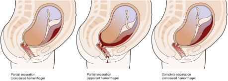
Abruptio placentae is the separation of placenta from the uterine wall before delivery of the fetus. The condition occurs in approximately 1% of pregnancies. There are three classes of abruption (Fig. 1) based on maternal and fetal status, including an assessment of uterine contractions, quantity of bleeding, fetal heart rate monitoring, and abnormal coagulation studies (fibrinogen, prothrombin time, partial thromboplastin time).

  • Grade I: Mild vaginal bleeding, uterine irritability, stable vital signs, reassuring fetal heart rate, normal coagulation profile (fibrinogen 450 mg/dl). Approximately half of abruptions are grade I.
  • Grade II: Moderate vaginal bleeding, hypertonic uterine contractions, orthostatic blood pressure measurements, unfavorable fetal status, fibrinogen 150 to 250 mg. Approximately a quarter of abruptions are grade II.
  • Grade III: Severe bleeding (may be concealed), hypertonic uterine contractions, overt signs of hypovolemic shock, fetal death, thrombocytopenia, fibrinogen <150 mg/dl. Approximately a quarter of abruptions are grade III.

Figure 1 Classification of placental abruption.

From Magowan BA: Clinical obstetrics & gynecology, ed 4, 2019, Elsevier.

Synonym

Premature separation of placenta

Placental abruption

ICD-10CM CODES
O45.8X1Other premature separation of placenta, first trimester
O45.8X2Other premature separation of placenta, second trimester
O45.8X3Other premature separation of placenta, third trimester
O45.8X9Other premature separation of placenta, unspecified trimester
O45.91Premature separation of placenta, unspecified, first trimester
O45.92Premature separation of placenta, unspecified, second trimester
O45.93Premature separation of placenta, unspecified, third trimester
Epidemiology & Demographics
Incidence (In U.S.)

9.6/1000 births; 80% occur before the onset of labor.

Prevalence

5% to 17%, some studies showing a 5- to 10-fold increase in risk; with two prior episodes, 25%.

Risk Factors

Hypertension (greatest association), trauma, polyhydramnios, multifetal gestation, smoking, use of cocaine, chorioamnionitis, preterm premature rupture of membranes. Table 1 summarizes placental abruption risk factors.

TABLE 1 Placental Abruption Risk Factors

  • Increasing parity or maternal age
  • Cigarette smoking
  • Cocaine abuse
  • Trauma
  • Maternal hypertension
  • Preterm premature rupture of membranes
  • Rapid uterine decompression associated with multiple gestation and polyhydramnios
  • Inherited or acquired thrombophilia
  • Uterine malformations or fibroids
  • Placental abnormalities or ischemia
  • Prior abruption

From Gabbe SG: Obstetrics, ed 6, Philadelphia, 2012, Saunders.

Physical Findings & Clinical Presentation

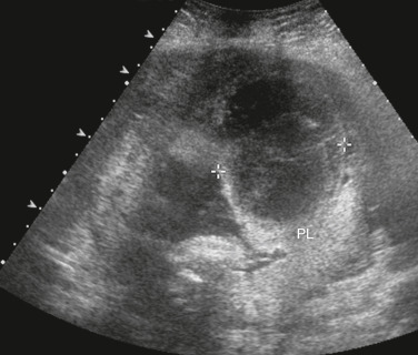
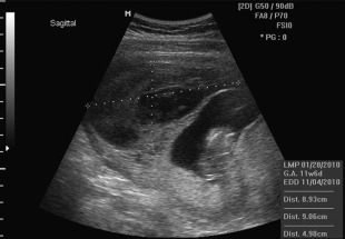
  • Triad of uterine bleeding (concealed or per vagina), hypertonic uterine contractions or signs of preterm labor, and evidence of fetal compromise exists.
  • More than 80% of cases have external bleeding; 20% of cases have no bleeding but have indirect evidence of abruption, such as failed tocolysis for preterm labor.
  • Tetanic uterine contractions are found in only 17%.
Etiology

  • Primary etiology: Unknown
  • Hypertension: Found in 40% to 50% of grade III abruptions
  • Rapid decompression of uterine cavity, as can occur in polyhydramnios or multifetal gestation
  • Blunt external trauma (motor vehicle accident, spousal abuse)

Diagnosis

Differential Diagnosis

  • Placenta previa
  • Cervical or vaginal trauma
  • Labor
  • Cervical cancer
  • Rupture of membranes
  • The differential diagnosis of vaginal bleeding in pregnancy is described in Section III
Workup

  • Placental abruption is primarily a clinical diagnosis that is supported by laboratory, radiographic (Fig. 2), and pathologic studies.
  • Initial assessment should evaluate for the source of bleeding, ruling out placenta previa that may contraindicate any type of vaginal examination (e.g., pelvic speculum examination).
  • Continuous fetal heart monitoring is indicated for all viable gestations (60% incidence of fetal distress in labor); may show early signs of maternal hypovolemia (late decelerations or fetal tachycardia) before overt maternal vital sign changes.
  • Actual amount of blood loss is often greater than initially perceived because of the possibility of concealed retroplacental bleeding and apparent “normal” vital signs. The relative hypervolemia of pregnancy initially protects the patient until late in the course of bleeding, when abrupt and sudden cardiovascular collapse can occur.

Figure 2 Placental abruption.

Transabdominal sonogram of the placenta (PL) with a hematoma (calipers) lifting the placenta away from the uterine wall.

From Rumack CM et al [eds]: Diagnostic ultrasound, ed 4, Philadelphia, 2011, Mosby.

Laboratory Tests

  • Baseline serum hemoglobin helps quantify blood loss and establish baseline values for serial comparisons during expectant management.
  • Coagulation profile: Platelets, fibrinogen, prothrombin, and partial thromboplastin time. Diffuse intravascular coagulation can develop with severe abruption. If fibrinogen is <150 mg/dl, estimated blood loss is approximately 2000 ml; if fibrinogen is <100 mg/dl, consider fresh frozen plasma to prevent further bleeding.
  • Type and antibody screen is important to identify Rh-negative patients who need Rh immune globulin.
Imaging Studies

Ultrasound should include fetal presentation and status, amniotic fluid volume, placental location, as well as any evidence of hematoma (retroplacental, subchorionic, or preplacental) (Fig. 3).

Figure 3 Ultrasonic image of a subchorionic abruption.

Courtesy K. Francois; from Gabbe SG: Obstetrics, ed 6, Philadelphia, 2012, Saunders.

Treatment

Acute General Rx

  • Stabilization of the mother is the first priority.
  • Treatment depends on gestational age of the fetus, severity of the abruption, and maternal status.
  • Initial assessment for signs of maternal hemodynamic compromise or hemorrhagic shock; large-bore intravenous access, with crystalloid fluid resuscitation using a replacement of 3 ml lactated Ringer solution for every 1 ml estimated blood loss.
  • Indwelling Foley catheter to monitor urine output and maternal volume status, with a goal of 30 ml/h urine output.
  • Assess fetal status and gestational age by sonogram and continuous fetal heart rate monitoring.
  • Because of the unpredictable nature of abruptions, cross-matched blood should be made available during the initial resuscitation period.
Chronic Rx

  • In the term fetus, delivery is indicated.
  • In the preterm fetus, consider betamethasone 12.5 mg IM q24h for two doses and then delivery, depending on the severity of the abruption and the likelihood of fetal complications from preterm birth.
  • Cesarean delivery should be reserved for cases of fetal distress or for standard obstetric indications. While cesarean delivery may be needed to stabilize the fetal and/or maternal status, the mother’s coagulation status may complicate the procedure and availability of blood products may be critical.
  • In cases of maternal stability and fetal prematurity, expectant management can occur in the setting of close follow-up, including regular evaluation of fetal growth and reassuring antenatal testing.
Disposition

Because of the unpredictable nature of abruptions, expectant management should occur only under controlled circumstances.

Referral

Abruptio placentae places mother and fetus in a high-risk situation and should be managed by a qualified obstetrician in a facility with capability for neonatal and maternal resuscitation, for supporting a preterm infant if delivery is indicated at an early gestational age, and for performing emergency cesarean deliveries.

Related Content

Abruptio Placentae (Patient Information)

Premature Labor (Related Key Topic)

Vaginal Bleeding during Pregnancy (Related Key Topic)