section name header

Basic Information

AUTHOR: Corey Elam Goldsmith, MD, FAAN

Definition

  • Optic atrophy refers to the degeneration of retinal ganglion axons of the optic nerve and a resulting picture of a pale optic nerve on fundoscopy.
  • It is a sign indicative of chronic optic nerve insult rather than a distinct disease entity.
Synonym

Unilateral/bilateral optic atrophy

ICD-10CM CODE
H47.2Optic atrophy
Epidemiology & Demographics
Predominant Sex

Depends on etiology

Predominant Age

Variable

Peak Incidence

Varies depending on cause

Physical Findings & Clinical Presentation

  • Optic atrophy is identified weeks to months after an initial insult such as optic neuritis, trauma, or ischemia. May occur insidiously with no acute inciting event in more genetic or toxic causes. Frequently, it is noted by the physician during a routine eye exam.
  • Asymmetry of disc color is often first subtle finding.
  • Temporal part of optic disc is pale initially; later, the entire disc becomes pale/white.
  • Optic disc pallor (Fig. E1) occurs 4 to 6 wk after optic nerve insult.
  • Unilateral lesion produces a relative afferent pupillary defect (RAPD): When swinging flashlight eye to eye, the abnormal pupil dilates to direct light.
  • Decreased visual acuity, blurred vision, visual field deficits (e.g., central scotoma), abnormal color vision (e.g., red desaturation).

Figure E1 Optic atrophy.

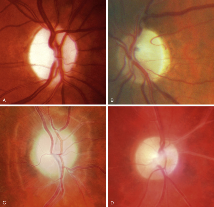
(A) Primary due to compression. (B) Primary due to nutritional neuropathy-note predominantly temporal pallor. (C) Secondary due to chronic papilledema-note prominent Paton lines. (D) Consecutive due to vasculitis.

Courtesy P. Gili, Fig. C. From Bowling B: Kanski’s clinical ophthalmology: a systemic approach, ed 8, Philadelphia, 2016, Elsevier.

Etiology

  • Inflammatory optic neuritis: MS, sarcoidosis, neuromyelitis spectrum disorder, infections (syphilis, CMV, HIV, Lyme disease, cat-scratch disease)
  • Vascular: Arteritic and nonarteritic ischemic optic neuropathy, central retinal artery occlusion, temporal arteritis, diabetes
  • Compression: Pituitary, orbital, or other tumors, idiopathic intracranial hypertension, meningioma, thyroid eye disease
  • Hereditary: Leber hereditary optic neuropathy, autosomal dominant optic atrophy, leukodystrophies, other mitochondrial diseases.
  • Nutritional, toxic, and metabolic: Amiodarone, isoniazid, tacrolimus, ethambutol, linezolid, vigabatrin, B12 deficiency, folate deficiency, copper deficiency, methanol, tobacco, alcohol1
  • Trauma
  • Radiation
  • Glaucoma (end-stage disease)

Diagnosis

Differential Diagnosis

Optic disc anomalies such as myelinated nerve fibers, “blonde” fundus

Workup

  • A detailed history to identify onset and course, exposure to risk factors such as toxic/nutritional factors, detailed family history, and associated findings such as headache, eye pain, proptosis, or neurologic deficits.
  • Ophthalmic examination to document deficits of visual acuity, color vision, contrast sensitivity, and visual fields must be performed. Relative afferent pupillary defect (RAPD) on swinging flashlight examination is sine qua non for unilateral optic nerve disease and must be documented. Absence indicates retinal disease or bilateral lesions.
  • General examination and neurologic examination can provide clues to an underlying cause.
Laboratory Tests

  • Serum B12, RPR, HIV
  • Autoimmune diseases: ESR, ANA, ACE, aquaporin-4 antibodies (NMO IgG)
  • Lumbar puncture if unknown cause or other associated neurologic deficits
Imaging Studies

  • Visual field testing can help characterize and localize the etiology.
  • Optical coherence tomography (OCT) to assess the thickness of the peripapillary retinal nerve fiber layer to establish and monitor.
  • MRI of the brain and orbits with contrast, fat suppression, and special (thin) cuts through orbits is necessary to identify compressive lesions in all patients with unexplained optic atrophy; especially important in patients with positive predictive factors for abnormal imaging (e.g., young age, progression, bilateral findings).2
  • If sarcoid is suspected, order chest X-ray and/or chest CT.

Treatment

Acute General Rx

Treat the underlying cause-discontinue identifiable toxins, use B12 replacement, neurosurgical intervention is necessary if tumor is found; consider IV steroids if there is evidence for active demyelinating disease.

Chronic Rx

The optic nerve does not regenerate, although symptoms often improve, especially if the provoking cause is removed.

Disposition

  • Visual loss usually occurs over weeks to months.
Referral

Ophthalmologist

Neurologist depending on the etiology

Pearls & Considerations

Comments

  • An experienced clinician should be able to identify pale optic discs and an RAPD.
  • Pupillary dilation with mydriatic agents (e.g., tropicamide) may be necessary to optimize funduscopic examination.
  • Patient education material can be obtained from the National Eye Institute, Department of Health and Human Services, 9000 Rockville Pike, Bethesda, MD 20814.

Related Content

  1. Grzybowski A. : Toxic optic neuropathies: an updated reviewActa Ophthalmol. ;93(5):402-410, 2015.
  2. Lee A.G. : The diagnostic yield of the evaluation for isolated unexplained optic atrophyOphthalmology. ;112(5):757-759, 2005.