section name header

Basic Information ⬇

AUTHOR: Khawja A. Siddiqui, MD

Definition

Subarachnoid hemorrhage (SAH) is defined as hemorrhage into the subarachnoid space surrounding the brain. This can be either nontraumatic or traumatic in nature. Cerebral aneurysms are the most common cause of nontraumatic SAH (up to 85%) and have the most devastating consequences. Other causes include venous bleed, which have less severe sequelae. Here we will focus on aneurysmal nontraumatic subarachnoid hemorrhage.

Synonyms

Subarachnoid bleed

SAH

ICD-10CM CODES
I60Subarachnoid hemorrhage
I60.1Subarachnoid hemorrhage from middle cerebral artery
I60.2Subarachnoid hemorrhage from anterior communicating artery
I60.3Subarachnoid hemorrhage from posterior communicating artery
I60.4Subarachnoid hemorrhage from basilar artery
I60.5Subarachnoid hemorrhage from vertebral artery
I60.7Subarachnoid hemorrhage from intracranial artery, unspecified
Epidemiology & Demographics
Incidence

Aneurysmal SAH: 6.67/100,000 persons worldwide and varies from 0.71 to 12.38 per 100,000 persons depending on geography. Annual incidence estimated at 500,000 cases worldwide.1

Predominant Sex & Age

Women in the fifth and sixth decades of life.2

Peak Incidence

Most aneurysmal SAH occurs in people who are between the ages of 55 and 60 yr.

Risk Factors

Although genetics seem to play a factor in aneurysm formation, lifestyle factors are more important for determining overall risk of rupture. These risk factors include smoking, hypertension, oral contraception, pregnancy, and sympathomimetic use.

Genetics

  • Several genes and medical conditions such as collagen vascular disease and autosomal dominant polycystic kidney disease have been associated with aneurysm formation, but their association with SAH remains controversial.3,4
  • Current guidelines recommend screening for aneurysms only if 2 or more first-degree relatives have a history of SAH or cerebral aneurysms.
  • Genetic screening is not recommended for patients with SAH.4
Physical Findings & Clinical Presentation

  • The primary symptom is a sudden, severe headache in >90% of cases. About 50% have a headache that is classically described as the “worst headache of my life” and reaches maximal intensity within 1 min-the so-called thunderclap headache. This headache may be associated with nausea/vomiting, neck pain, seizure, or complete loss of consciousness.
  • Most patients report a history of headaches during the weeks preceding the actual hemorrhage event. These are most likely sentinel bleeds that represent microhemorrhages.
  • A posterior communicating artery aneurysm may present as oculomotor (cranial nerve III) palsy, typically involving the pupillary fibers, even in a nonruptured setting.
  • The World Federation of Neurosurgeons (WFNS) score and the Hunt and Hess score are clinical scores that correlate with mortality (higher numbers indicating higher mortality) (Table 1).

Table 1 Most Commonly Used Clinical Grading Scales for Subarachnoid Hemorrhage

Hunt and Hess Scale
  • Grade 0: Asymptomatic
  • Grade 1: Mild headache and mild nuchal rigidity, no neurologic deficit
  • Grade 2: Moderate to severe headache but no neurologic deficit other than cranial nerve palsy
  • Grade 3: Drowsy, confused, or mild focal deficit
  • Grade 4: Stupor, moderate to severe hemiparesis, and early decerebrate posturing
  • Grade 5: Deep comatose, decerebrate posturing
World Federation of Neurological Surgeons Scale
Glasgow Coma ScaleMotor Deficit
Grade 015Absent
Grade 115Absent
Grade 213-14Absent
Grade 313-14Present
Grade 47-12Present or absent
Grade 53-6Present or absent

From Jankovic J et al: Bradley and Daroff’s neurology in clinical practice, ed 8, Philadelphia, 2022, Elsevier.

Etiology

  • Trauma.
  • About 85% of nontraumatic SAHs are caused by a ruptured berry aneurysm, whereas 10% do not reveal a bleeding source despite modern imaging techniques. About 5% are due to other causes, which include arteriovenous malformations (AVMs), tumors, vasculitis, reversible cerebrovascular vasoconstriction syndrome, cerebral sinus venous thrombosis, and coagulopathies.
  • Table 2 summarizes nonaneurysmal causes of subarachnoid hemorrhage.

Table 2 Nonaneurysmal Causes of Subarachnoid Hemorrhage

Trauma
Idiopathic perimesencephalic subarachnoid hemorrhage
Arteriovenous malformation
Intracranial arterial dissection
Cocaine and amphetamine use
Mycotic aneurysm
Pituitary apoplexy
Moyamoya disease
Central nervous system vasculitis
Sickle cell disease
Coagulation disorders
Primary or metastatic neoplasm

Causes are listed in approximate order of frequency.

From Goldman L, Shafer AI: Goldman-Cecil medicine, ed 26, 2019, Elsevier.

Diagnosis ⬆ ⬇

Differential Diagnosis

  • Other headache syndromes: Thunderclap headaches due to reversible cerebral vasoconstriction syndrome (often with recurrent thunderclap headaches), migraine headache, sexual headache, cough headache, exertional headache, secondary causes including, but not limited to, pituitary apoplexy or acute hydrocephalus.
Workup
Imaging Studies

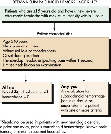
  • The Ottawa SAH Rule can assist in selecting patients who require further neuroimaging and workup (Fig. 1).
  • Computed tomography (CT) of the brain without contrast (Fig. 2) shows hemorrhage in around 95% of cases, especially during the acute phase (i.e., 24 to 48 hr) after the onset of bleeding. Box 1 describes a CT scan classification of subarachnoid hemorrhage. About 3% to 5% of SAH may be missed on initial CT of the head. MRI brain, specifically FLAIR sequence, is helpful in detecting subarachnoid blood if clinically suspected.
  • Lumbar puncture should be performed in all cases of suspected SAH with “normal CT of the head,” especially when clinical suspicion is high. The following suggest SAH:
    1. An RBC count of more than 100,000/m3 in tubes 1 AND 4. This is to differentiate from a traumatic tap in which there will be a drop in RBC count from tube 1 to 4.
    2. Presence of xanthochromia or bilirubin in the cerebrospinal fluid.
    3. SAH can also be excluded by the following two criteria: CSF RBC count <2000 × 106/L and no xanthochromia.
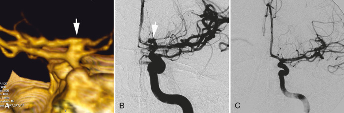
  • CT angiogram of the brain (Fig. E3 and Fig. E4).
  • Digital subtraction angiography with 3D processing when indicated is the gold standard for diagnosis of etiology in subarachnoid hemorrhage.
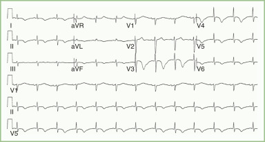
  • ECG may reveal diffuse repolarization abnormalities in precordial leads (Fig. E5).5
Figure 2 Subarachnoid Hemorrhage (SAH), Noncontrast Computed Tomography (CT), Brain Windows

Acute SAH appears white on noncontrast CT brain windows. A through C, Nonconsecutive axial slices, progressing from caudad to cephalad. In this case of diffuse SAH, note the presence of subarachnoid blood filling the sulci, as well as extending into the cisterns, Sylvian fissures, and even lateral ventricles. In A, blood (white) fills the suprasellar cistern. This star-shaped structure is normally filled with cerebrospinal fluid (CSF) (black). The quadrigeminal plate cistern is normally a smile-shaped black crescent, filled with CSF, but in this case it is filled with blood. Extremely bright calcifications in the choroid plexus of the posterior horns of the lateral ventricles are common, normal findings-do not mistake these for hemorrhage. Note their similarity in density to bone of the calvarium.

From Broder JS: Diagnostic imaging for the emergency physician, Philadelphia, 2011, Saunders.

BOX 1 Computed Tomography Scan Classification of Subarachnoid Hemorrhage (Fisher Scale)

  • Group 1: No blood detected
  • Group 2: Diffuse deposition or thin layer of blood, with all vertical layers of blood (interhemispheric fissure, insular cistern, ambient cistern) <1 mm thick
  • Group 3: Localized clots or vertical layers of blood 1 mm or greater in thickness
  • Group 4: Diffuse or no subarachnoid hemorrhage but with intraparenchymal or intraventricular clots

From Jankovic J et al: Bradley and Daroff’s neurology in clinical practice, ed 8, Philadelphia, 2022, Elsevier.

Figure 1 The Ottawa Subarachnoid Hemorrhage Rule.

!!flowchart!!

From Goldman L, Shafer AI: Goldman-Cecil medicine, ed 26, 2019, Elsevier.
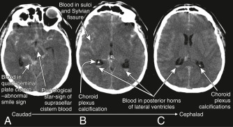
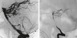
Figure E3 Baseline Angiogram Obtained Shortly after Subarachnoid Hemorrhage (Left) and Repeat Angiogram Obtained 7 Days Later (Right) Showing Severe Vasospasm of Basilar Artery, with Reduced Distal Flow

From Vincent JL et al: Textbook of critical care, ed 7, Philadelphia, 2017, Elsevier.

Figure E4 Patient with Hunt and Hess Grade 1 Subarachnoid Hemorrhage

A, Computed Tomographic Angiography, Posterior-Anterior View, Shows a Small-Domed, Broad-Necked Aneurysm at the Left Internal Carotid Artery Bifurcation (Arrow). Aneurysm Shape was Obscured by Venous Structures, and Relationship of Aneurysm Dome and Anterior Cerebral Artery was Unclear, So Digital Subtraction Catheter Angiography was Performed. B, Arterial Phase of Catheter Angiography, Anteroposterior View, Shows a Boot-Shaped, Very Small-Necked Aneurysm with a Clear Separation of the Left Anterior Cerebral Artery from the Aneurysm Dome (Arrow). C, Owing to the Small Neck Morphology Seen on the Catheter Angiogram, Endovascular Embolization Could Be Performed, and the Aneurysm was Completely Obliterated.

From Jankovic J et al: Bradley and Daroff’s neurology in clinical practice, ed 8, Philadelphia, 2022, Elsevier.

Figure E5 Electrocardiographic Changes in a Patient with an Acute Aneurysmal Subarachnoid Hemorrhage

Notice diffuse repolarization abnormalities in the precordial leads.

From Jankovic J et al: Bradley and Daroff’s neurology in clinical practice, ed 8, Philadelphia, 2022, Elsevier.

Laboratory Tests

  • Basic laboratory values, including CBC, chemistry panel, prothrombin time, partial thromboplastin time, and platelet count.
  • Serum troponin to evaluate for severe cardiac stress; elevated troponins indicate cardiac damage secondary to a catecholamine surge and can be associated with poor outcomes.
  • Patients with SAH are prone to developing cerebral salt wasting, resulting in hyponatremia. Sodium levels should be monitored frequently.

Treatment ⬆ ⬇

Nonpharmacologic Therapy

  • Airway, breathing, and circulation
  • Once stabilized, good neurologic exam
  • Cerebrospinal fluid (CSF) drainage may be required for patients who develop hydrocephalus and increased intracranial pressure. It is also recommended for patients with Hunt and Hess grade 3 or higher
Acute General Rx

  • Critical care management: Initial management strategies are geared toward stabilizing the patient and preventing re-hemorrhage and hydrocephalus. Re-hemorrhage occurs within 72 hr in up to 23% and is associated with very high mortality rates.
  • Blood pressure control: Tight blood pressure control is paramount before securing the aneurysm to protect against re-rupture. Blood pressure control can be achieved with the use of antihypertensive infusions such as intravenous nicardipine. A systolic blood pressure of less than 140 mm Hg is recommended. Placement of arterial line is recommended. After securing of the aneurysm, liberalization of blood pressure parameters is the standard.
  • Intracranial pressure control: Raised intracranial pressure occurs in more than 50% of patients with subarachnoid hemorrhage secondary to hydrocephalus, cerebral edema, cerebral infarction or other causes. Insertion of a ventricular catheter to treat acute hydrocephalus and maintain intracranial pressure <20 mm Hg can be lifesaving. Patients who are unable to be weaned may require permanent CSF diversion.
  • In cases of aneurysmal SAH, treatment focuses on occlusion/exclusion of the aneurysm to prevent rebleeding. Most aneurysms currently are treated endovascularly. The most common treatment methods are:
    1. Microsurgical clipping: Performed through a craniotomy by placing a clip around the neck of the aneurysm
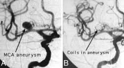
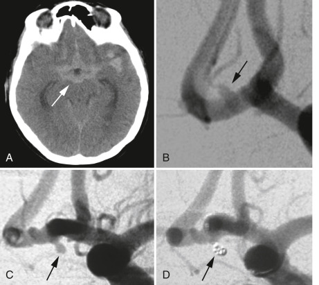
    2. Endovascular coiling (Fig. E6): Performed via digital subtraction angiography; it consists of deploying platinum coils inside the aneurysm (Fig. E7) or stents in the parent artery to cause thrombosis of the aneurysmal sac.
    3. Flow diverters and the Woven EndoBridge (WEB) device are also treatment options.
  • Vasospasm: Cerebral vasospasm is a morbid complication leading to cerebral ischemia, disability, and death after SAH. It typically develops between day 4 and 14 (but may occur up to day 21) after the hemorrhage, and it reaches a peak on day 6 to 8. Treatment strategies include:
    1. Induced hypertension with typical mean arterial pressure goals of 90 to 100 (after aneurysm is secured) and with euvolemia instead of hypervolemia, as the latter was found to lead to significant cardiopulmonary and hemodynamic complications. “Triple H” therapy-Hypertension, Hypervolemia, and Hemodilution-was originally employed to maintain cerebral perfusion, but it has fallen out of favor due to its many complications.
    2. Nimodipine (60 mg q4h or 30 mg q2h if blood pressure is low) has been shown to improve outcomes if it is administered between days 4 and 21 after the hemorrhage. Nimodipine has not been shown to reduce the incidence of vasospasm but does have a mortality benefit, likely acting as a neuroprotective agent.
    3. Intraarterial therapies such as intraarterial calcium channel blockers and balloon angioplasty may be employed as needed for symptomatic vasospasm.
  • Seizures occur in about 3% of patients during the acute phase. Use of prophylactic antiepileptics is controversial and not recommended, but patients presenting with seizures should be treated appropriately with anticonvulsants.
  • Pain control: Use short-acting and less-sedating medications (e.g., codeine, low-dose morphine).
  • Monitor and treat for cerebral salt wasting and any other electrolyte abnormalities or anemia.
  • Maintain normothermia.
  • Avoid hyperglycemia or hypoglycemia.
  • Management of SAH is complex and requires a multidisciplinary approach that includes expertise from neurology, critical care, neurosurgery, neuro-interventionalists, cardiologists, and endocrinologists, to name a few. A brief summary of the management is outlined above, but a detailed discussion is outside the scope of this text. The authors recommend supplementing this material with society guidelines such as those laid out by the Neurocritical Care Society.6

Figure E6 Angiogram Demonstrating Middle Cerebral Artery Aneurysm Before (A) and after Placement of Detachable Coils to Thrombose the Aneurysm (B)

From Vincent JL et al: Textbook of critical care, ed 7, Philadelphia, 2017, Elsevier.

Figure E7 Patient with Hunt and Hess Grade I.

A, Computed tomography (CT) of brain showed aneurysmal subarachnoid hemorrhage (aSAH) (white arrow), Fisher grade 4. B, At the time of admission, digital subtraction angiography showed no clear evidence of an aneurysm in the anterior communicating artery (Acomm) region. There is an infundibulum (arrow) of an Acomm perforator pointing superiorly. C, Digital subtraction angiography at day 7 shows a small 2 mm aneurysm (arrow) pointing inferiorly. D, After treatment, the aneurysm was completely obliterated using 1 platinum coil (arrow).

From Jankovic J et al: Bradley and Daroff’s neurology in clinical practice, ed 8, Philadelphia, 2022, Elsevier.

Chronic Rx

  • Management of reversible risk factors mentioned earlier (smoking, hypertension, drug use)
  • Management of neurologic disability through physical therapy and rehabilitation
Disposition

  • Outcomes after SAH have been improving over the years, with a 17% to 50% decrease in case fatality. The prehospital and 30-day mortality rates are still reported at around 15% to 33%.
  • Almost half of those who survive hospitalization have cognitive impairments or disability that affect their lifestyles.7
Referral

All patients should be managed at high-volume SAH centers, which are defined as having a case volume of more than 35 SAHs a year. Patients who present to alternative facilities should be transferred to a high-volume SAH center as soon as possible.

Pearls & Considerations ⬆ ⬇

Comments

  • “Thunderclap” headaches should be considered SAH until proven otherwise and evaluated by CT of the head with/without LP. MRI FLAIR sequence is also a helpful modality.
  • All SAH should be managed in a critical care setting (preferably neurocritical care unit) with neurosurgical care available.
  • Measures to prevent rebleeding include adequate control of blood pressure and aneurysm treatment with the use of coiling or clipping.
Prevention

Controlling some of the modifiable risk factors, especially smoking and blood pressure, may help to decrease the risk of aneurysmal rupture.

Patient & Family Education

  • SAH is a devastating condition, with most survivors developing significant neurologic or cognitive deficits. A good support system and an adequate physical and cognitive rehabilitation program may prove useful to survivors.
  • Screening may be useful for patients with two or more first-degree relatives with SAH.
Related Content

Subarachnoid Hemorrhage (Patient Information)

Related Content ⬆

    1. Hughes J.D. : Estimating the global incidence of aneurysmal subarachnoid hemorrhage: a systematic review for central nervous system vascular lesions and meta-analysis of ruptured aneurysmsWorld Neurosurg. ;115:430-447.e7, 2018.doi:10.1016/j.wneu.2018.03.220
    2. Macdonald R.L., Schweizer T.A. : Spontaneous subarachnoid haemorrhageLancet. ;389(10069):655-666, 2017.doi:10.1016/S0140-6736(16)30668-7
    3. Korja M., Kaprio J. : Controversies in epidemiology of intracranial aneurysms and SAHNat Rev Neurol. ;12(1):50-55, 2016.doi:10.1038/nrneurol.2015.228
    4. Thompson B.G. : Guidelines for the management of patients with unruptured intracranial aneurysms: a guideline for healthcare professionals from the American Heart Association/American Stroke AssociationStroke. ;46(8):2368-2400, 2015.doi:10.1161/STR.0000000000000070
    5. de Oliveira Manoel A.L. : Aneurysmal subarachnoid haemorrhage from a neuroimaging perspectiveCrit Care. ;18(6), 2014.doi:10.1186/s13054-014-0557-2
    6. Diringer M.N. : Critical care management of patients following aneurysmal subarachnoid hemorrhage: recommendations from the Neurocritical Care Society’s Multidisciplinary Consensus ConferenceNeurocrit Care. ;15(2):211-240, 2011.doi:10.1007/s12028-011-9605-9
    7. Nieuwkamp D.J. : Changes in case fatality of aneurysmal subarachnoid haemorrhage over time, according to age, sex, and region: a meta-analysisLancet Neurol. ;8(7):635-642, 2009.doi:10.1016/S1474-4422(09)70126-7