section name header

Basic Information

AUTHOR: R. Scott Hoffman, MD

Definition

The term conjunctivitis refers to an inflammation of the conjunctiva resulting from a variety of causes, including allergies and bacterial, viral, and chlamydial infections.

Synonyms

Red eye

Pink eye

Acute conjunctivitis

Subacute conjunctivitis

Chronic conjunctivitis

Purulent conjunctivitis

Pseudomembranous conjunctivitis

Papillary conjunctivitis

Follicular conjunctivitis

Newborn conjunctivitis

ICD-10CM CODES
H10.9Unspecified conjunctivitis
B30Viral conjunctivitis
H10.0Mucopurulent conjunctivitis
H10.1Acute atopic conjunctivitis
H10.4Chronic conjunctivitis
Epidemiology & Demographics
Incidence (In U.S.)

1.6% to 12% in newborns.

Prevalence (In U.S.)

  • Allergic conjunctivitis (Fig. E1), the most common form of ocular allergy, is usually associated with allergic rhinitis and may be seasonal or perennial.
  • Bacterial or viral conjunctivitis is often seasonal and can be extremely contagious.

Figure E1 Allergic conjunctivitis.

Arrow indicates area of chemosis in the conjunctivitis.

From Adkinson NF Jr et al [eds]: Middleton’s allergy principles and practice, ed 7, vol 2, Philadelphia, 2008, Mosby.

Predominant Age

Occurs at any age. Most cases in adults are due to viral infection. Children are more prone to develop bacterial conjunctivitis than viral forms. Table E1 summarizes manifestations of acute conjunctivitis in children

TABLE E1 Manifestations of Acute Conjunctivitis in Children

FEATURECLINICAL CHARACTERISTICS
BACTERIALVIRAL
Common pathogensHaemophilus influenzae (usually nontypable)Adenoviruses types 8, 19
Streptococcus pneumoniaeEnteroviruses
Moraxella catarrhalisHerpes simplex virus
Incubation24-72 h1-14 days
SYMPTOMS
PhotophobiaMildModerate to severe
Blurred visionCommon with dischargeIf keratitis is present
Foreign body sensationUnusualYes
SIGNS
DischargePurulent dischargeMucoid/serous discharge
Palpebral reactionPapillary responseFollicular response
Preauricular lymph nodeUnusual for acute (<10%)More common (20%)
ChemosisModerateMild
Hemorrhagic conjunctivaeOccasionally with pneumococcus or Haemophilus sp.Frequent with enteroviruses
Treatment (topical)Polymyxin B-trimethoprim orsulfacetamide 5% or erythromycin or moxifloxacin or gatifloxacinAdenovirus: Self-limited
Herpes simplex virus: Trifluridine 1% solution or ganciclovir 0.15% gel or acyclovir; ophthalmologic consultation
End of contagious period24 h after start of effective treatment7 days after onset of symptoms

From Marcdante KJ et al: Nelson essentials of pediatrics, ed 9, Philadelphia 2023, Elsevier.

Peak Incidence

More common in the spring and fall, when viral infections and pollens increase.

Physical Findings & Clinical Presentation

  • Infection and chemosis of conjunctivae with discharge. Gluing of the eyelids and no itching is more indicative of a bacterial cause (Fig. E2).
  • Cornea is clear or can be involved (certain bacteria, such as Neisseria, can rapidly develop into corneal infection).
  • Vision is often normal but can be blurred. Mucus and watering can cause fluctuating vision.
  • Fig. E3 illustrates the difference between papillary and follicular conjunctivitis.

Figure E2 Bacterial conjunctivitis.

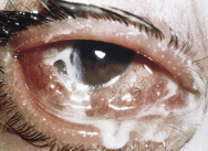
Purulent discharge and conjunctiva hyperemia suggest bacterial conjunctivitis. Viral conjunctivitis produces watery discharge, foreign body sensation, preauricular lymphadenopathy, and conjunctival follicles seen on slit lamp examination.

Reproduced with permission from the American Academy of Ophthalmology. From Goldman L, Schafer AI: Goldman’s Cecil medicine, ed 24, Philadelphia, 2012, Saunders.

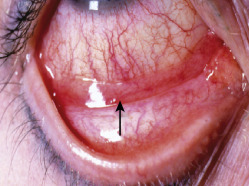
Figure E3 Conjunctivitis: Papillary versus follicular.

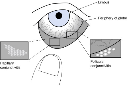
The erythema of conjunctivitis (shaded dark gray) is most intense on the inside surface of the eyelids (tarsal conjunctiva) and peripherally on the globe (near the fornices), whereas the erythema is less intense centrally near the limbus. In more severe conjunctivitis, the entire conjunctival surface (both tarsal and bulbar) is red. This pattern of erythema contrasts with iritis, which causes more intense erythema centrally around the limbus, a finding called circumlimbal flush or ciliary flush. In patients with conjunctivitis, the clinician should inspect the everted upper or lower lids, noting whether the inner membrane has its normal smooth surface or instead has small, uneven projections, which are characterized as either papillae or follicles. In this example, the clinician has used his thumb to gently evert the lower lid for inspection. Papillae (left bottom) are contiguous red vascular bumps; the center of each papilla contains a blood vessel. They are red on the surface and pale at the base. Papillae are often so tiny that the conjunctiva acquires a velvety appearance, and only magnification reveals their true nature. Other times, papillae may become large and produce a cobblestone appearance. Follicles (right bottom) are discrete 1- to 2-mm-diameter white bumps consisting of aggregates of lymphoid tissue; the center of each is avascular. They are pale on the surface and red at the base.

From McGee S: Evidence-based physical diagnosis, ed 4, Philadelphia, 2018, Elsevier.

Etiology

  • Bacterial: Haemophilus influenzae, Streptococcus pneumoniae, and Moraxella catarrhalis in children; Staphylococcus spp in adults. Gram-negative infections are more common in contact lens wearers. Gonococcal ophthalmia neonatorum is caused by Neisseria gonorrhoeae acquired by exposure of the neonatal conjunctivae to infected cervicovaginal secretions during delivery
  • Viral: Most common overall cause of infectious conjunctivitis
  • Chlamydial
  • Allergic
  • Traumatic (chemical or toxin exposure)
  • Chronic eyelid inflammation (blepharitis)

Diagnosis

Differential Diagnosis

  • Acute glaucoma (fixed pupil with headache may indicate acute angle closure)
  • Corneal lesions
  • Acute iritis (with pain and photophobia, blurred vision)
  • Episcleritis
  • Scleritis (more severe pain, local globe tenderness, and no drainage)
  • Canalicular obstruction (eye watering, inflammation near the tear punctum)
  • Table E2 compares allergic diseases of the eye; histologic and laboratory manifestations of allergic ocular disease are described in Table E3

TABLE E2 Allergic Diseases of the Eye

DiseaseClinical ParametersSigns and SymptomsDifferential Diagnosis
Seasonal allergic conjunctivitis (SAC)Occurs in sensitized individuals
Both females and males affected
Bilateral involvement
Seasonal allergens
Self-limiting
Ocular itching
Tearing (watery discharge)
Chemosis, redness
Often associated with rhinitis
Not sight-threatening
Infective conjunctivitis
Preservative toxicity (any eye drop with preservative)
Medicamentosa
Dry eye
Perennial allergic conjunctivitis
Vernal keratoconjunctivitis (VKC)
Atopic keratoconjunctivitis (AKC)
Perennial allergic conjunctivitis (PAC)Occurs in sensitized individuals
Both females and males affected
Bilateral involvement
Year-round allergens
Self-limiting
Ocular itching
Tearing (watery discharge)
Chemosis, redness
Often associated with rhinitis
Not sight-threatening
Infective conjunctivitis
Preservative toxicity
Dry eyeSAC/AKC/VKC
Atopic keratoconjunctivitis (AKC)Occurs in sensitized individuals
Peak incidence: 20-50 yr of age
Both females and males affected
Bilateral involvement
Seasonal or perennial allergens
Atopic dermatitis
Chronic symptoms
Severe ocular itching
Red, flaking periocular skin
Mucoid discharge, photophobia
Corneal erosions
Scarring of conjunctiva
Cataract (anterior subcapsular)
Sight-threatening
Contact dermatitis
Infective conjunctivitis Blepharitis
PemphigoidVKC/SAC/PAC/GPC
Vernal keratoconjunctivitis (VKC)Occurs in some sensitized individuals
Peak incidence: 3-20 yr of age
Males predominate (in 3:1 ratio)
Bilateral involvement
Warm, dry climate
Seasonal/perennial allergens
Chronic symptoms
Severe ocular itching
Severe photophobia
Thick, ropy discharge
Cobblestone papillae
Corneal ulceration and scarring
Sight-threatening
Infective conjunctivitis
BlepharitisAKC/SAC/PAC/GPC
Giant papillary conjunctivitis (GPC)Sensitization not necessary
Both females and males affected
Bilateral involvement
Prosthetic and contact lens exposure
Occurs anytime
Chronic symptoms
Nonseasonal occurrence
Mild ocular itching
Mild mucoid discharge
Giant papillae
Contact lens intolerance
Foreign body sensation
Protein buildup on contact lens
Not sight-threatening
Infective conjunctivitis
Preservative toxicitySAC/PAC/AKC/VKC

From Adkinson NF et al: Middleton’s allergy principles and practice, ed 8, Philadelphia, 2014, Saunders.

TABLE E3 Histopathologic and Laboratory Manifestations of Allergic Ocular Disease

DiseaseHistopathologic FeaturesLaboratory Manifestations
Seasonal/perennial allergic conjunctivitisMast cell/eosinophil infiltration in conjunctival epithelium and substantia propria
Mast cell activation
Upregulation of ICAM-1 on epithelial cells
Increased in tears:
Specific IgE antibody
Histamine
Tryptase
TNF-α
Atopic keratoconjunctivitisIncreased mast cells, eosinophils in conjunctival epithelium and substantia propria
Epithelial cell/goblet cell hypertrophy
Increased CD4/CD8 ratio in conjunctival epithelium and substantia propria
Increased collagen
Increased specific IgE antibody in tears
Depressed cell-mediated immunity
Increased IgE antibody and eosinophils in blood
Eosinophils found in conjunctival scrapings
Vernal keratoconjunctivitisIncreased mast cells, eosinophils in conjunctival epithelium and substantia propria
Eosinophil major basic protein deposition in conjunctiva CD4+ clones from conjunctiva found to have helper function for local production of IgE antibody
Increased collagen
Increased ICAM-1 on corneal epithelium
Increased specific IgE/IgG antibody in tears
Elevated histamine and tryptase in tears
Reduced serum histaminase activity
Increased serum levels of nerve growth factor and substance P
Giant papillary conjunctivitisGiant papillae
Conjunctival thickening
Mast cells in epithelium
No increased histamine in tears
Increased tryptase in tears

ICAM-1, Intercellular adhesion molecule 1; IgE, immunoglobulin E; IgG, immunoglobulin G; TNF-α, tumor necrosis factor-α.

From Adkinson NF et al: Middleton’s allergy principles and practice, ed 8, Philadelphia, 2014, Saunders.

Workup

  • History and physical examination
  • Visual acuity and eye examination
  • Reports of itching, pain, and visual changes
Laboratory Tests

Cultures are useful if not successfully treated with antibiotics; initial culture is usually not necessary because normal conjunctival flora interferes with helpful culture results.

Treatment

Nonpharmacologic Therapy

  • Warm compresses if infective conjunctivitis.
  • Cold compresses if allergic conjunctivitis.
  • Contact lenses should be taken out until an infection is completely resolved. Nondisposable lenses should be cleaned thoroughly as recommended by the manufacturer, and a new lens case should be used. Disposable contact lenses should be thrown away.
Acute General Rx

  • The majority of cases of bacterial conjunctivitis are self-limiting, and no treatment is necessary in uncomplicated cases. Antibiotic drops (e.g., levofloxacin, ofloxacin, ciprofloxacin, tobramycin, gentamicin ophthalmic solution, 1 or 2 drops q2-4h) are indicated for complicated bacterial conjunctivitis, in conjunctivitis caused by gonorrhea or chlamydia, and in bacterial conjunctivitis in contact lens wearers.
  • Caution: Be careful with ophthalmic corticosteroid treatment and avoid unless sure of diagnosis; corticosteroids can exacerbate infections and have been associated with increased intraocular pressure and cataract formation.
  • An oral antihistamine (e.g., cetirizine, loratadine, desloratadine, or fexofenadine) is effective in relieving itching.
  • Allergic conjunctivitis: Ophthalmic H1-antihistamines (e.g., cetirizine 0.24% ophthalmic solution) are effective for ocular itching and take effect within a few minutes and can be used as monotherapy for ocular symptoms but are generally ineffective for relief of erythema ophthalmic.1
  • Mast cell stabilizers (e.g., ketotifen, Patanol, Optivar, Elestat, olopatadine, azelastine) are also effective for allergic conjunctivitis but have a slower onset of action.
  • Bepotastine, alcaftadine, azelastine, epinastine, and ketotifen are H1-antihistamines and mast cell stabilizers effective for topical treatment of itching associated with allergic conjunctivitis. The topical NSAID ketorolac (0.5%, 1 drop qd) is also useful in allergic conjunctivitis. Table E4 describes topical ophthalmic medications for allergic conjunctivitis.
  • Antihistamine/decongestant combinations such as pheniramine/naphazoline (Visine A), available over the counter, are more effective than either agent alone but have a short duration and can result in rebound vasodilation with prolonged use. Others include Naphcon-A, Albalon-A, and Opcon-A.
  • Clinical manifestations and treatment of neonatal conjunctivitis is summarized in Table E5.

TABLE E4 Topical Ophthalmic Medications for Allergic Conjunctivitis

Drug and Trade NamesMechanism of Action and DosingCautions and Adverse Events
Azelastine hydrochloride 0.05%
Optivar
Antihistamine
Children 3 yr: 1 gtt bid
Not for treatment of contact lens-related irritation; the preservative may be absorbed by soft contact lenses. Wait at least 10 min after administration before inserting soft contact lenses.
Cetirizine 0.24% (Zerviate) ophthalmic solution
Emedastine difumarate 0.05%
Emadine
Antihistamine
Children 3 yr: 1 gtt qid
Soft contact lenses should not be worn if the eye is red. Wait at least 10 min after administration before inserting soft contact lenses.
Levocabastine hydrochloride 0.05%
Livostin
Antihistamine
Children 12 yr: 1 gtt bid-qid up to 2 wk
Not for use in patients wearing soft contact lenses during treatment.
Pheniramine maleateAntihistamine/vasoconstrictorAvoid prolonged use (>3-4 days) to avoid rebound symptoms. Not for use with contact lenses.
0.3% Naphazoline hydrochlorideChildren >6 yr: 1-2 gtt qid
0.025%
Naphcon-A, Opcon-A
Cromolyn sodium 4%
Crolom, Opticrom
Mast cell stabilizer
Children >4 yr: 1-2 gtt q4-6h
Can be used to treat giant papillary conjunctivitis and vernal keratitis. Not for use with contact lenses.
Lodoxamide tromethamine 0.1%
Alomide
Mast cell stabilizer
Children 2 yr: 1-2 gtt qid up to 3 mo
Can be used to treat vernal keratoconjunctivitis. Not for use in patients wearing soft contact lenses during treatment.
Nedocromil sodium 2% AlocrilMast cell stabilizer
Children 3 yr: 1-2 gtt bid
Avoid wearing contact lenses while exhibiting the signs and symptoms of allergic conjunctivitis.
Pemirolast potassium 0.1%
Alamast
Mast cell stabilizer
Children >3 yr: 1-2 gtt qid
Not for treatment of contact lens-related irritation; the preservative may be absorbed by soft contact lenses. Wait at least 10 min after administration before inserting soft contact lenses.
Epinastine hydrochloride 0.05%
Elestat
Antihistamine/mast cell stabilizer
Children 3 yr: 1 gtt bid
Contact lenses should be removed before use. Wait at least 15 min after administration before inserting soft contact lenses. Not for the treatment of contact lens irritation.
Ketotifen fumarate 0.025%
Zaditor
Antihistamine/mast cell stabilizer
Children 3 yr: 1 gtt bid q8-12h
Not for treatment of contact lens-related irritation; the preservative may be absorbed by soft contact lenses. Wait at least 10 min after administration before inserting soft contact lenses.
Olopatadine hydrochloride 0.1%, 0.2%, 0.7%
Patanol
Pataday
Pazeo
Antihistamine/mast cell stabilizer
Children 3 yr: 1 gtt bid (8 hr apart)
Children 2 yr: 1 gtt qd
Not for treatment of contact lens-related irritation; the preservative may be absorbed by soft contact lenses. Wait at least 10 min after administration before inserting soft contact lenses.
Alcaftadine 0.25%
Lastacaft
Antihistamine/mast cell stabilizer
Children >2 yr: 1 gtt bid q8-12h
Contact lenses should be removed before application; may be inserted after 10 min. Not for the treatment of contact lens irritation.
Bepotastine besilate 1.5%
Bepreve
Antihistamine/mast cell stabilizer
Children >2 yr: 1 gtt bid q8-12h
Contact lenses should be removed before application, may be inserted after 10 min. Not for the treatment of contact lens irritation.
Ketorolac tromethamine 0.5%
Acular
NSAID
Children 3 yr: 1 gtt qid
Avoid with aspirin or NSAID sensitivity. Use ocular product with caution in patients with complicated ocular surgeries, corneal denervation or epithelial defects, ocular surface diseases (e.g., dry eye syndrome), repeated ocular surgeries within a short period, diabetes mellitus, or rheumatoid arthritis; these patients may be at risk for corneal adverse events that may be sight-threatening. Do not use while wearing contact lenses.
Fluorometholone 0.1%, 0.25% suspension (0.1%, 0.25%) and ointment (0.1%)
FML, FML Forte, Flarex
Fluorinated corticosteroid
Children 2 yr, 1 gtt into conjunctival sac of affected eye(s) bid-qid. During initial 24-48 h, dosage may be increased to 1 gtt q4h. Ointment (1.3 cm in length) into conjunctival sac of affected eye(s) 1-3 times daily. May be applied q4h during initial 24-48 h of therapy
If improvement does not occur after 2 days, patient should be reevaluated. Patient should remove soft contact lenses before administering (contains benzalkonium chloride) and delay reinsertion of lenses for 15 min after administration. Close monitoring for development of glaucoma and cataracts.

bid, Two times daily; gtt, drops; NSAID, nonsteroidal antiinflammatory drug; q4-6h, every 4-6 h; qd, every day; qid, four times daily.

From Kliegman RM: Nelson textbook of pediatrics, ed 21, Philadelphia, 2020, Elsevier.

TABLE E5 Clinical Manifestations and Treatment of Neonatal Conjunctivitis

ETIOLOGIC AGENTCASES (%)DISCHARGE AND EXTERNAL EXAMINATIONTYPICAL AGE AT ONSETDIAGNOSISASSOCIATED MANIFESTATIONSTREATMENT
Chemical: silver nitrateVariable (1%), depending on use of silver nitrateWatery discharge1-3 daysNo organisms on smear or cultureNoneNone
Chlamydia trachomatis (inclusion conjunctivitis)2-40Scant discharge; mild swelling; hyperemia; follicular response; late corneal staining4-19 daysTest of discharge with a DIF test or PCR assayMay presage C. trachomatis pneumonia (at 3 wk to 3 mo of age)Erythromycin (oral)
Bacterial: Staphylococcus, Streptococcus, Pseudomonas, E. coli30-50Purulent moderate discharge; mild lid and conjunctival swelling; corneal involvement with risk for perforation2-7 daysGram stain; culture on blood agarTopical therapy
Neisseria gonorrhoeae<1Copious, purulent discharge; swelling of lids and conjunctivae; corneal involvement common; risk for perforation and corneal scar1-7 daysGram stain (gram-negative intracellular diplococci); culture on chocolate agar. Culture other sites including blood and CSFMay be associated with severe disseminated gonococcal infectionCeftriaxone
Hospitalize patient
HSV<1Clear or serosanguineous discharge; lid swelling; keratitis with cloudy cornea; dendrite formation3 days to 3 wkViral culture; DIF test, PCR assayMay be associated with disseminated perinatal HSV infectionAcyclovir (intravenous)

CSF, Cerebrospinal fluid; DIF, direct immunofluorescence; HSV, herpes simplex virus; PCR, polymerase chain reaction.

From Marcdante KJ et al: Nelson essentials of pediatrics, ed 9, Philadelphia 2023, Elsevier.

Chronic Rx

  • Depends on cause.
  • If allergic, nonsteroidals such as ketorolac and bromfenac ophthalmic solution; mast cell stabilizers such as Patanol and Zaditor (ketotifen) are useful for improving ocular itching in patients with allergic conjunctivitis.
  • If an infection, use antibiotic drops (see “Acute General Rx”).
  • Dry eyes need artificial tears, topical cyclosporine, or lacrimal duct plugs when indicated.
  • Chronic and recurrent conjunctivitis often occurs with blepharitis. Daily warm compresses and lid scrub treatment may help relieve symptoms.
Disposition

Follow carefully for the first 2 wk to ensure secondary complications do not occur. Otitis media can develop in 25% of children with H. influenzae conjunctivitis. Bacterial keratitis occurs in 30/1000 contact lens wearers.

Referral

To ophthalmologist if symptoms are refractory to initial treatment. Indications for urgent referral are severe eye pain or headache, photophobia, decreased vision, and contact lens use.

Pearls & Considerations

Comments

  • Red eyes are not simply conjunctivitis when the patient has significant pain or loss of sight. However, it is usually safe to treat pain-free eyes and the normal-seeing red eye with lid hygiene and topical treatment.
  • Use caution with patients wearing soft contact lenses, infants, and the elderly. ACUVUE Theravision with Ketotifen is a daily wear disposable, vision-correction soft drug-eluting contact lens that releases the H1-antihistamine Ketotifen for prevention of ocular itch due to allergic conjunctivitis in contact lens users.
  • Do not use steroids indiscriminately; use only when the diagnosis is certain.
  • Bacterial conjunctivitis is generally self-limiting. More than 60% of persons will improve with placebo within 2 to 5 days.
Related Content

Conjunctivitis (Patient Information)

Related Content

  1. Cetirizine ophthalmic solution (zerviate) for allergic conjunctivitisMed Left Drugs Ther. ;62, 2020.