section name header

Basic Information ⬇

AUTHOR: Relindis Azenwi Fru, MD

Definition

  • Sickle cell disease (SCD) is a hemoglobin synthesis disorder in which the substitution of valine for glutamic acid at position six of the beta-globin chain yields a variant molecule, hemoglobin S. In its deoxygenated form, hemoglobin S (deoxy HgbS) polymerizes into long strands, deforming red blood cells (RBCs) into a characteristic sickle shape. Chronic RBC membrane damage from this process locks cells into an abnormal sickle shape; along with abnormal adherence to vascular endothelium, this leads to obstruction in the microcirculation and causes painful crises-the hallmark of sickle cell disease.

SCD patients include those who are homozygous sickle cell hemoglobin (HbS) and those with one sickle hemoglobin gene inherited with other hemoglobin abnormalities, notably beta thalassemia (Hgb S/βo or Hgb S/β+ thalassemia) and hemoglobin C (HbSC). A comparison of sickle cell syndromes is summarized in Table 1.

TABLE 1 Comparison of Sickle Cell Syndromes

GENOTYPECLINICAL CONDITIONPERCENT HEMOGLOBINOTHER FINDING(S)
HB AHB SHB A2HB FHB C
SASickle cell trait55-6040-452-3--Usually asymptomatic
SSSickle cell anemia085-952-35-15-Clinically severe anemia; Hb F heterogeneous in distribution
S-β0 thalassemiaSickle cell β0-thalassemia070-803-510-20-Moderately severe anemia; splenomegaly in 50%; smear: hypochromic, microcytic anemia
S-β+ thalassemiaSickle cell β+-thalassemia10-2060-753-510-20-Hb F distributed heterogeneously; mild microcytic anemia
SCHb SC disease045-50--45-50Moderately severe anemia; splenomegaly; retinopathy; target cells
S-HPFHSickle-hereditary persistence of Hb F070-801-220-30-Often asymptomatic; Hb F is uniformly distributed

From Andreoli T et al: Cecil essentials of medicine, ed 7, Philadelphia, 2007, Saunders. In Marcdante KJ et al: Nelson essentials of pediatrics, ed 9, Philadelphia, 2023, Elsevier.

Synonyms

Sickle cell anemia

SCD

Hemoglobin S disease

ICD-10CM CODES
D57.1Sickle-cell disease without crisis
D57.20Sickle-cell/Hb-C disease without crisis
D57.211Sickle-cell/Hb-C disease with acute chest syndrome
D57.212Sickle-cell/Hb-C disease with splenic sequestration
D57.219Sickle-cell/Hb-C disease with crisis, unspecified
D57.3Sickle-cell trait
D57.40Sickle-cell thalassemia without crisis
D57.411Sickle-cell thalassemia with acute chest syndrome
D57.412Sickle-cell thalassemia with splenic sequestration
D57.419Sickle-cell thalassemia with crisis, unspecified
D57.80Other sickle-cell disorders without crisis
D57.811Other sickle-cell disorders with acute chest syndrome
D57.812Other sickle-cell disorders with splenic sequestration
D57.819Other sickle-cell disorders with crisis, unspecified
Epidemiology & Demographics

  • SCD is an autosomal-recessive disorder. Sickle cell disease affects approximately 100,000 Americans. One in 13 children born to African Americans has sickle cell trait, and approximately 1 in 365 black children born in the U.S. has sickle cell anemia. Among children born to Hispanic Americans, the incidence of SCD is 1 in 16,300 live births.
  • Sickle cell trait occurs in approximately 300 million people worldwide, with the highest prevalence of approximately 30% to 40% in sub-Saharan Africa, but also in the southern Mediterranean region (usually as Hgb S/βo or Hgb S/β+), parts of the Middle East, and India. In the U.S., it is found in nearly 10% of African Americans.
  • An estimated 2000 babies are born with sickle cell disease in the U.S. each year, and worldwide 275,000 infants are born with the disease annually.
  • There is no predominant sex.
Physical Findings & Clinical Presentation

  • Physical examination is variable depending on the degree of anemia and presence of acute vasoocclusive syndromes, as well as acute pulmonary, neurologic, cardiovascular, genitourinary, and musculoskeletal complications. Clinical manifestations of sickle cell disease are described in Table 2. Table 3 summarizes organ damage seen in sickle cell disease. Table 4 summarizes acute problems in sickle cell disease.
  • A study of the prevalence of pain in sickle cell patients found that they complained of pain on 55% of days surveyed; another study showed 40% to 80% complained. Chronic pain may be due to avascular necrosis of joints and is sometimes poorly explained.
  • There is no clinical laboratory finding that is pathognomonic of an episode of painful SCD crisis. The diagnosis is made solely on the basis of the medical history and physical examination. Elevated total bilirubin, reticulocytosis, and lactate dehydrogenase (LDH) are sometimes seen with crisis due to increased hemolysis. However, hemolytic anemia is typical for sickle cell disease at baseline. “Aplastic crisis” refers to crisis presenting with severe anemia and low reticulocyte count, usually caused by B19 parvovirus infection.
  • Bones are the most common site of pain. Dactylitis, or hand-foot syndrome (acute, painful swelling of the hands and feet), is the first manifestation of sickle cell disease in many infants. Irritability and refusal to walk are other common symptoms. After infancy, musculoskeletal pain can be symmetric, asymmetric, or migratory, and it may or may not be associated with swelling, low-grade fever, redness, or warmth.
  • In both children and adults, sickle vasoocclusive episodes are difficult to distinguish from osteomyelitis, septic arthritis, synovitis, rheumatic fever, or gout.
  • Cholecystitis presents with abdominal pain commonly in patients with chronic hemolysis. In general, localizing symptoms for infections should be evaluated because these can trigger sickle crisis. Adult patients with severe SCD have splenic atrophy, whereas patients with milder disease (e.g., Hgb S/C) may have splenic infarct-related pain.
  • Acute chest syndrome is a potentially life-threatening complication that manifests with chest pain, fever, wheezing, tachypnea, and cough and pulmonary infiltrates on imaging. Causes include infection (Mycoplasma, Chlamydia, viruses), infarction, and fat embolism; Fig. E1 illustrates the pathogenesis of the acute chest syndrome.
  • Musculoskeletal and skin abnormalities include leg ulcers (particularly on the malleoli) and limb-girdle deformities caused by avascular necrosis of the femoral and humeral heads. Osteonecrosis of the heads of the femur and humerus is found in nearly 50% of adults with Hgb S/S disease.
  • Endocrine abnormalities include delayed sexual maturation and late physical maturation, especially evident in boys.
  • Neurologic abnormalities on examination may include seizures and altered mental status. Strokes occur in about 10% of children and adults with sickle cell anemia and approximately 35% of children with sickle cell anemia have cerebrovascular disease.
  • Infections, particularly involving Salmonella, Staphylococcus aureus, Mycoplasma, and Streptococcus, are relatively common. Catheter-associated bacteremia should be considered if a vascular access device is present; these may present without fever.
  • Severe splenomegaly as a result of sequestration often occurs in children before splenic atrophy.

TABLE 2 Clinical Manifestations of Sickle Cell Disease∗

MANIFESTATIONCOMMENTS
AnemiaChronic, onset 3-4 mo of age; hemoglobin usually 6-10 g/dl
Aplastic crisisParvovirus infection, reticulocytopenia; acute and reversible; may need transfusion
Sequestration crisisMassive splenomegaly (may involve liver), shock; treat with transfusion
Hemolytic crisisMay be associated with G6PD deficiency
DactylitisHand-foot swelling in early infancy
PainMicrovascular painful vasoocclusive infarcts of muscle, bone, bone marrow, lung, intestines; chronic pain (nervous system sensitization)
Cerebrovascular accidents (overt and silent)Large and small vessel occlusion → thrombosis/bleeding (stroke); requires chronic transfusion; neurocognitive deficits
Acute chest syndromeInfection, asthma, atelectasis, infarction, fat emboli, severe hypoxemia, infiltrate, dyspnea, absent breath sounds; treated with transfusions, antibiotics, oxygen, bronchodilators
Chronic lung diseasePulmonary fibrosis, restrictive lung disease, cor pulmonale, pulmonary hypertension
PriapismCauses eventual impotence; treated with transfusion, oxygen, or corpora cavernosa-to-spongiosa shunt
OcularRetinopathy
Gallbladder diseaseBilirubin stones; cholecystitis
RenalHematuria, papillary necrosis, renal concentrating defect; nephropathy
CardiomyopathyHeart failure
SkeletalOsteonecrosis (avascular) of femoral or humeral head
Leg ulcerationSeen in older patients
InfectionsFunctional asplenia, defects in properdin system; pneumococcal bacteremia, meningitis, and arthritis; deafness from meningitis; Salmonella and Staphylococcus aureus osteomyelitis; severe Mycoplasma pneumonia
Growth failure, delayed pubertyMay respond to nutritional supplements
Psychosocial issuesDepression, anxiety, attention deficit hyperactivity disorder (ADHD)

G6PD, Glucose-6-phosphate dehydrogenase.

∗Clinical manifestations with sickle cell trait are unusual but include renal papillary necrosis (hematuria), sudden death on exertion, intraocular hyphema extension, and sickling in unpressurized airplanes.

From Marcdante KJ et al: Nelson essentials of pediatrics, ed 9, Philadelphia, 2023, Elsevier.

TABLE 3 Organ Damage Seen in Sickle Cell Disease

Organ or SystemInjury
SkinStasis ulcer
Central nervous systemCerebrovascular accident
EyeRetinal hemorrhage, retinopathy
CardiacCongestive heart failure
PulmonaryIntrapulmonary shunting, embolism, infarct, infection
VascularOcclusive phenomenon at any site
LiverHepatic infarct, hepatitis resulting from transfusion, hepatic sequestration, intrahepatic cholestasis
GallbladderIncreased incidence of bilirubin gallstones caused by hemolysis
SpleenAcute sequestration
UrinaryHyposthenuria, hematuria
GenitalDecreased fertility, impotence, priapism
SkeletalBone infarcts, osteomyelitis, aseptic necrosis
PlacentaInsufficiency with fetal wastage
LeukocytesRelative immunodeficiency
ErythrocytesChronic hemolysis

From Marx J et al: Rosen’s emergency medicine: concepts and clinical practice, ed 7, Philadelphia, 2010, Mosby.

TABLE 4 Acute Problems in Sickle Cell Disease

Dactylitis. Typically the bones of the hands and feet are affected, with fever and leukocytosis. It is often the first event in young children and can occur multiple times until the age 3 yr.
Painful crises. Typically occurs after the first few years in bones or occasionally abdominal viscera. Pain is caused by ischemia and can be very severe. Crises are associated with low-grade fever and mild leukocytosis compared with osteomyelitis, in which fever and leukocytosis are more pronounced. Pain relief with paracetamol, nonsteroidal antiinflammatory drugs, or opioids, as appropriate, should be instigated immediately. Supportive measures such as hydration, intravenously if necessary, and oxygen also help to reduce the duration of the pain crisis. Any precipitating cause such as infection should be treated.
Central nervous system events. Strokes occur in up to 17% of children and young adults. The pathogenesis is unclear, but angiography often shows occlusions or stenosis. Recurrence is likely unless a long-term transfusion program is initiated.
Acute chest syndrome. This is a common cause of death presenting with fever, tachypnea, chest pain, and leukocytosis, often with a sudden drop in hemoglobin. It can be difficult to differentiate among infection, infarction, and embolism. Common precipitating causes are pulmonary fat embolism and infections. Treatment is with transfusion (simple or exchange), antibiotics, and aggressive treatment of hypoxia.
Splenic sequestration. This occurs in children between ages 6 mo and 2 yr. It is caused by sudden trapping of red cells within the spleen, producing a sudden drop in hemoglobin and rapidly enlarging spleen, eventually leading to hypovolemic shock and death. Management includes early detection of the rapidly enlarging spleen and blood transfusion.
Priapism. Engorgement of the penis can be short-lived and self-terminating, or it can last in excess of 24 hr and may lead to impotence. Initial management is with fluids and analgesia, but persistent priapism (>12 hr) may need partial exchange transfusion and corporal aspiration.
Infections. Overwhelming infection with Streptococcus pneumoniae is the most common cause of death in children. Other common causes of infections in sickle cell disease include Haemophilus influenzae and Salmonella. A significant reduction in the number of deaths from sepsis has resulted from the routine use of vaccinations against these organisms and antibiotic prophylaxis. Malaria prophylaxis should be considered in endemic areas.

From Ryan ET et al: Hunter’s tropical medicine and emerging infectious diseases, ed 10, Philadelphia, 2019, Elsevier.

FIG E1 Pathogenesis of the acute chest syndrome.
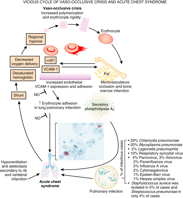
Three major triggers are associated with the development of acute chest syndrome: Infection, bone marrow fat embolization, and direct red cell intravascular sequestration causing lung injury and infarction. Lung injury results in ventilation-perfusion mismatch/shunt and hypoxemia, which leads to increased hemoglobin S polymerization and erythrocyte vaso-occlusion. This worsens bone marrow infarction and pulmonary vaso-occlusion to promote a vicious cycle. Fat embolization can be diagnosed by oil red O staining of pulmonary alveolar macrophages, revealing the characteristic red lipid inclusions (see inset). NO can inhibit steps in the pathologic process, but NO is scavenged by free hemoglobin released by hemolysis. α4β1, An integrin dimer; NO, nitric oxide; VCAM-1, vascular cell adhesion molecule-1.

From Broaddus VC et al: Murray & Nadel’s textbook of respiratory medicine, ed 7, Philadelphia 2022, Elsevier.

Diagnosis ⬆ ⬇

Differential Diagnosis

  • Thalassemia
  • Other hemolytic anemias
  • The differential diagnosis of patients presenting with a painful crisis is discussed in “Physical Findings”
Workup (Table 5

  • Screening of all newborns regardless of racial background is performed in the U.S. Screening can be performed with sodium metabisulfite reduction test (Sickledex test).
  • Hemoglobin electrophoresis will also confirm the diagnosis and is useful to identify hemoglobin variants such as fetal hemoglobin and hemoglobin C.
  • Patients will have evidence of hemolysis (elevated reticulocyte count, low haptoglobin, variable elevation of LDH and total bilirubin).
  • Chest x-ray often reveals typical vertebral body changes (“fish mouth” vertebrae) caused by chronic vasoocclusive injury to vertebral bodies.
  • For prenatal diagnosis, initial step is identification of parenteral globin gene mutation by DNA-based testing. If positive, then DNA-based testing of chorionic villus sampling or amniotic fluid cells is performed.

TABLE 5 Baseline Evaluations to Consider

Tests
Blood testsCBC with differential
Reticulocyte count
Hemoglobin HPLC or electrophoresis
LDH
Renal function tests
Liver function tests
Mineral panel
Serum iron, ferritin, TIBC
Vitamin D level
Hepatitis B sAg
Hepatitis C antibody
RBC alloantibody screen
RBC typing
D-dimera
C-reactive proteina
Brain natriuretic peptide
Urine and kidney testsUrinalysis
Renal ultrasonographyb
RadiologyMRI or MRA brain (adults)c or transcranial Doppler ultrasonography starting at age 2 yr (children)
Chest radiographyd
Hip or shoulder radiograph or MRI (or both)c
Bone density in teenagers and adults
Cardiology and pulmonaryEchocardiogram
NeurocognitiveNeurocognitive testingd

CBC, Complete blood count; HPLC, high-performance liquid chromatography; LDH, lactate dehydrogenase; MRA, magnetic resonance angiography; MRI, magnetic resonance imaging; RBC, red blood cell; sAg, surface antigen; TIBC, total iron-binding capacity.

a Consider following as surrogate markers after initiation of disease-modifying intervention.

b If hematuria with red blood cells in urine.

c As clinically indicated.

d If the patient has poor school performance, an abnormal memory, or abnormal MRI findings.

From Hoffman R et al: Hematology: basic principles and practice, ed 7, Philadelphia, 2018, Elsevier.

Laboratory Tests

  • Anemia (from chronic hemolysis), reticulocytosis, leukocytosis, and thrombocytosis are common. Hgb S/β0 and Hgb S/β+ thalassemia will have microcytosis; Hgb S/C is also typically microcytic. Hgb S/C may have normal or near-normal hematocrit but will have characteristic changes on peripheral smear (target cells).
  • Elevations of bilirubin, lactate dehydrogenase, and low haptoglobin are consistent with chronic hemolysis.
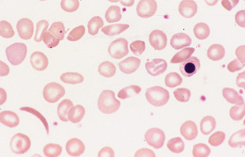
  • Peripheral blood smear may reveal sickle cells, target cells, poikilocytosis, and hypochromia (Fig. 2).
  • Elevated blood urea nitrogen and creatinine may be present in patients presenting acutely with dehydration or chronically with progressive renal insufficiency.
  • Urinalysis may reveal hematuria and proteinuria. Patients with SCD should be screened for microalbuminuria and proteinuria with spot urine testing by 10 yr of age.

Figure 2 Photomicrograph of a blood film.

Sickle cell anemia (homozygosity for hemoglobin S). Shown are a sickle cell, boat-shaped cells, a nucleated red cell, and target cells.

From Bain BJ et al: Dacie and Lewis practical haematology, ed 12, Philadelphia, 2017, Elsevier.

Imaging Studies (

  • Chest x-ray or noncontrast chest CT scan to evaluate acute and chronic lung changes is helpful.
  • Routine skeletal imaging is rarely helpful in acute crisis and should usually be reserved for pain that is not consistent with transient acute crisis.
  • MRI or bone scan is useful to address chronic avascular necrosis or, in the acute setting, osteomyelitis.
  • CT or MRI scan of brain is not indicated in asymptomatic adults and children with SCD but is often needed in patients with neurologic complications, such as transient ischemic attack, cerebrovascular accident, seizures, or altered mental status.
  • Transcranial Doppler ultrasonography (TCD) is used to identify children with sickle cell anemia who are at risk for stroke. There should be an annual screening starting at age 2 until age 16. Patients determined to be at risk (transcranial Doppler velocity ≥200 cm/s) should be enrolled in long-term transfusion programs. These transfusions are effective in reducing risk of stroke by >90%. In adults, magnetic resonance angiography (MRA) can be used instead of TCD to identify those at risk for stroke.
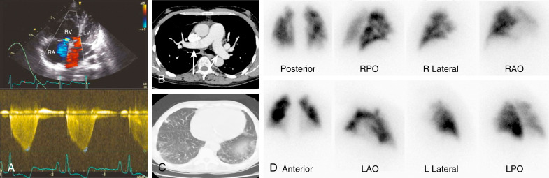
  • Doppler echocardiography should be performed in patients with unexplained respiratory symptoms to evaluate for pulmonary hypertension (Fig. E5), with right heart catheterization performed if abnormal. Screening for vasculopathy is done by estimating the tricuspid regurgitant jet velocity (TRV). Elevated values are predictive of early mortality. The prevalence of pulmonary hypertension when right heart catheterization is performed is approximately 6% in adults with sickle cell disease.

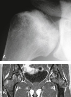
Figure E3 Sickle Cell Disease

A, Infarction of the Humeral Head Has Led to the Separation of an Osteochondral Fragment of the Subarticular Bone with Adjacent Reactive Medullary Sclerosis. B, Coronal T1-Weighted Image of the Proximal Femora Showing Patchy Hypointensity Within the Marrow Resulting from Marrow Hyperplasia and Bilateral Femoral Head Osteonecrosis.

From Adam A et al: Grainger & Allison’s diagnostic radiology, ed 5, 2007, Churchill Livingstone. In Grant LA: Grainger & Allison’s diagnostic radiology essentials, ed 2, Philadelphia, 2019, Elsevier.

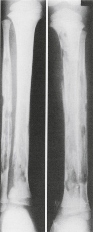
Figure E4 Sickle cell disease with salmonella osteomyelitis.

Extreme destructive changes in the long bones have been caused by infection superimposed upon infarction. Numerous sequestra are present.

From Sutton D: Textbook of radiology and imaging, ed 7, 1998, Churchill Livingstone. In Grant LA: Grainger & Allison’s diagnostic radiology essentials, ed 2, Philadelphia, 2019, Elsevier.

FIG E5 Imaging features of sickle cell disease and pulmonary hypertension.

(A)Top, Echocardiographic apical four-chamber view of the heart, illustrating severe right ventricular (RV) and right atrial (RA) dilation and moderate tricuspid regurgitation (blue Doppler). Bottom, Doppler tracing from a patient with severe pulmonary hypertension reveals a jet velocity of greater than 4 m/sec. (B) Axial chest computed tomography scan showing enlargement of pulmonary arteries (arrows) in severe pulmonary hypertension. (C) Chest computed tomography showing mild pulmonary fibrosis typical of patients with sickle cell disease and pulmonary hypertension. (D) Perfusion scan demonstrating patchy areas of abnormal perfusion. Ventilation scans were normal (not shown). LAO, Left anterior oblique; LPO, left posterior oblique; LV, left ventricle; RAO, right anterior oblique; RPO, right posterior oblique.

From Broaddus VC et al: Murray & Nadel’s textbook of respiratory medicine, ed 7, Philadelphia 2022, Elsevier.
Treatment ⬆ ⬇

Nonpharmacologic Therapy

  • Patients should be instructed to avoid conditions that may precipitate sickling crisis, such as extremes of cold and heat and dehydration.
  • Maintain adequate hydration (PO or IV).
  • Correct hypoxia when present.
Acute General Rx

  • Initiate aggressive IV and oral hydration; most patients in crisis have evidence of dehydration. Use hypo-osmolar fluids, such as LR or 0.45 normal saline, for intravenous hydration during acute crisis in the hospitalized patient. Normal saline has adverse biochemical effects on sickled cells and may lead to increase pain.
  • L-arginine 1000 mg/kg TID, decreases narcotic use by approximately 54% in the hospitalized patient during a vaso-occlusive crisis.
  • Aggressively diagnose and treat suspected infections such as urinary infection, respiratory infections, or catheter-associated line infections. Table 6 summarizes bacteria and viruses that most frequently cause serious infection in patients with sickle cell disease.
  • Provide pain relief during the vaso-occlusive crisis (Table 7). Most patients will have a treatment history that can guide dosing. Opiate management is complicated by high levels of tolerance in patients often treated over many years. Patient-controlled analgesic pumps are often helpful; caution should be used when employing continuous infusions, which are usually not necessary or helpful. Morphine and hydromorphone are most commonly used. Meperidine is rarely used now and generally discouraged because of neurologic side effects, although some patients state a preference for it.
  • Oral diphenhydramine is used to control pruritus, which is commonly associated with opiate analgesics; preferred to IV therapy in guidelines.
  • Tables 8 and 9 summarize overall strategies for the management of acute chest syndrome.
  • Follow oxygenation in patients presenting with chest pain or respiratory symptoms; assess for evolving acute chest syndrome if oxygenation deteriorates.
  • Urology evaluation for priapism.

TABLE 8 Overall Strategies for the Management of Acute Chest Syndrome

Prevention
Incentive spirometry and periodic ambulation in patients admitted for sickle cell pain, surgery, or febrile episodes
Watchful waiting in any hospitalized child or adult with sickle cell disease (pulse oximetry monitoring and frequent respiratory assessments)
Cautious use of intravenous fluids
Intense education and optimum care of patients who have sickle cell anemia and asthma
Diagnostic Testing and Laboratory Monitoring
Blood cultures, if febrile
Nasopharyngeal samples for viral culture (respiratory syncytial virus, influenza), depending on clinical setting
Complete blood counts every day and appropriate chemistries
Continuous pulse oximetry
Chest radiographs for persistent or progressive illness
Treatment
Blood transfusion (simple or exchange) depending on clinical features; consider maintaining an active type and crossmatch
Supplemental O2 for drop in pulse oximetry by 4% over baseline, or values <90%
Empirical antibiotics (third-generation cephalosporin and macrolide)
Continued respiratory therapy (incentive spirometry and chest physiotherapy as necessary)
Bronchodilators and corticosteroids for patients with asthma
Optimum pain control and fluid management

From Kliegman RM: Nelson textbook of pediatrics, ed 21, Philadelphia, 2020, Elsevier.

TABLE 9 Treatment of the Acute Chest Syndrome

Oxygen therapy to maintain arterial hemoglobin oxygen saturation >92%
Pain control and incentive spirometry to reduce chest wall splinting and pulmonary atelectasis
Close clinical observation
  • Monitor Po2/Fio2 ratio
  • Particular attention to worsening respiratory function
Asthma therapy if indicated
Empirical antibiotics
  • Cover typical and atypical respiratory pathogens
  • Consider regional and seasonal risk of methicillin-resistant Staphylococcus aureus
  • Anticipate influenza A or B infections and treat/prevent accordingly
Transfusion therapy
  • Main indication for transfusion therapy in ACS is worsening respiratory function
  • Simple transfusion is as effective as erythrocytapheresis in the usual patient
  • Patients with high initial hemoglobin concentrations (≥9 g/dl) or patients with more severe disease should receive erythrocytapheresis
  • Transfused blood should be matched to Rh, C, E, and Kell antigens, and transfusion records documenting history of prior alloantibodies should be obtained

ACS, Acute chest syndrome; Fio2, fractional concentration of oxygen in inspired gas; Po2, partial pressure of oxygen.

From Broaddus VC et al: Murray & Nadel’s textbook of respiratory medicine, ed 7, Philadelphia, 2022, Elsevier.

TABLE 7 Recommended Dose and Interval of Analgesics Necessary to Obtain Adequate Pain Control in Patients With Sickle Cell Disease

Dose/RateComments
Severe to Moderate Pain
MorphineParenteral: 0.1-0.15 mg/kg every 3-4 h
Recommended maximum single dose, 10 mg
PO: 0.3-0.6 mg/kg every 4 h
Drug of choice for pain; lower doses in elderly adults and infants and in patients with liver failure or impaired ventilation
MeperidineParenteral: 0.75-1.5 mg/kg every 2-4 h
Recommended maximum dose, 100 mg
PO: 1.5 mg/kg every 4 h
Increased incidence of seizures; avoid in patients with renal or neurologic disease and those who receive MAOIs
HydromorphoneParenteral: 0.01-0.02 mg/kg every 3-4 h
PO: 0.04-0.06 mg/kg every 4 h
OxycodonePO: 0.15 mg/kg/dose every 4 h
KetorolacIM: Adults: 30 or 60 mg initial dose followed by 15-30 mg; children: 1 mg/kg load followed by 0.5 mg/kg every 6 hEqual efficacy to 6 mg MS; helps narcotic-sparing effect; not to exceed 5 days; maximum, 150 mg first day, 120 mg maximum on subsequent days; may cause gastric irritation
ButorphanolParenteral: Adults: 2 mg every 3-4 hAgonist-antagonist; can precipitate withdrawal if given to patients who are being treated with agonists
Mild Pain
CodeinePO: 0.5-1 mg/kg every 4 h
Maximum dose, 60 mg
Mild to moderate pain not relieved by aspirin or acetaminophen; can cause nausea and vomiting
AspirinPO: Adults: 0.3-6 mg every 4-6 h; children: 10 mg/kg every 4 hOften given with a narcotic to enhance analgesia; can cause gastric irritation; avoid in febrile children
AcetaminophenPO: Adults: 0.3-0.6 g every 4 h; children: 10 mg/kgOften given with a narcotic to enhance analgesia
IbuprofenPO: Adults: 300-400 mg every 4 h; children: 5-10 mg/kg every 6-8 hCan cause gastric irritation
NaproxenPO: Adults: 500 mg/dose initially and then 250 every 8-12 h; children: 10 mg/kg/day (5 mg/kg every 12 h)Long duration of action; can cause gastric irritation
IndomethacinPO: Adults: 25 mg every 8 h; children: 1-3 mg/kg/day given 3 or 4 timesContraindicated in psychiatric, neurologic, renal diseases; high incidence of gastric irritation; useful in gout

IM, Intramuscular; MAOI, monoamine oxidase inhibitor; MS, morphine sulphate; PO, oral.

Adapted from Charache S et al: Effect of hydroxyurea on the frequency of painful crises in sickle cell anemia: investigators of the multicenter study of hydroxyurea in sickle cell anemia, N Engl J Med 332:1317, 1995. In Hoffman R et al: Hematology: basic principles and practice, ed 7, Philadelphia, 2018, Elsevier.

TABLE 6 Bacteria and Viruses That Most Frequently Cause Serious Infection in Patients With Sickle Cell Disease

MicroorganismType of InfectionComments
Streptococcus pneumoniaeSepticemiaCommon despite prophylactic penicillin and pneumococcal vaccine
MeningitisLess frequent than in years past
PneumoniaRarely documented except in infants and young children
Septic arthritisUncommon
Haemophilus influenzae type bSepticemia
Meningitis
PneumoniaMuch less common in recent years because of immunization with conjugate vaccine
Salmonella speciesOsteomyelitis
SepticemiaMost common cause of bone and joint infection
Escherichia coli and other gram-negative enteric pathogensSepticemia
Urinary tract infection
OsteomyelitisFocus sometimes not apparent
Staphylococcus aureusOsteomyelitisUncommon
Mycoplasma pneumoniaePneumoniaPleural effusions; multilobe involvement
Chlamydia pneumoniaePneumonia
Parvovirus B19Bone marrow suppression (aplastic crisis)High fever common; rash and other organ involvement infrequent
Hepatitis viruses (A, B, and C)HepatitisMarked hyperbilirubinemia

Data from Buchanan GR, Glader BE: Benign course of extreme hyperbilirubinemia in sickle cell anemia: analysis of six cases, J Pediatr 91:21, 1977. From Hoffman R et al: Hematology: basic principles and practice, ed 7, Philadelphia, 2018, Elsevier.

Chronic Rx

  • Hydroxyurea increases hemoglobin F levels, reduces the incidence of vasoocclusive complications, and improves survival. It is helpful in patients with Hgb S/S and S/βo thalassemia; its value in other variants is less certain. There is strong evidence to support use of hydroxyurea therapy in children ages 9 mo and older to decrease the frequency of vasoocclusive crises and acute chest syndrome. Hydroxyurea is also strongly recommended for adults with three or more crises during any 12-mo period, with SCD pain or chronic anemia interfering with daily activities, or with severe or recurrent episodes of acute chest syndrome. It should be stopped in pregnancy, and contraception counseling should be given to all patients.
  • Recommended starting doses for hydroxyurea are 15 mg/kg/day in adults and 5 to 10 mg/kg/day in patients with renal disease. A recent randomized study has shown markedly superior efficacy with a dose escalation strategy of up to 30 mg/kg/day compared with traditional dosing of 20 mg/kg/day.
  • Pharmaceutical grade L-glutamine is approved for use to decrease crisis symptoms in patients with Hgb S/S and Hgb S/βo thalassemia; benefits include decrease in acute crisis episodes, reduced hospitalization, and decrease in episodes of acute chest syndrome. Dosing is 5 to 15 g tid, taken with food or 8 oz of cold or room-temperature fluid. Constipation, nausea, headache, and abdominal discomfort were common side effects, occurring in approximately 15% to 20% of patients. It may work with or without concomitant use of hydroxyurea. Caution may be warranted in prescribing L-glutamine to patients with clinically significant renal or hepatic dysfunction.
  • Replace folic acid (1 mg PO daily) to replace increased utilization of folic acid stores from chronic hemolysis. Patients also have mineral and vitamin deficiencies (calcium; zinc; and vitamins A, C, D, and E) and may need oral supplementation.
  • Chronic pain management represents an enduring challenge, made more difficult by the current opiate addiction and overdose epidemic. In one study, patients reported pain in 55% of days. Review of guidelines for safe opiate prescribing is strongly recommended and management with pain management specialists is also strongly recommended.
  • Indications for PRBC transfusion in SCD are described in Table 10. Urgent exchange transfusion for acute chest syndrome with progressive hypoxia (arterial oxygen saturation <90%) or multiorgan failure may be lifesaving. Simple transfusion may be adequate in milder cases with a target Hgb of 10 g/dl. Transfusion therapy is appropriate for patients with stroke or at high risk by transcranial Doppler study (see earlier) if possible. Transfusion has shown to be beneficial in children with silent infarcts by MRI in preventing progression. Transfusion to Hgb 10 g/dl is recommended in anemic patients undergoing general anesthesia to reduce crisis and respiratory complications after surgery. Patients on chronic transfusion therapy should receive RBC matched at C, E, and K antigens to avoid alloimmunization. Transfusion is not recommended for asymptomatic anemia. Serum ferritin level should be monitored quarterly. Iron overload due to blood transfusions (transfusional hemosiderosis) can be treated with chelating agents (deferoxamine [SC infusion], deferasirox [PO], and deferiprone [PO]).
  • Annual screening for proteinuria is recommended. ACE inhibitor therapy should be started for microalbuminuria in adults with SCD to prevent progression of renal injury. Progressive renal injury may cause worsening anemia and responds to erythropoietin.
  • Annual retinopathy screening should be performed beginning at age 10, especially for patients with Hgb S/C variant, in whom proliferative retinopathy occurs in about 30% to 50%. It is less common in Hgb S/S and other variants.
  • Gene therapy for SCD patients represents a novel approach. Clinical trials with lentiviral vector-mediated addition of an antisickling β-globin gene into autologous hematopoietic stem cells are ongoing with encouraging early results in terms of reduction of sickle cell crises and correction of the biologic hallmarks of the disease. A level of erythrocyte fetal hemoglobin (HbF) comprising alpha and gamma globins may ameliorate the hemolytic anemia of sickle cell disease by mitigating sickle hemoglobin polymerization and erythrocyte sickling. BCL11A is a repressor of gamma-globin expression and HbF production in adult erythrocytes. Its down-regulation is a promising therapeutic strategy for induction of HbF. Gene therapy trials with the use of LentiGlobin have shown sustained production of HbAT87Q in most red cells, leading to reduced hemolysis and complete resolution of severe vaso-occlusive events.1
  • Allogeneic stem cell transplantation can be curative in young patients with symptomatic SCD.
  • Crizanlizumab, an antibody against the adhesion molecule p-selectin, revealed a significantly lower rate of sickle cell-related pain crisis than placebo and was associated with a low incidence of adverse events in a recent study. It has been approved for the prevention of vasoocclusive crises in adults and pediatric patients ages 16 yr and older.
  • Voxelotor, an inhibitor of sickle hemoglobin (HbS) polymerization, significantly increases hemoglobin levels and reduces markers of hemolysis and is approved for the therapy of adults and children over age 12 yr.
  • Penicillin V 125 mg PO bid should be administered by age 2 mo and increased to 250 mg bid by age 3 yr. Penicillin prophylaxis can be discontinued after age 5 yr, except in children who have had splenectomy.
  • Table 11 summarizes disease-modifying treatments to consider.

TABLE 11 Disease-Modifying Treatments to Considera

Robust clinical dataPenicillin prophylaxis
Streptococcus pneumoniae vaccination
Hydroxyurea
Chronic exchange transfusion
Iron chelation for chronic iron overloadb
Limited clinical dataDaily multivitamin without iron or folate supplementation and vitamin D replacementc
Haemophilus influenzae vaccination
Influenza vaccination
Erythropoietin
Phlebotomy
ExperimentalHb F reactivation with decitabine, histone deacetylase inhibitors, or imids
Erythropoietin for chronic relative reticulocytopenia
Nutritional supplements and antioxidants (e.g., glutamine, zinc, multivitamins)
N-acetylcysteine

Hb F, Fetal hemoglobin.

a See text for specific indications and limitations.

b Best data from thalassemia patient experience.

c Risks minimal; therefore it is generally done.

From Hoffman R et al: Hematology: basic principles and practice, ed 7, Philadelphia, 2018, Elsevier.

TABLE 10 Indications for Transfusion in Sickle Cell Disease

DurationConsensusMethodGoal∗
Stroke, acuteSingle+ExHbS <30%
Stroke, ongoing careChronic+EitherHbS <30%
High-velocity transcranial DopplerChronic+EitherHbS <30%
ACS, initial episodeSingle+Dir >ExHgb 10
ACS, recurrent6-12 mo+Either
Pulmonary hypertensionChronic+Either
Multiorgan failureSingle+Ex
Major surgerySingle+DirHgb 10
Acute anemiaSingle+Dir
Recurrent spleen sequestrationChronic+
Sepsis/meningitisSingle+Dir
Severe chronic pain6-12 mo+
Congestive heart failureChronic+
Silent infarct with abnormal neuropsychologyChronic–
Pregnancy–
Anemia/renal failureChronic–
Leg ulcers6-12 mo–
Severe growth delay–
Severe eye disease–
Priapism–

ACS, Acute chest syndrome; Dir, direct; Ex, exchange; Hb, hemoglobin type; Hgb, hemoglobin concentration; +, consensus reached; –, consensus not reached.

∗Goal of transfusion if a consensus has been reached.

From Fuhrman BP et al: Pediatric critical care, ed 4, Philadelphia, 2011, Saunders.

Referral

  • Hospitalization for pain crisis unresponsive to oral analgesics, or involving fever, respiratory symptoms, or vomiting and diarrhea.
  • Optimal management requires coordination with hematology, blood banking, pain management, and psychosocial counseling and support.
  • Referral for patients with organ-specific complications, notably pulmonary hypertension, acute/chronic kidney disease, and ophthalmologic complications.
  • Referral to an ophthalmologist for an annual dilated retinal examination beginning at 10 yr of age.

Pearls & Considerations ⬆ ⬇

Comments

  • The average life span of individuals with sickle cell trait is similar to that of the general population. It may be associated with hematuria, often painless, and rhabdomyolysis under extreme conditions. Chronic pain and acute pain are not typical of sickle cell trait. It is also associated with a higher incidence of renal medullary cancer. Chronic problems in sickle cell disease are summarized in Table 12.
  • Regular immunizations, especially pneumococcal vaccination, are recommended. The prophylactic administration of penicillin soon after birth and the timely administration of pneumococcal and Haemophilus influenzae type b vaccines have resulted in a significant decline in the incidence of these infections. The heptavalent conjugated pneumococcal vaccine (Prevnar) should be administered from 2 mo of age. The 23-valent unconjugated pneumococcal vaccine (Pneumovax) is given from age 2 yr and can be boosted once 3 yr later. Influenza vaccination can be given after 6 mo of age.
  • In patients with SCD presenting with acute crisis, intravenous hydration with hypotonic saline is favored over normal saline. Normal saline can have adverse biochemical effects on sickle cell and possibly lead to increased pain.
  • Pulmonary hypertension is a complication of chronic hemolysis and is associated with a high risk of death. It can be detected by Doppler echocardiography in more than 30% of adult patients with sickle cell disease. Cardiac catheterization will confirm the diagnosis. It is resistant to hydroxyurea therapy.
  • Malnutrition can lead to poor clinical outcomes in patients with SCD. Identifying patients at risk might improve outcomes.
  • Vitamin D deficiency is common and should be treated to prevent adverse skeletal outcomes.
  • Metformin increases fetal hemoglobin (HbF) levels in patients with SCD. A recent study revealed that metformin use in SCD patients with diabetes is associated with fewer episodes of consequential SCD complications and with lower health care utilization.
  • Exposure to systemic corticosteroids is associated with a fourfold excess risk for hospitalization for a vaso-occlusive episode (VOE).2

TABLE 12 Chronic Problems in Sickle Cell Disease

Growth and developmentReduced height and weight
Pubertal delay
Cognitive impairment (recurrent small strokes)
LocomotorOsteonecrosis of humeral and femoral heads
Chronic leg ulcers
CardiovascularMyocardial infarction
Left and right ventricular dilatation
PulmonaryPulmonary fibrosis
Pulmonary hypertension
Cor pulmonale
GenitourinaryRenal papillary necrosis-hematuria and tubular defects
Chronic renal failure
Frequent urinary tract infections in women
Impotence (secondary to priapism)
OcularProliferative retinopathy (30% of patients)
Blindness (especially in SC disease)
Retinal detachment

SC, Sickle cell.

From Ryan ET et al: Hunter’s tropical medicine and emerging infectious diseases, ed 10, Philadelphia, 2019, Elsevier.

Related Content

Sickle Cell Anemia (Patient Information)

Suggested Readings ⬆ ⬇

  1. Ataga K.I. : Crizanlizumab or the prevention of pain crises in sickle cell diseaseN Engl J Med. ;376:429-439, 2017.
  2. Badawy S.M., Payne A.B. : Association between clinical outcomes and metformin use in adults with sickle cell disease and diabetes mellitusBlood Adv. ;3:3297-3306, 2019.
  3. Carden M.A. : Extracellular fluid tonicity impacts sickle red blood cell deformability and adhesionBlood. ;130(24):2654-2663, 2017.
  4. DeBraun M.R. : Controlled trial of transfusions for silent cerebral infarcts in sickle cell anemiaN Engl J Med. ;371:699-710, 2014.
  5. Esrick E.B. : Post-transcriptional genetic silencing of BCL11A to treat sickle cell diseaseN Engl J Med. ;384(3):205-215, 2021.
  6. John C.C. : Hydroxyurea dose escalation for sickle cell anemia in Sub-Saharan AfricaN Engl J Med. ;382(26):2524-2533, 2020.
  7. Magrin E. : Lentiviral and genome-editing strategies for the treatment of β-hemoglobinopathiesBlood. ;134(15):1203-1213, 2019.
  8. Morris C.R. : A randomized, placebo-controlled trial of arginine therapy for the treatment of children with sickle cell disease hospitalized with vaso-occlusive pain episodesHaemotologica. ;98(9):1375-1382, 2013.
  9. Nevitt S.J. : Hydroxyurea (hydroxycarbamide) for sickle cell diseaseCochrane Database Syst Rev. ;4, 2017.
  10. Niihara Y. : A phase 3 trial of l-glutamine in sickle cell diseaseN Engl J Med. ;379(3):226-235, 2018.
  11. Parent F. : A hemodynamic study of pulmonary hypertension in sickle cell diseaseN Engl J Med. ;365:44-53, 2011.
  12. Pier F. : Sickle cell diseaseN Engl J Med. ;376, 2017.
  13. Sii-Felice K. : Hemoglobin disorders: lentiviral gene therapy in the starting blocks to enter clinical practiceExp Hematol. ;64:12-32, 2018.
  14. Vichinsky E. : A phase 3 randomized trial of Voxelotor in sickle cell diseaseN Engl J Med. ;381:509-519, 2019.
  15. Yawn B.P., John-Sowah J. : Management of sickle cell disease: recommendations from the 2014 Expert Panel ReportAm Fam Physician. ;92(12):1069-1076, 2015.

Related Content ⬆

    1. Kanter J. : Biologic and clinical efficacy of LentiGlobin for sick cell diseaseN Engl J Med. ;386(7):617-628, 2022.
    2. Walter O. : Risk of vaso-occlusive episode after exposure to corticosteroids in patients with sickle cell diseaseBlood. ;139(26):3771-3777, 2022.