section name header

Basic Information ⬇

AUTHOR: Daniel K. Asiedu, MD, PhD, FACP

Definition

Infection caused by the species of the genus Candida. All the different Candida species can cause disease, but infections caused by Candida albicans are the most common. Candida species are ubiquitous and are the most common fungal pathogens affecting mankind. Cutaneous candidiasis comprises superficial Candida infections of the skin and mucosal membranes.

Cutaneous candidiasis can be classified into two subgroups: Cutaneous candidiasis syndromes and chronic mucocutaneous syndromes.

Cutaneous candidiasis syndromes include:

  • Generalized cutaneous candidiasis
  • Intertrigo
  • Candida folliculitis
  • Paronychia/onychomycosis
  • Perianal candidiasis
  • Erosio interdigitalis blastomycetica
  • Balanitis

Chronic mucocutaneous syndromes include:

  • Oropharyngeal candidiasis
  • Esophageal candidiasis
  • Vulvovaginal candidiasis
  • GI candidiasis (gastric/intestines/perianal)
  • Candida cystitis
Synonyms

Yeast infection

Candidosis

Moniliasis

Oidiomycosis

ICD-10CM CODES
B37.2Candidiasis of skin and nail
B37.8Candidiasis, unspecified
B37.89Other sites of candidiasis
Epidemiology & Demographics

  • Candida species: It is the most common fungal infection in immunocompromised people.
  • Most females (75%) experience an episode of vulvovaginal candidiasis in their lifetime.
Incidence

Estimated to be 50 cases per 100,000 persons

Prevalence

Colonizes more than 50% of U.S. population

Predominant Sex & Age

  • Female >male
  • No predominant age, but neonates and the elderly (adults >65 yr) are susceptible to Candida colonization and to getting mucocutaneous candidiasis
Risk Factors

Risk factors that allow Candida infection include:

  • Age >65 yr
  • Females in the third trimester
  • Defects in the mucocutaneous barrier (e.g., wounds, burns, ulcerations)
  • Decreased/defective granulocytes/monocytes
  • Diseases of white blood cells (e.g., chronic granulomatous disease)
  • Complement deficiency
  • Certain diseases associated with cell-mediated immunity (e.g., HIV, DM)
  • Use of certain medications (e.g., broad-spectrum antibiotics, high doses of corticosteroids)
  • Increased skin pH due to panty liners and occlusive attire
  • Chronic mucocutaneous candidiasis: (CMC) is characterized by susceptibility to Candida infection of skin, nails (Fig. E1), and mucous membranes. Patients with recessive CMC and autoimmunity have mutations in the autoimmune regulator AIRE. Mutations in the CC domain of STAT1 underlie autosomal-dominant CMC and lead to defective Th1 and Th17 responses, which may explain the increased susceptibility to fungal infections. Some patients have autosomal recessive polyglandular autoimmune syndrome type 1 (also autoimmune polyendocrinopathy-candidiasis-ectodermal dystrophy [APECED] syndrome)
  • Risk factors for oropharyngeal candidiasis are summarized in Box E1

Figure E1 Hand and nail involvement in chronic mucocutaneous candidiasis.
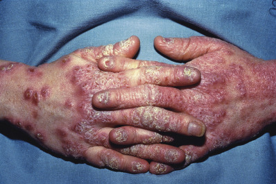
From James WD et al: Andrews’ diseases of skin, ed 12, Philadelphia, 2016, Saunders.

BOX E1 Oropharyngeal Candidiasis: Risk Factors

Local Factors (Mucosal Barrier Function)

  • Heavy smoking
  • Foreign bodies (dentures and nasogastric tubes)
  • Radiation-induced mucositis
  • Inhalational and topical corticosteroid use
  • Xerostomia
  • Mucosal tumors
Systemic Factors

  • Immunosuppression, age, virus or retrovirus, chemotherapy, and corticosteroid use
  • Diabetes mellitus
  • Intrinsic immunodeficiency
  • Myelodysplasia/leukemia
  • Antibiotic use

From Flint PW et al: Cummings otolaryngology, head and neck surgery, ed 7, Philadelphia, 2021, Elsevier.

Anatomic sites predisposed to Candida infection include:

  • Axilla
  • Beneath the breast, abdominal fold, intertriginous areas
  • Periungual creases
  • Inguinal creases
  • Back and buttocks of bedridden persons
Physical Findings & Clinical Presentation

There are several clinical presentations of cutaneous candidiasis. A few are presented here.

  • Cutaneous candidiasis
    1. Presents as erythematous, sometimes shiny with flakes and fluid lesions at the edge of the redness (satellite pustules). It is itchy, and the skin becomes inflamed. Pustules may be present in candidiasis of the scrotal and perineal skin.
  • Gastrointestinal tract candidiasis
    1. Oropharyngeal candidiasis [thrush]
      1. Usually seen in diabetics, after exposure to inhaled steroids, broad-spectrum antibiotics, chemotherapy, radiation to head and neck, and in immunosuppressed individuals (e.g., patients with a history of HIV infection). It is also seen in some patients who wear dentures. Symptoms include:
        1. White, thick patches on the oral mucosa (Fig. E2), tongue, palate, or oropharynx or under dentures
        2. Dysphagia, mouth soreness, and pain on eating and swallowing
        3. Tongue burning
        4. Loss of taste
      2. Physical examination shows:
        1. Erythema of the buccal mucosa
        2. White patches on buccal cavity surfaces (described previously)
        3. Transverse fissuring
  • Esophageal candidiasis
      1. Most common in patients with:
        1. Hematologic cancers
        2. HIV/AIDS (it is an AIDS-defining illness)
      2. History of oropharyngeal candidiasis. Symptoms include:
        1. Odynophagia (pain on swallowing), hallmark of the disease
        2. Dysphagia
        3. Epigastric pain
        4. Retrosternal pain
      3. Physical examination shows:
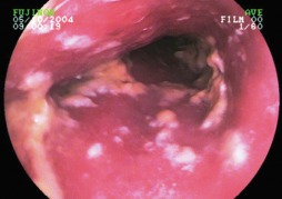
        1. Affects mainly the distal one third of the esophagus. Endoscopy shows areas of the erythema and edema; scattered white patches or ulcers
  • Perianal candidiasis
      1. Skin maceration
      2. Itching
      3. Frequently extends to the perineum
  • Paronychia/onychomycosis
    1. Fungal infection of the nail and surrounding tissues
    2. Associated with diabetes mellitus and immersion of hands or feet in water
    3. History: Pain and redness around and beneath the nail and nail bed
    4. Physical exam: Inflammation around the toenail. There may also be nail thickening and discoloration (dystrophic nails). Nail loss may also occur
  • Respiratory tract candidiasis
    1. Usually seen in hospitalized patients.
    2. About 25% of outpatients have their respiratory tract colonized by Candida species.
  • Genitourinary tract candidiasis
    1. Vulvovaginal candidiasis
      1. Most common form of mucosal candidiasis. Recurrent vulvovaginal candidiasis (chronic yeast infection) is defined as four or more acute symptomatic yeast infections per year. This affects about 138 million women annually worldwide.
      2. Risk factors include increased estrogen level (e.g., contraceptive pill), steroid or antibiotic use, diabetes, HIV infection, intrauterine device (IUD), and diaphragm use.
      3. It causes itching, curdy white discharge, and occasionally dysuria, dyspareunia, and vaginal irritation.
      4. On examination the mucosa may be inflamed with vulvar and vaginal erythema, and vaginal discharge, classically described as white and curdy.
      5. Painful erythema or itchy penile inflammation may occur in male sexual partners of affected females.
    2. Candida balanitis
      1. Usually acquired through sexual contact with a partner who has vulvovaginal candidiasis. Common in diabetic patients and uncircumcised individuals. The dominant species is C. albicans.
      2. Symptoms include penile pruritus and white patches on penis. There could be severe burning and itching. Infection could also spread to the perineum.
      3. Physical exam: Dry, erythematous, and scaly patches on penis and sometimes on the thighs, scrotum, gluteal folds, and buttocks. Papules, vesicles, or pustules and ulceration may also be present.
  • Others
    1. Erosio interdigitalis blastomycetica: Denudating/macerating area commonly seen in third web space.
    2. Mastitis: Injured nipples in lactating women can lead to infections, including Candida infections.
    3. Candida folliculitis: Pustulous nodules in hairy areas.
    4. Intertrigo: This occurs in folds of the skin and creases (Fig. E3). It is characterized by erosions, exudation, oozing, and maceration.
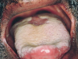
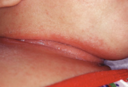
Figure E2 Oral candidiasis.

From Swartz, MH: Textbook of physical diagnosis, ed 7, Philadelphia, 2014, Elsevier.

Figure E3 Intertriginous candidiasis of the neck.

From Kliegman RM et al: Nelson textbook of pediatrics, ed 19, Philadelphia, 2011, Saunders.

Etiology

The most common cause of cutaneous candidiasis is Candida albicans.

Diagnosis ⬆ ⬇

Differential Diagnosis

  • Intertrigo
  • Seborrheic dermatitis
  • Psoriasis
  • Bacterial vaginosis
    1. Contact dermatitis
Workup

  • Mucocutaneous and cutaneous candidiasis:
    1. Obtain scrapings from the skin, oral, and vaginal mucosa or nails.
    2. The presence of hyphae/pseudohyphae or budding yeast cells on wet smear, as well as confirmation by culture, is the recommended procedure to diagnose cutaneous candidiasis.
    3. KOH smears are helpful.
  • Respiratory candidiasis:
    1. Sputum Gram stain: Shows yeast cells
    2. Sputum cultures
    3. Lung biopsy: Establishes the diagnosis
  • Gastrointestinal candidiasis:
    1. Upper endoscopy with or without biopsy (Fig. E4)

Figure E4 Endoscopic appearance of esophageal candidiasis.

Courtesy Dr. B. Rembacken, Leeds, U.K.

Treatment ⬆

Pharmacologic Therapy

  • Cutaneous candidiasis:
    1. Decrease/prevent moisture in area
    2. Apply antifungal agents (nystatin powder or cream with an azole or ciclopirox [e.g., clotrimazole, econazole, miconazole])
  • Gastrointestinal candidiasis:
    1. Oropharyngeal candidiasis
      1. Treat with either
        1. Oral topical antifungal agent (e.g., nystatin 1:100,000 U/ml, 5 ml swish and swallow qid) OR
    2. Systemic oral azoles (e.g., fluconazole 100 mg qd ×14 days)
      1. In HIV-positive patients, use high doses of fluconazole (100 to 200 mg PO qd for 7 to 14 days), itraconazole, or posaconazole
  • Candida esophagitis:
    1. Treat with systemic fluconazole for 2 to 3 wk
    2. Treat with intravenous (IV) fluconazole if patient is unable to take oral medication
  • Genitourinary tract candidiasis:
    1. Vulvovaginal candidiasis: Treatment options for acute cases include:
      1. A single dose of oral fluconazole (fluconazole 150 mg PO × 1 dose) OR
      2. Topical antifungal agent
      3. Oteseconazole capsule (Vivjoa) was recently approved by the FDA for the prevention of recurrent yeast infection in females who are NOT of reproductive age
    2. For chronic or recurrent cases, treat with fluconazole 150 mg qd × 3 doses and then 150 mg/wk for 6 mo
  • Chronic mucocutaneous candidiasis:
    1. Treatment with azoles is effective (e.g., fluconazole 100 to 400 mg daily)
    2. When patient improves, follow with maintenance treatment with same azole for life
Follow-Up Care

  • Mucocutaneous candidiasis:
    1. Patient should be instructed to call or follow up if symptoms persist, recur, or worsen.
    2. For recurrent infections:
      1. Check HIV antibodies.
      2. Check FBS, HbA1C.
      3. Rule out hematologic malignancy or solid organ malignancy.
      4. Refer to infectious disease specialist if no etiology is found.
Prevention

  • Maintaining dry environment (e.g., by wearing cotton underwear)
  • Decreased use of antibiotic
  • No douching
Related Content

Candidiasis (Patient Information)