section name header

Basic Information

AUTHORS: Mitchell Powell, MD and Lydia Sharp, MD

Definition

Myasthenia gravis (MG) is an autoimmune disorder affecting postsynaptic neuromuscular transmission, most commonly mediated by antibodies directed against the nicotinic acetylcholine receptor (AChR) of the neuromuscular junction. Anti-AChR antibodies cause a decrease in functional postsynaptic ACh receptors, resulting in fatigable weakness. A small percentage of MG patients lack AChR antibodies, and a subset of these patients possess antibodies against muscle-specific tyrosine kinase (MuSK) or low-density lipoprotein receptor-related protein 4 (LRP4), which both affect pre- and postsynaptic function of the neuromuscular junction.1,2 Finally, a portion of AChR antibody-negative myasthenia patients do not possess any detectable antibodies, and this group of patients is appropriately termed “seronegative.” A classification of MG is described in Table 1.

TABLE 1 Osserman Classification Used by the Myasthenia Gravis Foundation of America to Standardize Clinical Symptoms1

Class IAny ocular muscle weakness
Class IIOcular muscle weakness of any severity, MILD limb weakness
Class IIaPredominantly limb and/or axial muscle weakness
Class IIbPredominantly bulbar and/or respiratory muscle weakness
Class IIIOcular muscle weakness of any severity, MODERATE weakness of other muscles
Class IIIaPredominantly limb and/or axial muscle weakness
Class IIIbPredominantly bulbar and/or respiratory muscle weakness
Class IVOcular muscle weakness of any severity, SEVERE weakness of other muscles
Class IVaPredominantly limb and/or axial muscle weakness
Class IVbPredominantly bulbar and/or respiratory muscle weakness
Class VIntubation with or without mechanical ventilation

1 Jaretzki A et al: Myasthenia gravis: recommendations for clinical research standards. Task Force of the Medical Scientific Advisory Board of the Myasthenia Gravis Foundation of America, Neurology 55:16-23, 2000.

From Parrillo JE, Dellinger RP: Critical care medicine: principles of diagnosis and management in the adult, ed 5, Philadelphia, 2019, Elsevier.

Synonym

MG

ICD-10CM CODES
G70.00Myasthenia gravis without (acute) exacerbation
G70.01Myasthenia gravis with (acute) exacerbation
P94.0Transient neonatal myasthenia gravis
Epidemiology & Demographics
Incidence (In U.S.)

8 to 10 cases annually per 1 million persons. It is the most common disorder of neuromuscular junction transmission.

Prevalence (In U.S.)

150 to 250 cases per 1 million persons.

Predominant Sex

Females are affected more often than males (3:2) in adults; they are equally affected in the elderly.

Peak Incidence

Female, second to third decades; male, sixth to eighth decades.

Genetics

Increased frequency of HLA-B8, DR3.

Physical Findings & Clinical Presentation

  • The hallmark of MG is weakness worsened with exercise and improved with rest.
  • Generalized weakness involving proximal muscles, the diaphragm, and neck extensors is common.
  • Weakness is confined to eyelids and extraocular muscles in approximately 15% of patients (Figs. 1, E2, 3, and Box 1). This is referred to as ocular myasthenia gravis.
  • Bulbar symptoms of ptosis, diplopia, dysarthria, and dysphagia are common.
  • Reflexes, sensation, and coordination remain normal.
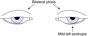
Figure 1 Myasthenia gravis.

Myasthenia gravis may mimic any ocular disorder causing diplopia, although most often it mimics weakness of the superior rectus muscle or medial rectus muscle (i.e., difficulty with sustained elevation or adduction of the eye, respectively). Clues to the diagnosis of myasthenia gravis are associated ptosis, fluctuating course, and normal pupils.

From McGee S: Evidence-based physical diagnosis, ed 4, Philadelphia, 2018, Elsevier.

Figure 3 Typical myasthenic facies.
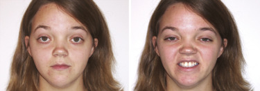
At rest (left), there is slight bilateral lid ptosis, which is partially compensated by asymmetric contraction of the frontalis muscle, raising the right eyebrow. During attempted smile (right), there is contraction of the medial portion of the upper lip and horizontal contraction of the corners of the mouth without the natural upward curling, producing a “sneer.”

From Jankovic J et al: Bradley and Daroff’s neurology in clinical practice, ed 8, Philadelphia 2022, Elsevier.

BOX 1 Ocular Findings in Myasthenia Gravis

  1. Weakness usually involves one or more ocular muscles without overt pupillary abnormality.
  1. Weakness is typically variable, fluctuating, and fatigable.
  1. Ptosis that shifts from one eye to the other is virtually pathognomonic of MG.
  1. With limited ocular excursion, saccades are superfast, producing ocular “quiver.”
  1. After downgaze, upgaze produces lid overshoot (“lid twitch”)
  1. Pseudo-internuclear ophthalmoplegia-limited adduction, with nystagmoid jerks in abducting eye.
  1. In asymmetric ptosis, covering the ptotic eye may relieve contraction of the opposite frontalis.
  1. Passively lifting a ptotic lid may cause the opposite lid to fall: “Enhanced ptosis” or “curtain sign.”
  1. Edrophonium may improve only some of several weak ocular muscles; others may actually become weaker.
  1. 10.Edrophonium may relieve asymmetric ptosis and produce retraction of the opposite lid from frontalis contraction.
  1. 11.The opposite lid may droop further as the more involved lid improves after edrophonium.
  1. 12.Cold applied to the eye may improve lid ptosis: “Ice-pack test” (see Fig. E5).

From Jankovic J et al: Bradley and Daroff’s neurology in clinical practice, ed 8, Philadelphia, 2022, Elsevier.
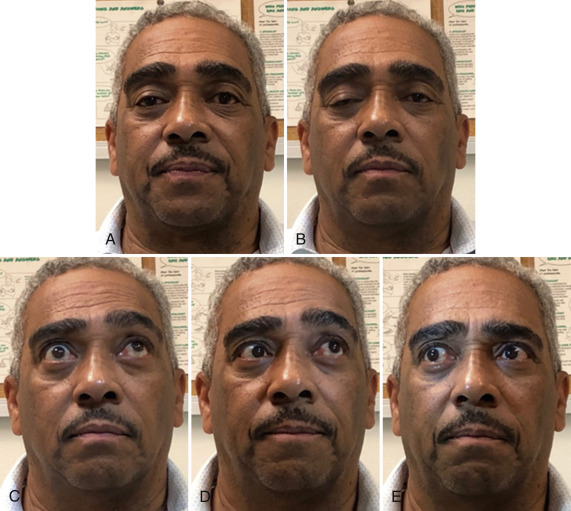
Figure E2 Ocular Motility Abnormalities in Myasthenia Gravis Due to Weakness of Multiple Periocular Muscles in Both Eyes

(A and B) Progressive right lid ptosis during sustained forward gaze from fatigable weakness of the right levator palpebrae. (C) Attempted upward gaze. There is incomplete superior movement of both eyes, worse in the right eye. Note the asymmetric furrowing of the forehead and elevation of the eyebrows. (D) On left lateral gaze, there is skew deviation with incomplete medial movement of the right eye and incomplete abduction of the left eye. (E) On right lateral gaze, there is incomplete movement of both eyes from weakness of right lateral rectus and left medial rectus muscles.

From Jankovic J et al: Bradley and Daroff’s neurology in clinical practice, ed 8, Philadelphia 2022, Elsevier.

Etiology

An antibody-mediated decrease in nicotinic AChRs in the postsynaptic neuromuscular junction results in defective neuromuscular transmission and subsequent muscle weakness and fatigue. Early-onset MG is associated with HLA-B8.1, whereas late-onset MG is associated with HLA-DQB1, HLA-DQA1, and HLA-DRB1.3

Some myasthenia gravis patients present with MuSK or LRP4 antibodies instead of AChR antibodies. The MuSK antibody is present in about 40% to 50% of the subset of individuals who lack the AChR antibody, and the LRP4 antibody is present in a fraction of the remainder.1 Both MuSK and LRP4 antibodies interrupt the process of AChR aggregation at the neuromuscular junction. Anti-MuSK patients have a similar syndrome to AChR MG, although they may have more bulbar weakness, facial tongue and proximal muscle atrophy, and either lack of or paradoxic response to pyridostigmine. Anti-LRP4 patients generally have milder symptoms that favor ocular findings. However, serious cases can still yield severe clinical symptoms.1 Both MuSK and LRP4 antibody MG have not been shown to have any association with thymoma.4

Less than 10% of myasthenia gravis patients are seronegative for AChR, MuSK, and LRP4 antibodies. This syndrome is termed seronegative myasthenia gravis and presents with a higher proportion of entirely ocular symptoms. Generally, this group of patients responds well to classic MG therapies, including pyridostigmine, steroids, immunosuppression, and thymectomy.

Serologic and clinical presentation of myasthenia gravis subgroups are summarized in Table 2.

TABLE 2 Serologic and Clinical Presentation of Myasthenia Gravis Subgroups1-8

AChR
Antibody
MuSK AntibodyLRP4 AntibodyStriated Muscle Antigens Titin Antibody RyR AntibodyAge at Onset
Sex
Clinical
Findings
Response to TherapyResponse to ThymectomyPrognosis
Ocular MG50%-75%NoneNoneNoneOlder
Male
OcularGoodGoodGood (but 50% can develop generalized MG in 2 yr)
Early-onset generalized MG80%-85% (high titer)NoneNoneNone-rare<50 yrGeneralRarely needs immunosuppressionGoodLess severe
Low mortality
Late-onset generalized MGNone-rareNoneNone50%
54% titin
33% RyR
50 yrGeneralOften requires immunosuppressionPoorSevere
Thymomatous-associated
MG
Positive, nearly 100% (low titer)NoneNone95%
50% titin
47% RyR
Ranges:
Older but <40 yr if RyR
General
If RyR: Ocular, bulbar, respiratory weakness
Often requires immunosuppressionFairRyR: Invasive, malignant thymoma
Severe
Higher mortality
Generalized MG
MuSK positive
None100%NoneNoneYounger femaleFacial, bulbar, neck, respiratory weakness paraspinal, esophageal musclesPoor response to AChE inhibitorsPoor (no thymic changes)Severe, progressive course
Generalized MG LRP4 positiveNoneNone100%NoneYounger femaleGeneralGoodNot enough dataFair
MG with thymic hyperplasia89%NoneNoneNoneYounger femaleGeneralGoodGoodGood
ACh antibody negativeNegativeNegativeNegativePresentYounger femaleGeneralPoorFair

ACh, Acetylcholine; AChE, acetylcholinesterase; AChR, acetylcholine receptor; MG, myasthenia gravis; MuSK, muscle-specific tyrosine kinase; LRP4, low-density lipoprotein receptor-related protein 4; RyR, ryanodine receptor.

1 Romi F et al: Myasthenia gravis patients with ryanodine receptor antibodies have distinctive clinical features, Eur J Neurol 14:617-620, 2007.

2 Romi F et al: Myasthenia gravis: clinical, immunological, and therapeutic advances, Acta Neurol Scand 111:134-141, 2005.

3 Akaishi T et al: Response to treatment of myasthenia gravis according to clinical subtype, BMC Neurol 16:225, 2016.

4 Gilhus NE, Verschuuren JJ: Myasthenia gravis: subgroup classification and therapeutic strategies, Lancet Neurol 14:1023-1036, 2015.

5 Hong Y et al: Autoantibody profile and clinical characteristics in a cohort of Chinese adult myasthenia gravis patients, J Neuroimmunol 298:51-57, 2016.

6 Gilhus NE et al: Myasthenia gravis-autoantibody characteristics and their implications for therapy, Nat Rev Neurol 12:259-268, 2016.

7 Roberts PF et al: Thymectomy in the treatment of ocular myasthenia gravis, J Thorac Cardiovasc Surg 122:562-568, 2001.

8 Rivner MH et al: Clinical features of LRP4/agrin-antibody-positive myasthenia gravis: a multicenter study, Muscle Nerve 62(3):333-343, 2020. Available from https://doi.org/10.1002/mus.26985.

From Parrillo JE, Dellinger RP: Critical care medicine: principles of diagnosis and management in the adult, ed 5, Philadelphia, 2019, Elsevier.

Diagnosis

Differential Diagnosis

Lambert-Eaton myasthenic syndrome, botulism, medication-induced myasthenia, chronic progressive external ophthalmoplegia, congenital myasthenic syndromes, thyroid disease, basilar meningitis, intracranial mass lesion with cranial neuropathy, Miller-Fisher variant of Guillain-Barré syndrome

Workup

  • Edrophonium (Tensilon) test (Fig. E4): Useful in MG patients with ocular symptoms, although uncommonly used now. Cardiac monitoring and atropine ready at the bedside are essential.
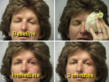
  • Patients with MG may also have a positive ice-pack test (Fig. E5).
  • Repetitive nerve stimulation: Successive stimulation shows decrement of muscle action potential in clinically weak muscle; may be negative in up to 50%.
  • Single-fiber electromyography: Highly sensitive; abnormal in up to 95% of patients.
  • Serum AChR antibodies MuSK and/or LRP4 antibodies.

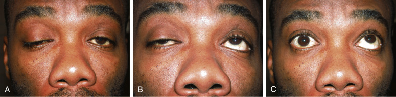
Figure E4 Positive edrophonium test in myasthenia gravis.

(A) Asymmetric ptosis in the primary position. (B) Defective upgaze. (C) Following injection of edrophonium, there is marked bilateral improvement of ptosis and modest improvement of left upgaze.

From Kanski JJ et al: Clinical ophthalmology: a systematic approach, ed 7, Philadelphia, 2010, Saunders.

Figure E5 Ice-pack test in myasthenia gravis.

Before testing (A) there is ptosis of both upper lids, more marked on the left. An ice pack is placed over the ptotic eye for 2 min (B). On removal of the ice pack, the ptosis is improved (C) and gradually returns (D).

From Jankovic J et al: Bradley and Daroff’s neurology in clinical practice, ed 8, Philadelphia, 2022, Elsevier.

Laboratory Tests

  • Forced vital capacity (FVC) is the most useful test for assessing neuromuscular respiratory status. Patients with an FVC of <20 ml/kg are at high risk of respiratory failure and should be monitored in an ICU setting. Although the decision of when to intubate is a clinical one, FVC falling below 10 to 15 ml/kg generally requires intubation.
  • CT scan with contrast of anterior chest to look for thymoma (about 10% of patients) or thymic hyperplasia (about 80% of patients). Prevalence increases with age.
  • Thyroid-stimulating hormone and free T4 to rule out thyroid disease.

Treatment

Nonpharmacologic Therapy

  • Patient education to facilitate recognition of worsening symptoms and impress need for medical evaluation at onset of clinical deterioration
  • Avoidance of selected drugs (Table 3) known to provoke exacerbations of MG (β-blockers, aminoglycoside and quinolone antibiotics, penicillamine, interferons, class I antiarrhythmics [procainamide, quinidine, etc.])
  • Prompt treatment of infections, diet modification, and speech evaluation with dysphagia

TABLE 3 Medications to Avoid in Myasthenia Gravis1

Medications to AvoidExamplesRecommendation for AvoidanceMechanism of Weakness
Antibiotics
AminoglycosidesGentamycin
Streptomycin
ContraindicatedBlocks ACh receptor, prevents release of ACh
Tobramycin
Amikacin
Less likely problematic
AntimalarialsQuinine
Chloroquine
ContraindicatedPresynaptic blockage of voltage-dependent sodium channels and postsynaptic potentiation of depolarization
MacrolidesErythromycin, Tetracycline
Azithromycin
RelativeAffects presynaptic transmission
FluoroquinolonesMoxifloxacin
Ciprofloxacin
Levofloxacin
Ofloxacin
FDA black box warningUnknown
KetolideTelithromycinFDA black box warningUnknown
PolymyxinsContraindicatedPresynaptic and postsynaptic effects
Cardiovascular
AntiarrhythmicsProcainamide
Propafenone
ContraindicatedDecreases the release of AChSodium influx blocker
β-BlockersPropranolol
Atenolol
Ophthalmic timolol
RelativeUnclear-may be at the neuromuscular junction or muscle membrane
Calcium channel blockerVerapamil
Amlodipine
RelativePresynaptic and postsynaptic blockade of L-type calcium channels
Anticonvulsants
PhenytoinRelativeDepressed postsynaptic response to ACh; inhibition of calcium channel; increase in muscle membrane threshold
CarbamazepineRelativeTriggers immune response
GabapentinRelativeBinds voltage-gated calcium channel
Chemotherapeutic Agents
Doxorubicin
Etoposide
Cisplatin
RelativeUnknown
Others
InterferonRelativeAutoantibody production
Neuromuscular blocking agentsAtracurium
Cisatracurium
Vecuronium
RelativeBlocks ACh receptors
StatinsRelativeUnknown
CorticosteroidsRelativeDirect blocking of ACh receptor through ionic channels
Botulinum toxinContraindicatedImpairs synaptic transmission
MagnesiumRelativeImpairs synaptic transmission
PenicillamineContraindicatedBinds the ACh receptor; induces antibodies to receptor

ACh, Acetylcholine; FDA, Food and Drug Administration.

1 Ahmed A, Simmons Z: Drugs which may exacerbate or induce myasthenia gravis: a clinician’s guide, Internet J Neurol 10:1-8, 2008.

From Parrillo JE, Dellinger RP: Critical care medicine: principles of diagnosis and management in the adult, ed 5, Philadelphia, 2019, Elsevier.
Acute General Rx (Table E4

  • Symptomatic treatment with acetylcholinesterase inhibitors:
    1. Pyridostigmine 30 to 60 mg PO q4 to 6h initially; onset of effect is 30 min, duration 4 h. May be titrated up to 120 mg every 4 h. GI upset is common with higher doses and may respond to hyoscyamine.
  • Immunosuppressive treatment with corticosteroids is first-line treatment, and is often transitioned to a nonsteroidal immunosuppressive agent such as azathioprine or mycophenolate to avoid the significant side effects of long-term steroid use:
    1. Prednisone initiated at 10 to 20 mg daily titrated by 5-mg increments to effect or dose of 1 mg/kg/day with improvement in 2 to 4 wk and maximal response by 3 to 6 mo.
    2. Azathioprine initiated at 50 mg daily titrated to 2 to 3 mg/kg/day with clinical effect in 6 to 12 mo. Azathioprine is not recommended in patients with no thiopurine methyltransferase activity.
    3. Mycophenolate mofetil 500 mg twice a day titrated to 2 g/day with clinical effect in 3 to 6 mo, but can be up to 12 mo.
  • Targeted immunotherapy for myasthenia gravis allows quicker and more effective treatment with fewer side effects. Multiple new therapies are currently being researched and/or in clinical trials, including Fc receptor inhibitors, complement inhibitors, IL-6 inhibitors, chimeric antigen receptor (CAR) and chimeric autoantibody receptor (CAAR) T cell therapy, and hematopoietic stem cell transplantation.5 A current list of some alternative agents is below:
    1. Efgartigimod alfa given at 10 mg/kg for 4 wk in weekly infusions with clinical effect within 1 mo. This drug is an Fc receptor inhibitor that promotes IgG degradation and was approved by the FDA in 2021. Further research is needed to determine the long term benefit of this drug, but it can be considered as a glucocorticoid-sparing bridge until slower-acting agents such as azathioprine and mycophenolate take effect.6
    2. Ravulizumab initiated as IV infusion at 2.4-3 g (weight based) followed by 3-3.6 g (weight based) 2 wk after loading dose, followed by every 8 wk afterwards. This drug is a human monoclonal antibody that binds complement C5 similar to eculizumab but with a more convenient dosing schedule. It was FDA approved for AChR antibody positive patients in 2022.7
    3. Eculizumab initiated at 900 mg IV weekly for four doses, then 1200 mg on the 5th wk and every 2 wk afterwards. Like ravulizumab, this drug is a complement inhibitor approved for adult patients who are acetylcholine-antibody positive and have severe symptoms despite current immunotherapy use.
    4. Cyclosporine initiated at 5 mg/kg/day with clinical effect within 1 to 2 mo. Note a faster onset of effect than azathioprine or mycophenolate, but also a less tolerable side effect profile due to concerns for renal toxicity and drug interactions.
    5. Tacrolimus initiated at 0.1 mg/kg/day divided into two doses then titrated to plasma concentration of 7 to 8 ng/ml with clinical effect within 6 to 12 mo. This drug is better tolerated than cyclosporine but still has potential for serious side effects.
    6. Rituximab initiated at 1 g every 2 wk for two doses, OR 375 mg/m2 weekly for four doses. Generally used in refractory MG or earlier in MuSK antibody MG. Redosing interval varies per protocol and by clinical response of the patient.
  • Plasmapheresis and IV immunoglobulin (IVIG) are short-term options for immunotherapy during an exacerbation. There is no significant difference in efficacy between IVIG and plasmapheresis.8
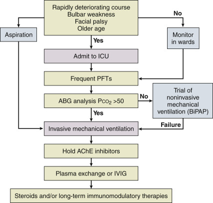
  • Mechanical ventilation is lifesaving in the setting of a myasthenic crisis. Consider elective intubation if forced vital capacity is <10 to 15 ml/kg.
  • Fig. E6 illustrates a flowchart for the management of myasthenic crisis.

Figure E6 Clinical flowchart for the management of myasthenic crisis.

ABG, Arterial blood gas; AChE, acetyl cholinesterase; BiPAP, bilevel positive airway pressure; ICU, intensive care unit; IVIG, intravenous immunoglobulin; PFT, pulmonary function test.

From Parrillo JE, Dellinger RP: Critical care medicine, principles of diagnosis and management in the adult, ed 4, Philadelphia, 2014, Elsevier.

Table E4 Therapeutic Agents Used in Myasthenia Gravis

AgentInitial DoseMaintenance DoseOnset of ActionMajor Adverse EventsMonitoringComments
Pyridostigmine30-60 mg tid60-120 mg tid to 5×/day, adjusted based on symptoms, typically not to exceed 480 mg/day15-30 minStomach cramps, nausea, vomiting, diarrhea, muscle twitching and cramps, sweating, salivation, blurred visionUse the minimal amount that produces clinical improvement; this is best achieved by using a dose that produces observable improvement after most administrationsCan counter muscarinic adverse events with anticholinergic agents (i.e., glycopyrrolate, hyoscyamine sulfate, propantheline, diphenoxylate HCl with atropine, or loperamide)
PrednisoneOption 1: 10-20 mg/day, increasing daily dose by 5 mg daily equivalent every week until treatment goal achieved
Option 2: Start at 50-80 mg/day; this approach may require inpatient hospitalization (see text for details)
Slow alternate day taper after treatment goal achieved for several days (see text for details). Taper more slowly once 10 mg/day dose equivalent. Continuing a low dose long-term can help to maintain the treatment goal2-4 wkHypertension, diabetes, weight gain, bone loss, cataracts, GI ulcers, glaucoma, neuropsychiatric symptoms, growth retardation in children, hypothalamic-pituitary axis suppressionHbA1c every few months, blood pressure checks, bone density monitoring, eye exam for glaucoma and cataractsAdminister in single morning dose; temporary worsening is seen in up to 50% of patients, starting on high doses and in some patients on lower doses; IVIg or PLEX may prevent steroid-induced worsening
Azathioprine50 mg/dayIncrease by 50 mg increments every 1-2 wk to target of 2.5-3 mg/kg/day2-10 mo for initial response. Up to 24 mo for maximum benefitFever, abdominal pain, nausea, vomiting, anorexia, leukopenia, hepatotoxicity, skin rashCBC, LFTs 1-4 times in first mo, then monthly to every third month. Regular dermatologic examinations if taken chronically10% of patients cannot tolerate because of flulike reaction; major drug interaction with allopurinol; TPMT enzyme testing can be performed, if available, before starting treatment to identify patients at high risk of bone marrow suppression
Cyclosporine100 mg bidIncrease slowly as needed to 3-6 mg/kg/day on bid schedule.1-3 moHirsutism, tremor, gum hyperplasia, hypertension, hepatotoxicity, nephrotoxicity, PRESCBC, LFTs, BUN/Cr monthly ×3, then every 3 mo; monitor trough drug levelsBioequivalence differs between preparations, so avoid brand switching when possible; grapefruit juice may increase blood level; high potential for drug-drug interactions
Mycophenolate mofetil500 mg bid1000-1500 mg bid2-12 moDiarrhea, vomiting, leukopenia, teratogenicity (black box warning)CBC weekly for 4 wk, every 2 wk for 4 wk, then monthly to every 3rd mo; REMS program when used in women of childbearing ageDiarrhea may resolve by change to tid dosing
Efgartigimod alfa10mg/kg qwk x 4 wkEfficacy of repeated cycles unclear1 moHeadache, infectionsBMPApproved for AChR Ab MG only. Possible glucocorticoid-sparing bridge
Ravulizumab2.4-3g IV3-3.6g IV 2 wk then q8wk afterwards1-2 moUpper respiratory infections, leukopenia, transaminitis, meningococcal meningtitisCBC, LFTs; monitor for signs of meningitisEasier dosing than Exulizumab. Approved for AChR Ab MG only, when continued symptoms despite other therapies.
Meningococcal vaccination at least 2 wk prior-if not possible, then antimicrobial prophylaxis recommended
Eculizumab900 mg IV weekly × 4 wk1200 mg IV q2wk1-2 moUpper respiratory infections, leukopenia, transaminitis, meningococcal meningtitisCBC, LFTs; monitor for signs of meningitisApproved for AChR Ab MG only, when continued symptoms despite other therapies.
Meningococcal vaccination at least 2 wk prior-if not possible, then antimicrobial prophylaxis recommended
Cyclophosphamide(1) Oral: 50 mg/day(2) IV: 500 mg/m2 monthlyOral: Increase by 50 mg/wk to maintenance dose of 2-3 mg/kg/day2-6 moAlopecia, leukopenia, nausea and vomiting, skin discoloration, anorexia, hemorrhagic cystitis, malignancyCBC, BUN/Cr, electrolytes, LFTs, urinalysis every 2-4 wkIV pulse therapy may be less toxic because cumulative dose is lower
Tacrolimus3-5 mg/day or 0.1 mg/kg/dayIncrease dosing as needed for response following trough levels (see last column)1-3 moHyperglycemia, hypertension, headache, hyperkalemia, nephrotoxicity, diarrhea, nausea, vomiting, PRESBUN/Cr, glucose, K+; trough drug levels every few weeks initially, then less frequentlyInsulin-dependent diabetes mellitus developed in 20% of postrenal transplant patients; trough levels of 8-9 ng/ml may be effective
Methotrexate5-15 mg weekly for 2 wkIncrease by 5 mg every 2 wk to a maximum dose of 15-25 mg weekly2-6 moLeukopenia, mouth ulceration, nausea, diarrhea, headaches, hair loss, hepatotoxicity, pulmonary fibrosis, rare nephrotoxicity, teratogenicityCBC, LFTs monthly initially, then at least every 3 mo. Monitor periodically for interstitial lung disease, a rare occurrence with doses used for immunotherapyConsider folic acid 5 mg/day to reduce toxicity. Absolutely contraindicated in pregnancy
Intravenous immunoglobulin (IVIg)2 g/kg over 2-5 days0.4-1 g/kg every 4 wk; can attempt to decrease frequency over time1-2 wkHeadache, aseptic meningitis, nephrotoxicity, ischemic events, fluid overload, leukopenia, thrombocytopeniaBUN/Cr every month, decreasing to every 3rd mo over timeIgA level before starting treatment may be useful to identify congenital IgA deficiency, a contraindication to IVIg use; avoid in patients with recent thrombotic/ischemic event. Use sucrose-free formulation for patients at risk of renal toxicity

BUN/Cr, Blood urea nitrogen/creatinine; CBC, complete blood count; GI, gastrointestinal; IgA, immunoglobulin A; IV, intravenous; LFTs, liver function tests; PLEX, plasma exchange; REMS, risk evaluation and mitigation strategy; TPMT, thiopurine s-methyltransferase.From Sanders DB et al: International consensus guidance for the management of myasthenia gravis: executive summary, Neurology 87:419-425, 2016.

Surgical Rx

  • In thymomatous MG, thymectomy is indicated in all patients. If the tumor cannot be surgically resected, chemotherapy can be considered for prevention of local invasion and symptom relief.
  • For nonthymomatous AChR-antibody positive MG, thymectomy improves clinical outcomes and reduces the need for steroids sustained over at least a 5-yr period. Surgical referral should be considered in patients <60 yr old without significant medical comorbidities even if no frank thymic tissue is seen on imaging, especially if they are refractory to therapy.9,10
  • MuSK and LRP4 myasthenia are not associated with thymic pathology and generally thymectomy is not beneficial for these patients.11
Disposition

Course of disease is highly variable. Mortality rate has decreased from 75% to 4.5% over the past four decades.

Referral

  • Referral to a general neurologist or neuromuscular specialist is appropriate.
  • Surgical referral for thymectomy in selected cases (see “Surgical Rx”).

Pearls & Considerations

Related Content

Myasthenia Gravis (Patient Information)

Related Content

  1. Rivner M.H. : Clinical features of LRP4/agrin-antibody-positive myasthenia gravis: a multicenter study Available from Muscle Nerve. ;62(3):333-343, 2020.https://doi.org/10.1002/mus.26985
  2. Ghazanfari N. : Muscle specific kinase: organiser of synaptic membrane domains Available from Int J Biochem Cell Biol. ;43(3):295-298, 2011.https://doi.org/10.1016/j.biocel.2010.10.008
  3. Muñiz-Castrillo S. : Associations between HLA and autoimmune neurological diseases with autoantibodies Available from Auto immun Highlights. ;11(1), 2020.https://doi.org/10.1186/s13317-019-0124-6
  4. Gilhus N.E. : Available from Myasthenia gravis, N Engl J Med. ;375(26):2570-2581, 2016.https://doi.org/10.1056/NEJMra1602678
  5. Menon D., Bril V. : Pharmacotherapy of generalized myasthenia gravis with special emphasis on newer biologicals Available from Drugs. ;82(8):865-887, 2022.https://doi.org/10.1007/s40265-022-01726-y
  6. Howard J.F. : Safety, efficacy, and tolerability of efgartigimod in patients with generalised myasthenia gravis (ADAPT): a multicentre, randomised, placebo-controlled, phase 3 trial Available from Lancet Neurology. ;20(7):526-536, 2021.https://doi.org/10.1016/S1474-4422(21)00159-9
  7. Vu T. : Terminal complement inhibitor ravulizumab in generalized myasthenia gravis Available from NEJM Evidence. ;1(5), 2022.https://doi.org/10.1056/EVIDoa2100066
  8. Barth D. : Comparison of IVIg and PLEX in patients with myasthenia gravisNeurology. ;76:2017-2023, 2011.
  9. Aydin Y. : Thymectomy in myasthenia gravis Available from Eurasian J Med. ;49(1):48-52, 2017.https://doi.org/10.5152/eurasianjmed.2017.17009
  10. Cataneo A.J.M. : Thymectomy in nonthymomatous myasthenia gravis-systematic review and meta-analysis Available from Orphanet J Rare Dis. ;13, 2018.https://doi.org/10.1186/s13023-018-0837-z
  11. Guptill J.T. : Anti-MuSK antibody myasthenia gravis: clinical findings and response to treatment in two large cohorts Available from https://doi.org/10.1002/mus.22006 Muscle Nerve. ;44(1):36-40, 2011.