section name header

Basic Information

AUTHOR: Bharti Rathore, MD

Definition

Autoimmune hemolytic anemia (AIHA) is a condition in which binding of autoantibodies and/or complement to red blood cells (RBCs) results in premature clearance of RBCs. Primary AIHA comprises approximately 50% of cases, whereas secondary AIHA is usually associated with diseases or drugs. The classification of the hemolytic anemias is described in Table 1.

TABLE 1 Classification of the Hemolytic Anemias

Acquired
Environmental factors
  • Antibody: Immunohemolytic anemias
  • Mechanical trauma: TTP, HUS, heart valve
  • Toxins, infectious agents: Malaria, etc.
Membrane defects
  • Paroxysmal nocturnal hemoglobinuria
  • Spur cell anemia
  • Hereditary spherocytosis, etc.
Congenital
Defects of cell interior
  • Hemoglobinopathies: Sickle cell, thalassemia
  • Enzymopathies: G6PD deficiency, etc.

G6PD,Glucose-6-phosphate dehydrogenase; HUS, hemolytic-uremic syndrome; TTP, thrombotic thrombocytopenic purpura.

From Goldman L, Schafer AI: Goldman’s Cecil medicine, ed 24, Philadelphia, 2012, Saunders.

Synonyms

AIHA

Autoimmune hemolytic anemia

Cold agglutinin disease

CAHA

Cold hemagglutinin disease

CHAD

CAIHA

Drug-induced hemolytic anemia

Warm autoimmune hemolytic anemia

WAHA

WAIHA

ICD-10CM CODES
D59.0Drug-induced autoimmune hemolytic anemia
D59.1Other autoimmune hemolytic anemias
Epidemiology & Demographics

The annual incidence is 1 to 3 cases per 100,000 persons, with 10% mortality; most common in women <50 yr.

Physical Findings & Clinical Presentation

  • Most common presentation is dyspnea and fatigue.
  • Pallor, jaundice may be present.
  • Tachycardia with a flow murmur may be present if anemia is pronounced.
  • Patients with intravascular hemolysis may present with dark urine and back pain.
  • Hepatomegaly and/or lymphadenopathy suggests an underlying lymphoproliferative disorder or malignancy; splenomegaly may indicate hypersplenism as a cause of hemolysis.
  • Chronic process with multiple relapses typically seen during the course of the disease.
Etiology

  • Warm antibody mediated: Almost always Immunoglobulin G (IgG) antibodies only (often idiopathic or associated with leukemia, lymphoma [Table 2], thymoma, myeloma, viral infections [Table 3], babesiosis, and collagen-vascular disease). In warm AIHA, antibodies have the highest affinity to the antigen at 37° C
  • Cold antibody mediated: IgM and complement in majority of cases (often idiopathic; at times associated with infections, lymphoma, or cold agglutinin disease)
  • Interleukin-33 may have a role in promoting increasing RBC autoantibodies in AIHA
  • Drug induced (Table 4): Three major mechanisms:
    1. Antibody directed against Rh complex (e.g., methyldopa)
    2. Antibody directed against RBC-drug complex (hapten-induced, e.g., penicillin)
    3. Antibody directed against complex formed by drug and plasma proteins; the drug-plasma protein-antibody complex causes destruction of RBCs (innocent bystander, e.g., quinidine)

TABLE 4 Drug-Induced Autoimmune Hemolytic Anemia

DrugRisk FactorsAIHA OnsetType of AIHAResponse to TreatmentDiseases Treated
MethyldopaNot knownDelayedWAIHAResolution after withdrawalHypertension
IFN-αPretherapeutic positive DATDelayed (8-11 mo)WAIHAResolution spontaneous or after steroidsHepatitis C; hematologic malignancies
EfazulimabNot knownMany moWAIHAResolution after withdrawalArthritis (rare)
EtanerceptNot knownDelayedCAIHAResolution after rituximabRheumatoid arthritis (rare)
Fludarabine
Cladribine
Pentostatin
CLL
Pretherapeutic positive
DAT result
Early (median, 3-4 cycle) or delayedWAIHA
Mixed AIHA
Half of AIHA resolve after steroidsCLL
Lymphomasa
AMLa
BendamustineCLLNo or only very low risk of AIHACLL
Lymphomas
ChlorambucilCLLDelayed onsetWAIHACLL
EculizumabPatients with incomplete responseAfter treatmentCAIHAPNH
LenalidomideDuring treatmentWAIHAResolution after withdrawalOne case treated for lymphoma
Checkpoint inhibitors (anti-CTLA4, anti-PD1/PD1L)During treatmentWAIHASolid tumors, Hodgkin lymphoma

AIHA, Autoimmune hemolytic anemia; AML, acute myeloid leukemia; CAIHA, cold autoimmune hemolytic anemia; CLL, chronic lymphocytic leukemia; DAT, direct antiglobulin test; IFN, interferon; PNH, paroxysmal nocturnal hemoglobinuria; WAIHA, warm autoimmune hemolytic anemia.

a No or very low risk.

From Hoffman R et al: Hematology, basic principles and practice, ed 7, Philadelphia, 2018, Elsevier.

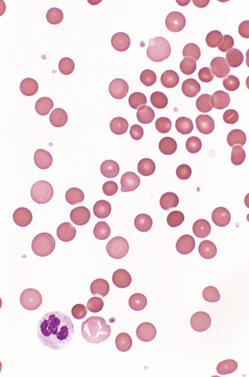
TABLE 2 Secondary Autoimmune Hemolytic Anemia in Malignancies

MalignancyPrevalenceWAIHAsCAIHAs
MGUSVery lowNoneAll
All NHL0.23%-2.6%
CLL4.3%-9%90%10%
SMZL10%
LPL3%-5%NoneMost
Angioimmunoblastic T-cell lymphoma13%
Hodgkin lymphoma0.19%-1.7%AllNone
Ovarian teratomaVery lowAllNone
Solid tumorsVery low

CAIHAs, Cold antibody autoimmune hemolytic anemias; CLL, chronic lymphocytic leukemia; LPL, lymphoplasmacytic lymphoma; MGUS, monoclonal gammopathy with unknown significance; NHL, non-Hodgkin lymphoma; SMZL, splenic marginal zone lymphoma; WAIHAs, warm antibody autoimmune hemolytic anemias.

From Hoffman R et al: Hematology, basic principles and practice, ed 7, Philadelphia, 2018, Elsevier.

TABLE 3 Autoimmune Hemolytic Anemia After Infections

InfectionWAIHACAIHA (Specificities)
Respiratory tract infections (unspecified)--+ (DL)/PCH
Viral infections (specific)EBV+/–+ (anti-i)
CMV++/– (anti-i)
Parvovirus (B19)+ (often with PRCA)+/– (DL)
Varicella+/–+ (anti-Pr, anti-I, anti-DL)
Rubella-+ (anti-Pr1)
Monotypic IgM
HIV++ (anti-I, anti-i, anti-Pr)
Bacterial infections (specific)Mycoplasma+/–+ (anti-I, anti-Pr)
Brucellosis+/–+ (anti-I)
Haemophilus influenzae+ (DL)
Parasitic infections (specific)Visceral leishmaniosis+-

-, Not reported; +, predominant type of autoimmune hemolytic anemia; +/–, single or few cases reported; CAIHA, cold antibody autoimmune hemolytic anemia; CMV, cytomegalovirus; EBV, Epstein-Barr virus; HIV, human immunodeficiency virus; IgM, immunoglobulin M; PCH, paroxysmal cold hemoglobinuria; PRCA, pure red blood cell aplasia; WAIHA, warm antibody autoimmune hemolytic anemia.

From Hoffman R et al: Hematology, basic principles and practice, ed 7, Philadelphia, 2018, Elsevier.

Diagnosis

Differential Diagnosis

  • Hemolytic anemia caused by membrane defects (acquired: Paroxysmal nocturnal hemoglobinuria, spur cell anemia, Wilson disease; inherited: Spherocytosis, elliptosis), hemoglobinopathies, and enzyme deficiencies (G6PD, pyruvate kinase)
  • Non-immune-mediated (microangiopathic hemolytic anemias, hypersplenism, cardiac valve prosthesis, giant cavernous hemangiomas, march hemoglobinuria, physical agents, infections, heavy metals, certain drugs [nitrofurantoin, sulfonamides, ribavirin])
  • Chronic lymphocytic leukemia (CLL) can cause direct antiglobulin test (DAT) positivity (15% of CLL cases) without AIHA. In a series of patients treated with fludarabine and cyclophosphamide, only 30% of patients with DAT+ disease developed AIHA after therapy; conversely, 85% of patients with AIHA were previously positive for DAT
Workup

Evaluation consists primarily of laboratory evaluation to confirm hemolysis and exclude other causes of the anemia. Although most cases of AIHA are idiopathic, potential causes should always be sought.

Laboratory Tests

  • The basic features of hemolytic anemia are reticulocytosis (rarely reticulocytopenia with concurrent bone marrow suppression), low haptoglobin levels, elevated indirect bilirubin, and elevated lactate dehydrogenase (LDH).
  • Initial laboratory tests: Complete blood count (anemia), reticulocyte count (elevated), liver function studies (elevated indirect bilirubin, LDH), evaluation of peripheral smear (Fig. E1).
  • A direct antiglobulin test (DAT, Coombs test) is initially performed with a polyspecific antibody to detect IgG or complement C3d bound to RBCs.1 If the DAT is positive, the diagnosis of autoimmune hemolytic anemia (AIHA) is confirmed. A positive direct Coombs test indicates presence of antibodies or complement on the surface of RBCs. A positive indirect Coombs test indicates presence of freely circulating anti-RBC antibodies in the patient’s serum.
  • If the DAT is positive with IgG alone or with IgG + C3d, the AIHA is most likely due to warm antibody (WAIHA), whereas if the DAT is positive with C3d only, it is most likely caused by a cold antibody (CAIHA).
  • Positive DAT findings and characteristic features of autoantibody in AIHA are summarized in Table 5. Typical serologic features of the different types of drug-induced hemolytic anemia of immunologic origin are summarized in Table 6.
  • Hepatitis A/B/C serology, HIV, antinuclear antibody.
  • Urinary tests may reveal hemosiderinuria or hemoglobinuria (documenting intravascular hemolysis).

TABLE 5 Positive Direct Antiglobulin Test Findings, Characteristic Features of Autoantibody in Autoimmune Hemolytic Anemia

Warm AIHACHADMixed-AIHAPCHIgA AIHA
DATIgG or IgG + C3 or IgG + C3 + IgMC3 or C3 + IgMIgG + C3 + IgMC3Neg with polyspecific reagent
Pos with anti-IgA
Antibody characteristic
1) Antibody subclassIgGIgMIgG + IgMIgGIgA
1) SpecificityApparent Rh specificity (common)I, i, PrP
1) Thermal reactivity (in vitro)Optimal at 37°C (98.6° F) by IAT0-30° C (32-86° F) by saline agglutinationCombined0-24° C (32-75.2° F), biphasic in nature
1) Antibody titer at 4° C (39.2° F)Not applicable256Usually <64 but can be >256Usually <32
AutoagglutinationNoCommonCommonLess commonNo

AIHA, Autoimmune hemolytic anemia; C, complement; CHAD, cold hemagglutinin disease; DAT, direct antiglobulin test; IAT, indirect antiglobulin test; Ig, immunoglobulin; Neg, negative; PCH, paroxysmal cold hemoglobinuria; Pos, positive. The determination of autospecificity is not required for diagnosis of CHAD, but confirmation of high thermal amplitude (i.e., cold autoagglutinin reacting at or above 30° C [86° F]) is essential.

From Bain BJ et al: Dacie and Lewis practical haematology, ed 12, Philadelphia, 2017, Elsevier.

TABLE 6 Serologic Features of the Different Types of Drug-Induced Hemolytic Anemia of Immunologic Origin

MechanismPrototype drugDATIAT
Drug-dependent antibodyNo drugSerum + drugEluate + drug
C activationQuin(id)ineCNegCNeg
No C activationPenicillinIgGNegIgGIgG
Autoantibodyα-MethyldopaIgGIgGNANA

C, Complement; DAT, direct antiglobulin test; IAT, indirect antiglobulin test; IgG, immunoglobulin G; NA, not applicable; Neg, negative.

Occasionally also IgG.

From Bain BJ et al: Dacie and Lewis practical haematology, ed 12, Philadelphia, 2017, Elsevier.

Figure E1 In warm antibody hemolytic anemia, numerous spherocytes are seen.

From Jaffe ES et al: Hematopathology, Philadelphia, 2011, Saunders.

Imaging Studies

  • Chest x-ray examination
  • Computed tomography scans of chest, abdomen, and pelvis if underlying lymphoproliferative disorder is suspected

Treatment

Nonpharmacologic Therapy

  • Discontinuation of any potentially offending drugs
  • Plasmapheresis and exchange transfusion for severe life-threatening cases only
  • Avoid cold exposure in patients with cold antibody AIHA
Acute General Rx

  • Warm AIHA:
    1. Initially, prednisone 1 to 2 mg/kg/day in divided doses is used. A sustained response is achieved in 80% to 90% cases after which tapering is initiated (after 2 to 3 wk) and completed over 3 to 6 mo.
    2. Based on the results of two clinical trials that demonstrated improved outcomes and longer duration of response, the combined upfront use of steroids and rituximab is another option for up-front clinical use.2
    3. For patients relapsing or refractory to initial steroid therapy alone, rituximab is used instead of splenectomy in most cases with response rates of 80%, which are achieved in 2 to 3 wk and are persistent in up to 60% of cases at 3 yr.2,3
    4. Splenectomy is nowadays typically used in patients refractory to both corticosteroids and rituximab.
    5. Immunosuppressive drugs (mycophenolate, cyclosporine, cyclophosphamide) and/or immunoglobulins are recommended after failure of the above approaches to produce an adequate remission.
    6. Danazol, used in conjunction with corticosteroids, may be useful in some cases of warm AIHA.
  • Cold AIHA:
    1. Corticosteroids are generally ineffective, and response rates are much lower than those seen with warm AIHA, and they should not be used to treat cold agglutinin disease.
    2. Therapy of underlying lymphoproliferative disorder or infection is recommended.
    3. In primary cold agglutinin disease, the use of bendamustine and rituximab chemotherapy is the standard approach in fit patients; rituximab alone is used in elderly patients.2
    4. In severe cases, intravenous immunoglobulin therapy or plasmapheresis can be used as a temporary bridge until more effective therapies achieve durable responses.
    5. Complement-directed therapy with eculizumab can also be considered in severe cases.
    6. Sutimlimab, a humanized monoclonal antibody, selectively targets the C1s protein in the complement pathway, which is a complex serine protease responsible for activating this pathway.4 Patients who received sutimlimab show rapidly halted hemolysis, increased hemoglobin levels, and reduced fatigue.
    7. Splenectomy is not effective as the sensitized RBCs are mainly removed in the liver.
  • Table 7 summarizes treatment options for primary and secondary warm autoimmune hemolytic anemia and cold autoimmune hemolytic anemia. Second-line treatment options after steroids are described in Table 8.

TABLE 7 Treatment Options for Primary and Secondary Warm Autoimmune Hemolytic Anemia and Cold Autoimmune Hemolytic Anemia

Disease or ConditionFirst LineSecond LineBeyond Second LineLast Resort
Primary AIHASteroids (rituximab)Splenectomy
Rituximab
Azathioprine, MMF, cyclosporine, cyclophosphamideHigh-dose cyclophosphamide, alemtuzumab
B- and T-cell NHLSteroidsChemotherapy +/– rituximab (splenectomy in SMZL)Other anti-CD23 antibodies/ibrutinib
Hodgkin lymphomaSteroidsChemotherapy
Solid tumorsSteroids
Surgery
Ovarian dermoid cystOvariectomy
SLESteroidsAzathioprineMMFRituximab
Autologous SCT
Ulcerative colitisSteroidsAzathioprineTotal colectomy
CVIDSteroids + IgG replacement
ALPDSteroidsMMFSirolimus
Wiskott-Aldrich syndromeSteroidsAllogeneic SCT
Allogeneic SCTSteroidsRituximabSplenectomy
T-cell infusion
Organ transplantationReduction of immune suppression, steroids
Drug inducedWithdrawalSteroids
Primary CADProtection from cold exposureRituximab
Chlorambucil
Fludarabine + rituximabEculizumab, bortezomib
PCHSupportive treatment (postinfectious)Rituximab (chronic)

AIHA, Autoimmune hemolytic anemia; ALPD, autoimmune lymphoproliferative disorders; CAD, cold agglutinin disease; CVID, common variable immune deficiency; IgG, immunoglobulin G; MMF, mycophenolate mofetil; NHL, non-Hodgkin lymphoma; PCH, paroxysmal cold hemoglobinuria; SCT, stem cell transplantation; SLE, systemic lupus erythematosus; SMZL, splenic marginal zone lymphoma.

Early second-line treatment because of known poor response to steroids.

Off-label use in single cases.

From Hoffman R et al: Hematology, basic principles and practice, ed 7, Philadelphia, 2018, Saunders.

TABLE 8 Second-Line Treatment Options After Steroids

TreatmentDosing and ApplicationSide EffectsPrecautions
Splenectomy (acute)Preferentially laparoscopicInfections, thrombosisPostoperative thromboprophylaxis
Splenectomy (long term)-Infections
Venous thrombosis
Vaccination, patient information
Rituximab375 mg/m2 on days 1, 8, 15, and 22 IVInfusional reactions InfectionsPremedication with antihistamines (and steroids)
Danazol200-400/day POHepatotoxicityNone
CyclophosphamidePO or IV
Dose adjusted to neutrophil count
Neutropenia
Mutagenesis
Neutrophil count monitoring, bladder protection after high doses
Azathioprine2.0-3.0 mg/kg/day PO
Dose adjusted to neutrophil count
NeutropeniaNeutrophil count monitoring; avoid interaction with other drugs (e.g., allopurinol)
MMF1-2 × 1 g/day POGastrointestinal
CyclosporinePO
Dose adjusted to blood levels (target 200-400 ng/ml)
Nephrotoxicity
Gum hyperplasia
Monitoring of CyA levels and creatinine
AlemtuzumabSC (variable doses)NeutropeniaAntiinfectious prophylaxis
Complement inhibition with eculizumab, TNT009InfectionsVaccination

CyA, Cyclosporine A; IV, intravenous; MMF, mycophenolate mofetil; PO, oral; SC, subcutaneous.

From Hoffman R et al: Hematology, basic principles and practice, ed 7, Philadelphia, 2018, Elsevier.

Disposition

Prognosis is generally good unless anemia is associated with underlying disorder with a poor prognosis (e.g., leukemia, myeloma).

Referral

  • Hematology referral in all cases of AIHA
  • Surgical referral for splenectomy in refractory cases of warm AIHA

Pearls & Considerations

Comments

  • The direct antiglobulin test demonstrates the presence of antibodies or complement on the surface of RBCs and is the hallmark of autoimmune hemolysis.
  • Warm AIHA is often associated with autoimmune diseases, whereas cold AIHA often follows viral infections (e.g., mononucleosis) and Mycoplasma pneumoniae infections.
  • HIV can induce both warm and cold AIHA.
  • Hemolytic anemia is a common autoimmune complication of hematopoietic stem cell transplantation occurring in up to 6% of patients as a late complication (median 202 days); it presents as either warm or cold AIHA.

Related Content

    1. Hill A. : Autoimmune hemolytic anemiaHematology Am Soc Hematol Educ Program. ;30(1):382-389, 2018.
    2. Jäger U. : Diagnosis and treatment of autoimmune hemolytic anemia in adults: recommendations from the First International Consensus MeetingBlood Rev. ;41, 2020.
    3. Barcellini W. : How I treat warm autoimmune hemolytic anemiaBlood. ;137(10):1283-1294, 2021.
    4. Röth A. : Sutimlimab in cold agglutinin diseaseN Engl J Med. ;384(14):1323-1334, 2021.