section name header

Basic Information

AUTHOR: Glenn G. Fort, MD, MPH

Definition

Mediastinitis is an infection involving the connective mediastinal tissue that fills the interpleural spaces and surrounds the mediastinal organs. It can be acute or chronic.

Synonyms

Fibrosing mediastinitis

Sclerosing mediastinitis

Granulomatous mediastinitis

ICD-10CM CODE
J98.5Mediastinitis
Epidemiology & Demographics

Acute mediastinitis (Box E1) occurs most frequently as a postoperative infection after a median sternotomy and can be a life-threatening infection. Most infections are bacterial in nature. Causes and clinical settings for mediastinitis are summarized in Table E1.

Chronic mediastinitis is a chronic form of infection in the mediastinum characterized by an invasive and compressive inflammatory infiltrate. It is mostly caused by fungi and some bacteria.

TABLE E1 Acute Mediastinitis: Causes and Clinical Settings

Perforation of a Thoracic Viscus
Forceful vomiting (Boerhaave syndrome)
Direct penetrating trauma to esophagus or trachea
Foreign body ingestion or aspiration
Iatrogenic complication of instrumentation: Endoscopy, bronchoscopy, intubation or airway management, transesophageal echocardiography, central venous catheter placement
Erosion of carcinoma or necrotizing infection
Direct Extension From Outside Mediastinum
Descending necrotizing mediastinitis from pharyngeal, odontogenic origin
Pancreatitis
Pneumonia or empyema
Osteomyelitis of rib, vertebrae, or paraspinous abscess
Mediastinitis After Cardiothoracic Surgery
Spontaneous Mediastinitis
Hematogenous seeding, usually by Streptococcus
Hemorrhagic mediastinitis of inhalational anthrax

From Broaddus VC et al: Murray & Nadel’s textbook of respiratory medicine, ed 7, Philadelphia, 2022, Elsevier.

BOX E1 Classification of Mediastinitis

Acute Mediastinitis

  1. Due to traumatic perforation of the esophagus
    1. Spontaneous or postemetic
    2. Foreign body-associated
    3. Instrumentation or surgery
  2. Due to extension of infection from adjacent structures
    1. Infection of the head and neck
    2. Infections of lungs, pleura, lymph nodes, or pericardium
    3. Subphrenic infection
    4. Vertebral osteomyelitis
    5. Hemotogenous dissemination
  3. Postoperative
    • Chronic Mediastinitis

From Cherry JD et al: Feigen and Cherry’s textbook of pediatric infectious diseases, ed 8, Philadelphia, 2019, Elsevier.

Incidence

Incidence of postoperative mediastinitis ranges from 0.4% to 5%.

Risk Factors

Mediastinal infections have four possible sources:

  • Direct contamination as seen in trauma or surgery (e.g., open heart, esophageal)
  • Hematogenous or lymphatic spread
  • Extension of infection from the neck or retroperitoneum
  • Extension from the lung, pleura, or chest wall
Physical Findings & Clinical Presentation

  • Patients with acute mediastinitis present with acute onset of fever, tachycardia, chest pain, dysphagia, or respiratory distress. There may be signs of sternal wound infection or cellulitis and/or crepitus and edema of the chest wall.
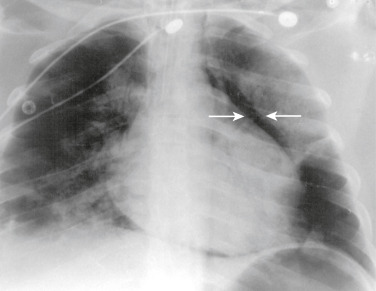
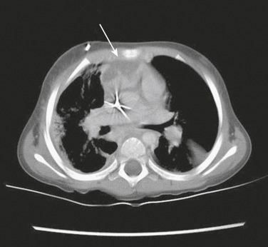
  • Patients with chronic mediastinitis are mostly asymptomatic until symptoms develop related to invasion or obstructions of structures within the mediastinum or adjacent to the mediastinum, such as cough, dyspnea, wheezing, chest pain, dysphagia, or hemoptysis. Complications of chronic or sclerosing mediastinitis include:
  • Superior vena cava syndrome. Histoplasma is the most common nonmalignant cause of this syndrome, marked by edema of face, neck, and torso; neck vein distention; and headache
  • Pulmonary venous or arterial obstruction
  • Esophageal obstruction, cor pulmonale, constructive pericarditis
  • Thoracic duct obstruction
Etiology (Table E2

Acute mediastinitis:

  • Related to head and neck infections or esophageal perforation
    1. Anaerobic bacteria: Peptostreptococci, Veillonella, Fusobacterium, Actinomyces, Prevotella, Eubacterium, Bacteroides
    2. Aerobic bacteria: Streptococcus, Staphylococcus, Corynebacterium, Moraxella, enteric gram-negative rods
    3. Fungi: Candida albicans
  • Related to cardiothoracic surgery
    1. Gram-positive bacteria: Staphylococcus aureus, Staphylococcus epidermidis, Enterococcus, Streptococcus
    2. Gram-negative bacteria: Escherichia coli, Enterobacter, Klebsiella, Proteus, Pseudomonas, other Enterobacteriaceae
    3. Fungi: Candida albicans

Chronic mediastinitis:

  • Histoplasma capsulatum, a dimorphic fungus, is the most common and can cause mediastinal granuloma or fibrosing mediastinitis. A leakage of fungal antigens from lymph nodes into the mediastinal space is believed to cause a hypersensitivity reaction and subsequent exuberant fibrotic response.
  • Other: Mycobacterium tuberculosis, Nocardia, actinomycosis, aspergillosis.

TABLE E2 Microbiology of Mediastinitis

Organisms Frequently Recovered in Mediastinitis Secondary to Infection of the Head and Neck or Esophageal Perforation
Anaerobic
Gram-positive cocci-Peptostreptococcus spp.
Gram-positive bacilli-Actinomyces, Eubacterium, Lactobacillus
Gram-negative cocci-Veillonella
Gram-negative bacilli-Bacteroides spp., Fusobacterium spp., Prevotella spp., Porphyromonas spp.
Aerobic or Facultative
Gram-positive cocci-Streptococcus spp., Staphylococcus spp.
Gram-positive bacilli-Corynebacterium
Gram-negative cocci-Moraxella
Gram-negative bacilli-Enterobacteriaceae, Pseudomonas spp., Eikenella corrodens
Fungi-Candida albicans
Representative Organisms Recovered in Mediastinitis Secondary to Cardiothoracic Surgery, With Representative Rate and Range
Gram-Positive Cocci
Staphylococcus aureus, 25% (7.1%-66.7%)
Staphylococcus epidermidis, 30% (6%-45.5%)
Enterococcus spp., 10% (8%-18.8%)
Streptococcus spp., 2% (0%-18.2%)
Gram-Negative Bacilli
Escherichia coli, 5% (0%-12.5%)
Enterobacter spp., 10% (4%-21.4%)
Klebsiella spp., 3% (0%-21.1%)
Proteus spp., 2% (0%-7.1%)
Other Enterobacteriaceae, 2% (0%-20%)
Pseudomonas spp., 2% (0%-54%)
Fungi
C. albicans, <2 (0%-20.5%)
Polymicrobial, 10% (0%-40%)
Others Occasionally Reported
Acinetobacter, Salmonella spp., Legionella spp., Bacteroides fragilis, Corynebacterium spp., Burkholderia cepacia, Mycoplasma hominis, Candida tropicalis, Aspergillus spp., Nocardia spp., Kluyvera, Gordonia sputi, Mycobacterium fortuitum, Mycobacterium chelonae, Rhodococcus bronchialis
Other Unusual Causes of Mediastinitis
Anthrax, brucellosis, actinomycosis, paragonimiasis, Streptococcus pneumonia

From Bennett JE et al: Mandell, Douglas, and Bennett’s principles and practice of infectious diseases, ed 8, Philadelphia, 2015, Saunders.

Diagnosis

Differential Diagnosis

For chronic mediastinitis:

  • Tumors that can also cause superior vena cava syndrome (e.g., Hodgkin and non-Hodgkin lymphomas, mesothelioma)
  • Sarcoidosis
  • Behçet syndrome
  • Mediastinal fibrosis associated with radiation
  • Silicosis
Laboratory Tests

  • CBC with differential, C-reactive protein, and procalcitonin can point to bacterial infection.
  • Obtain cultures: Aerobic and anaerobic bacteria and fungi, intraoperatively or of any purulent drainage.
  • Pathologic examination: Distinguish between cancer and infection for chronic mediastinitis and allow for specific fungal stains on tissues.
Imaging Studies

Chest x-ray: Can show diffuse mediastinal widening or evidence of mediastinal abscess, including gas bubbles or fluid level. Pneumomediastinum (Fig. E1) or pneumothorax can be seen with esophageal perforation (Table E3).

Chest computed tomography (CT) (Fig. E2): Can show the same as x-ray but is more sensitive in determining degree of mediastinal involvement and may guide drainage procedures for treatment or diagnosis.

MRI may be superior to CT for sclerosing mediastinitis.

Figure E1 Mediastinitis.

Chest x-ray shows large pneumomediastinum and pneumopericardium (arrows) in a patient with mediastinitis.

From Bennett JE et al: Mandell, Douglas, and Bennett’s principles and practice of infectious diseases, ed 8, Philadelphia, 2015, Saunders.

Figure E2 Contrast-Enhanced Computed Tomography Scan Shows a 3 cm × 3 cm Heterogeneous Enhancing Abscess (Arrow) in the Anterior Mediastinum of a Child Acutely Ill with Streptococcus Pneumoniae Bacteremia and Pneumonia with Empyema

Cultures from the mediastinal abscess at the time of surgical drainage were sterile.

From Cherry JD et al: Feigin and Cherry’s textbook of pediatric infectious diseases, ed 8, Philadelphia, 2019, Elsevier.

TABLE E3 Risk Factors for Surgical Site Infection/Mediastinitis Postcardiac Surgery

Preoperative Risk FactorsOperative Risk FactorsPostoperative Risk Factors
Increasing age
Diabetes
Staphylococcus aureus nasal colonization
Previous sternotomy
COPD
Peripheral vascular disease
Class 3-4 angina
Renal failure requiring hemodialysis
History of endocarditis
Cigarette smoking
Low cardiac output
Concurrent infection
Prolonged preoperative hospitalization
Preoperative use of a ventricular assist device
Emergent surgery
Heart transplant
Increasing complexity of surgery
Use of internal thoracic arteries in CABG
Prolonged operative time
Hair removal by razor, not clippers
Inappropriate timing of antibiotics
Prolonged time on cardiopulmonary bypass
High core temperature during bypass (>38° C; 100.4° F)
Need for reexploration
Prolonged ICU stay
Need for mechanical ventilation >48 hr
Lack of perioperative glucose control
Placement of tracheostomy
Postoperative myocardial infarction
Receipt of multiple blood products
Postoperative low cardiac output state

CABG, Coronary artery bypass grafting; COPD, chronic obstructive pulmonary disease; ICU, intensive care unit. From Bennett JE et al: Mandell, Douglas, and Bennett’s principles and practice of infectious diseases, ed 8, Philadelphia, 2015, Saunders.

Treatment

Nonpharmacologic Therapy

Surgery remains the gold standard treatment of mediastinitis for optimal drainage and debridement.

  • Open techniques: Debridement of infected tissue and open packing of the wound with delayed closure or use of vacuum-assisted closure for acute mediastinitis
  • Closed techniques: Debridement of infected tissues, closure of the sternum, and postoperative irrigation through drainage tubes for acute mediastinitis
Acute General Rx

  • Intravenous antibiotics: Also a cornerstone of therapy but without surgery may fail. Broad-spectrum antibiotics should be used until cultures are finalized. Combination of piperacillin-tazobactam or meropenem plus vancomycin offers good initial coverage for acute mediastinitis. Other options include ciprofloxacin or cefepime for gram-negative rods, linezolid for gram-positive bacteria, metronidazole for anaerobic bacteria.
  • Therapy is 2 to 3 wk, but some cases may require 4 to 6 wk.
Chronic Rx

There is no definitive cure for chronic fibrosing or sclerosing mediastinitis. Antifungal agents and steroids generally do not work. The goal of therapy is to palliate symptoms by relieving airway, vascular, or esophageal obstruction. Surgery in patients with extensive fibrosis has high morbidity and mortality.

Disposition

Patients may need extensive wound care and possible vacuum-assisted closure and prolonged intravenous antibiotics.

Referral

  • Thoracic surgeon and/or head and neck surgeon for surgery and debridement
  • Infectious diseases consultant for antibiotic selection and long-term management

Pearls & Considerations

Comments

Histoplasma capsulatum is a dimorphic fungus found commonly in bird and bat fecal material and is most prevalent in the Ohio and Mississippi river valleys of the United States.

Prevention

Antibiotic prophylaxis should be given within 60 min before incision for surgeries requiring sternotomy. Options include cefazolin 1 g intravenous (IV) if <80 kg and 2 g if >80 kg, or cefuroxime 1.5 g IV. If the patient is penicillin allergic or has a history of methicillin-resistant S. aureus (MRSA) infection or surgery is to be done in a hospital where MRSA infection is common, use vancomycin 1 g IV.

Suggested Readings

  1. Abu-Omar Y. : European Association for Cardio-Thoracic Surgery expert consensus statement on the prevention and management of mediastinitisEur J Cardio Thorac Surg. ;51(1):10-29, 2017.
  2. Pastene B. : Mediastinitis in the intensive care unit patient: a narrative reviewClin Microbiol Infect. ;26(1):26-34, 2020.
  3. Peikert T. : Fibrosing mediastinitis: clinical presentation, therapeutic outcomes, and adaptive immune responseMedicine. ;90(412), 2018.