section name header

Basic Information

AUTHOR: Glenn G. Fort, MD, MPH

Definition

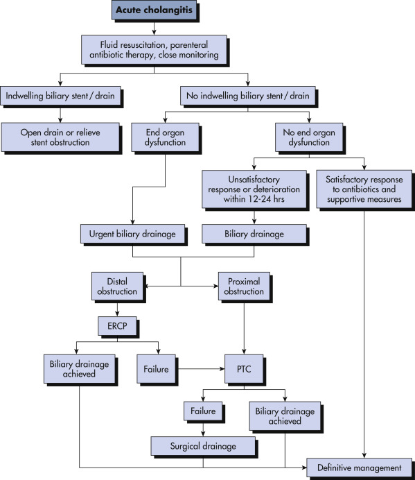
Cholangitis refers to an inflammation and/or infection of the hepatic and common bile ducts associated with obstruction of the common bile duct.

Synonyms

Biliary sepsis

Ascending cholangitis

Suppurative cholangitis

ICD-10CM CODE
K83.0Cholangitis
Epidemiology & Demographics
Incidence (In U.S.)

Complicates approximately 1% of cases of cholelithiasis

Prevalence (In U.S.)

2 cases/1000 hospital admissions

Predominant Sex

  • Females, for cholangitis secondary to gallstones
  • Males, for cholangitis secondary to malignant obstruction and HIV infection
Predominant Age

Seventh decade and older; unusual <50 yr of age

Peak Incidence

Seventh decade

Physical Findings & Clinical Presentation

  • Usually acute onset of fever, abdominal pain (RUQ), and jaundice (Charcot triad)
  • All signs and symptoms in only 50% to 85% of patients
  • Often, dark coloration of the urine resulting from bilirubinuria
  • Complications:
    1. Bacteremia (50%) and septic shock
    2. Hepatic abscess and pancreatitis
Etiology

Obstruction of the common bile duct causing rapid proliferation of bacteria in the biliary tree

  • Most common cause of common bile duct obstruction: Stones, usually migrated from the gallbladder
  • Other causes: Prior biliary tract surgery with secondary stenosis, tumor (usually arising from the pancreas or biliary tree), and parasitic infections from Ascaris lumbricoides or Fasciola hepatica
  • Iatrogenic after contamination of an obstructed biliary tree by endoscopic retrograde cholangiopancreatoscopy (ERCP) or percutaneous transhepatic cholangiography (PTC)
  • Primary sclerosing cholangitis (PSC)
  • HIV-associated sclerosing cholangitis: Associated with infection by cytomegalovirus, Cryptosporidium, Microsporidia, and Mycobacterium avium complex

Diagnosis

Differential Diagnosis

  • Biliary colic
  • Acute cholecystitis
  • Liver abscess
  • Peptic ulcer disease (PUD)
  • Pancreatitis
  • Intestinal obstruction
  • Right kidney stone
  • Hepatitis
  • Pyelonephritis
Workup

  • Blood cultures
  • CBC
  • Liver function tests
  • Procalcitonin
Laboratory Tests

  • Usually, elevated white blood cell count with a predominance of polymorphonuclear forms
  • Elevated alkaline phosphatase and bilirubin in chronic obstruction
  • Elevated transaminases in acute obstruction
  • Positive blood cultures in 50% of cases, typically with enteric gram-negative aerobes (e.g., Escherichia coli, Klebsiella pneumoniae), enterococci, or anaerobes
Imaging Studies

  • Ultrasound:
    1. Allows visualization of the gallbladder and bile ducts to differentiate extrahepatic obstruction from intrahepatic cholestasis
    2. Insensitive but specific for visualization of common duct stones
  • Computed tomography (CT) scan:
    1. Less accurate for gallstones
    2. More sensitive than ultrasound for visualization of the distal part of the common bile duct
    3. Also allows better definition of neoplasm
  • ERCP:
    1. Confirms obstruction and its level
    2. Allows collection of specimens for culture and cytology
    3. Indicated for diagnosis if ultrasound and CT scan are inconclusive
    4. May be indicated in therapy (see “Treatment”)

Treatment

Nonpharmacologic Therapy

Biliary decompression:

  • May be urgent in severely ill patients or those unresponsive to medical therapy within 12 to 24 h
  • May also be performed semielectively in patients who respond
  • Options:
    1. ERCP with or without sphincterotomy or placement of a draining stent
    2. Percutaneous transhepatic biliary drainage for the acutely ill patient who is a poor surgical candidate
    3. Recently, endoscopic ultrasound-guided biliary drainage has been proven as an alternative to percutaneous transhepatic biliary drainage in specialized centers when ERCP fails or is not available
    4. Surgical exploration of the common bile duct
Acute General Rx

  • Nothing by mouth
  • Intravenous hydration
  • Broad-spectrum antibiotics directed at gram-negative enteric organisms, anaerobes, and enterococcus such as carbapenems (meropenem: 1 g q8h or imipenem: 500 mg intravenous [IV] q6h if life threatening), piperacillin/tazobactam: 3.375 or 4.5 g IV q6h, or ampicillin-sulbactam, or ticarcillin-clavulanate; if infection is nosocomial, post-ERCP, or the patient is in shock, broaden antibiotic coverage
  • Fig. 1 illustrates a clinical algorithm for management of acute cholangitis
Figure 1 Management Algorithm for Patients with Acute Cholangitis

The Priority is Prompt Biliary Drainage, and Some Patients May Require Surgical Drainage Without Attempted Eus Biliary Decompression. ERCP, Endoscopic Retrograde Cholangiopancreatography; Eus, Endoscopic Ultrasound; Ptc, Percutaneous Transhepatic Cholangiography.

!!flowchart!!

From Cameron JL, Cameron AM: Surgical therapy, ed 12, Philadelphia, 2017, Elsevier.

Chronic Rx

Repeated decompression may be necessary, particularly when obstruction is related to neoplasm.

Disposition

  • Excellent prognosis if obstruction is amenable to definitive surgical therapy; otherwise relapses are common.
Referral

  • To biliary endoscopist if obstruction is from stones or a stent needs to be placed
  • To interventional radiologist if external drainage is necessary
  • To a general surgeon in all other cases
  • To an infectious disease specialist if blood cultures are positive or the patient is in shock or otherwise severely ill

Pearls & Considerations