AUTHORS: James P. Reichart, MD and Nelson Kopyt, DO
Pyelonephritis is an ascending infection of a bacterial pathogen that infects the renal pelvis and kidney. It primarily presents as a urinary tract infection (UTI) characterized by painful urination (dysuria) with associated flank pain/tenderness, nausea, vomiting, and/or fever. Older adults may also present with failure-to-thrive, unexplained anorexia, other organ system decompensation, or generalized deterioration.
|
Pyelonephritis is common in the U.S. with overall rates of 15 to 17 cases/10,000 women and 3 to 4 cases/10,000 men. Pyelonephritis accounts for 9.1% to 31% of severe sepsis cases annually, depending on geographic area. Average annual mortality is 16.1%, ranging from 5% for patients <25 yr to 43% for ages >64 yr.
Women are 5 times more likely to be hospitalized than men until the age of 65 yr. In men beyond the age of 65 yr, the difference in prevalence narrows. Risk factors associated with pyelonephritis in healthy women are sexual intercourse (≥3 times wk over the previous 30 days), a new sex partner in the past year, use of spermicide, UTI during the past 12 mo, mother with a history of UTI, diabetes mellitus, and urinary incontinence. Urinary tract obstruction is the most important risk factor for a complicated UTI.
Congenital urologic structural disorders associated with vesicoureteral reflux predispose individuals to infections at an early age (<5 yr) and produce renal scarring in most male and some female patients. Pyelonephritis may produce an Ask-Upmark kidney (segmental renal hypoplasia) that is found more often in young female patients with severe hypertension.
Diagnosis established by clinical presentation, history, and physical examination.
Pyelonephritis is suspected in cases of lower urinary tract symptoms (e.g., urinary frequency, urgency, and dysuria) and frequently accompanied by any of the following:
Physical examination may elicit costovertebral angle tenderness with flank pain, a nearly universal finding. Its absence suggests an alternative diagnosis. Patients presenting with nephrolithiasis/ureterolithiasis usually do not present with costovertebral angle tenderness. Usually, abdominal or suprapubic tenderness is present.
Ascending infections from intestinal bacteria that colonize the perineum and vulvae in women account for most infections. Less commonly, bacteria, viruses, or fungal pathogens may produce hematogenously induced pyelonephritis.
In older adults, E. coli is less common (60%). Patients with diabetes develop infections from Klebsiella species, Enterobacteriaceae, Clostridioides species, or Candida species.
During the past decade, community-acquired bacteria (particularly E. coli) that produce extended-spectrum beta-lactamases have emerged as a cause of acute pyelonephritis worldwide. The most common risk factors for these uropathogens include frequent visits to health care centers, recent use of antimicrobials (e.g., cephalosporins and fluoroquinolones), older age, immunosuppression, recurrent pyelonephritis, nephrolithiasis, and comorbid conditions such as diabetes mellitus and recurrent UTIs.
Differential diagnosis includes the following:
Obtained from and conducted on a clean-catch voided or catheterized specimen, if unable to void or cooperate. Dipstick and microscopic examination must be performed on a fresh specimen for preservation of formed elements (e.g., cells, casts, and microorganisms). Most cases demonstrate pyuria and positive leukocyte esterase in association with a positive blood reaction and microhematuria. Leukocyte casts are generally of kidney origin but may be absent.
Historically, clean, midstream cultures are obtained from all patients suspected of having acute pyelonephritis to guide antibiotic therapy. However, a clean-catch specimen may not be necessary. Recent evidence demonstrates no significant difference in the number of contaminated or unreliable culture results when urine is collected with or without preparatory cleansing. Obtain a catheterized urine sample if the patient is unable to void, uncooperative, or has an altered mental status. There is no difference in colony counts or organisms between catheterized and midstream voiding samples.
More than 95% of acute pyelonephritis cases exhibit >105 colony forming units of a single bacterium per milliliter of urine. However, it is important to obtain an accurate history regarding the timing of culture acquisition and prior antibiotic administration. A negative culture with classic clinical and radiologic findings does not rule out acute pyelonephritis, as shown by a prospective study, in which only 23.5% of 196 patients with clinical and radiologic evidence of acute pyelonephritis had demonstrated positive urine cultures. A urine Gram stain may aid in the choice of empiric antimicrobial therapy pending urine culture. If gram-positive cocci are seen, consider Enterococcus species or S. saprophyticus as causative.
Posttreatment urinalysis and culture are unnecessary if symptomatic improvement occurs, but these studies should be obtained when symptomatic improvement does not occur after 2 or 3 days of antibiotic treatment, or if symptoms recur within 2 wk of treatment. Urinary tract imaging is recommended in such cases.
Cultures are obtained from hospitalized patients but may not be routinely required in uncomplicated cases. Approximately 15% to 30% of patients with acute pyelonephritis are bacteremic. Older adults and individuals with complicated acute pyelonephritis are more likely to develop bacteremia and sepsis.
Urine cultures yield a causative organism in nearly all cases of acute pyelonephritis. Therefore, a positive blood culture may be diagnostically redundant. However, in unclear cases, or when an alternative diagnosis to acute pyelonephritis is considered, blood cultures should be obtained.
The primary imaging modalities used in patients with pyelonephritis are computed tomography (CT), MRI, and ultrasound. Most uncomplicated cases of acute pyelonephritis do not require imaging studies unless symptoms do not improve, recurrence occurs, or if the patient has prolonged fever (>72 h) or persistent bacteremia. Abdominal radiographs (i.e., kidney, ureter, and bladder x-ray [KUB]) are of limited utility in acute pyelonephritis, unless staghorn calculi are present. Retrograde or antegrade pyelography may be helpful in severe obstruction that is not evident after noninvasive evaluations. Voiding cystourethrography demonstrates vesicoureteral reflux and generally is conducted routinely only in children.
Recommendations for radiologic tests:
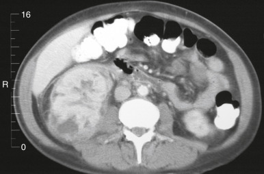
Figure 1 Acute pyelonephritis: Contrast material-enhanced computed tomography (CT) scan.
The heterogeneous CT nephrogram shows the diffuse involvement of the right kidney. Stranding and some fluid are visible in the perinephric space with thickening of Gerota fascia.
From Skorecki K et al: Brenner & Rectors the kidney, ed 10, Philadelphia, 2016, Elsevier.
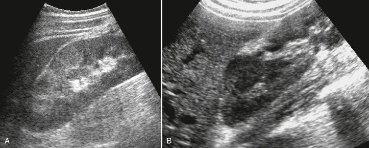
Figure 2 Acute pyelonephritis.
A, Subtle focal increased echogenic areas are seen in the anterior cortex of the right kidney. B, Single focal hypoechoic area is seen in the upper pole of the kidney in another patient.
From Rumack CM et al: Diagnostic ultrasound, ed 4, Philadelphia, 2011, Elsevier.
Although the risk of contrast-induced nephropathy has declined substantially, exert caution during contrast administration in patients with chronic kidney disease or for those taking metformin. When evaluating kidney function, diagnostic decision-making must include estimated glomerular filtration rate trends, not serum creatinine levels, especially in older adults with reduced muscle mass. Patients with acute pyelonephritis and acutely elevated baseline serum creatinine concentrations may warrant CT imaging to rule out obstruction. If the risk of radiocontrast media administration outweighs its benefits, consider MRI or retrograde or antegrade pyelography.
The purpose of imaging is to identify underlying structural abnormalities such as occult obstruction from a stone or abscess and serious complications such as emphysematous pyelonephritis (EPN). In a prospective study of 213 patients with acute pyelonephritis, there were no differences in frequency of fever, leukocytosis, C-reactive protein, pyuria, urine cultures, and duration of symptoms before hospitalization for positive or negative CT. Accordingly, systematic CT or MRI is not required to exclude an anatomic abnormality. Such abnormalities cannot be predicted based on clinical, biochemical, or culture parameters.
EPN is a necrotizing infection that produces intraparenchymal kidney gas visualized by renal imaging. This disorder is associated with high mortality. Risk factors include diabetes mellitus and/or urinary tract obstruction. Gas-forming bacteria, most commonly E. coli, produce gas typically restricted within the Gerota fascia. Studies suggest an overall EPN mortality rate of 19%, reporting significant treatment success rates with percutaneous drainage and antibiotics (66%) and with nephrectomy (90%). EPN must be differentiated from a renal abscess, which can also be associated with a gas collection. With drainage and antibiotic treatment, a renal abscess has a favorable prognosis.
A basic metabolic profile and CBC with differential count are required to estimate kidney function in patients with suspected acute pyelonephritis. If the diagnosis is in doubt, other laboratory tests may be appropriate to clarify the differential diagnosis (e.g., lipase, transaminase, and β-hCG levels).
Close outpatient follow-up is possible for patients with minimal GI symptoms and the ability to maintain fluid intake and oral medications. Prompt antibiotic therapy prevents progression of infection and must be initiated following acquisition of appropriate cultures. Begin empiric therapy based on risk of adverse effects, local community bacterial profiles, and resistance rates. Antibiotics are revised after urine culture results are available.
The concept of requiring long-term treatment for acute pyelonephritis has been questioned. Women with acute pyelonephritis were randomized to PO treatment with ciprofloxacin 500 mg bid for 7 days or 14 days, and 27% of these patients experienced bacteremia from E. coli. No differences in the cure rates were found (87% and 96%, respectively).
Because of the high prevalence of resistance to PO beta-lactam antibiotics and TMP-SMX, these agents usually are reserved for cases in which susceptibility results are known. Additional factors (e.g., allergy history, potential drug-drug interactions, drug availability) may require empiric treatment with these agents before susceptibility testing results are known. In this circumstance, a long-acting, broad-spectrum parenteral drug (e.g., ceftriaxone 1 g or gentamicin 5 mg/kg) may be administered as a one-time dose or longer, until sensitivities of the organism are known. If the local prevalence of fluoroquinolone resistance to E. coli exceeds 10%, an initial IV dose of ceftriaxone or gentamicin is recommended, followed by an oral fluoroquinolone regimen.
Significant clinical improvement during appropriate empiric antibiotic therapy should occur within 48 to 72 h. If improvement does not occur, a complication of acute pyelonephritis or an alternative diagnosis such as an abscess, EPN, or an obstructing calculus should be considered. Any unexpected change in the clinical picture warrants immediate investigation with a CT scan.
Hospitalization is indicated for the following reasons:
Inpatient care includes supportive care, monitoring of culture results, adjustment of antibiotic regimen, and IV volume repletion as required. IV antibiotics are continued until defervescence occurs and clinical improvement occurs. Next, there is conversion to a PO antibiotic regimen for a total duration of 10 to 14 days.
Cortical abscesses historically required surgical drainage; however, using current antibiotics is commonly sufficient for cure.
Chronic pyelonephritis may lead to formation of struvite stones (magnesium ammonium phosphate stones). Formation requires infection with a urease-producing organism such as Proteus or Klebsiella. Symptoms directly attributable to struvite stones are uncommon. Typically, patients will present with symptoms of a UTI, mild flank pain, or hematuria. The stone may grow rapidly over a period of weeks to months if treatment is inadequate. Medical treatment for struvite stones is often ineffective and only indicated when surgery is not an option. The most common surgical intervention is percutaneous nephrolithotomy. Open surgery, once the gold standard, is now rarely used.
Surgical intervention is generally recommended in patients with newly discovered stones or in patients with a solitary kidney or two equally functioning kidneys. Nephrectomy is a reasonable option in patients with a nonfunctional kidney, particularly when chronic infection is present.
Xanthogranulomatous pyelonephritis (XGP) is a rare variant of chronic pyelonephritis with destruction of renal parenchyma.
Chronic nephrolithiasis, especially in high-risk populations such as patients with diabetes or immunosuppressed patients, can predispose individuals and promote complicated infections leading to XGP and ENP. These are very rare disorders that are difficult to diagnose, and an unrecognized renal tumor can be hidden behind a suspected diagnosis of XGP and ENP.