section name header

Clinical Algorithms

Abdominal Abscess

ICD-10CM #Varies with specific location of abscess

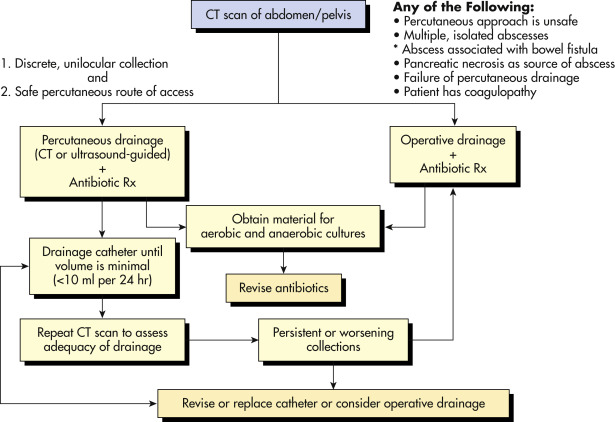
Figure 1 Approach to Management of Intraabdominal Abscesses Including Indications for Consideration of Percutaneous Versus Operative Drainage

!!flowchart!!

CT, Computed tomography; Rx, treatment.

From Parrillo JE, Dellinger RP: Critical care medicine, principles of diagnosis and management in the adult, ed 5, Philadelphia, 2019, Elsevier.
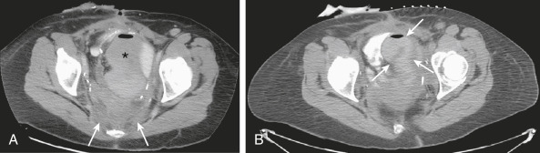
Figure 2 A, Axial Computed Tomography (CT) Image Shows an Apparently Rim-Enhancing Structure Containing Gas (Asterisk) in the Deep Pelvis Adjacent to Tethered Bowel Loops in a Patient with Prior Pelvic Irradiation

The structure could represent an abscess or a dilated loop of small bowel. The presacral inflammation (arrows) is related to radiation change. B, CT image obtained 2 h later shows ingested oral contrast in this structure (arrows) confirming that this is a bowel loop rather than an abscess.

From Feldman M et al: Sleisenger and Fortran’s gastrointestinal and liver disease, ed 10, Philadelphia, 2016, Elsevier.
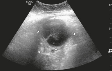
Figure 3 Abdominal Ultrasound of a Typical Abscess (Arrowheads) Demonstrating Central Decreased Echogenicity, Thickened Wall, and Debris Arising Anterior to the Descending Colon (Arrow) in a Patient with Diverticulosis Compatible with a Diverticular Abscess

From Feldman M et al: Sleisenger and Fortran’s gastrointestinal and liver disease, ed 10, Philadelphia, 2016, Elsevier.
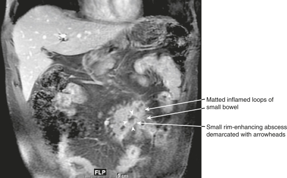
Figure 4 Coronal Magnetic Resonance Image with Gadolinium Contrast of a Patient with Crohn Disease Showing a Small Rim-Enhancing Collection (Arrowheads) Interposed Between Several Loops of Inflamed Bowel (Arrows) Compatible with an Interloop Abscess

Interloop abscesses are not amenable to percutaneous drain placement.

From Feldman M et al: Sleisenger and Fortran’s gastrointestinal and liver disease, ed 10, Philadelphia, 2016, Elsevier.
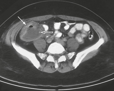
Figure 5 Axial Computed Tomography Demonstrating a Right Lower Quadrant Abscess (Closed Arrow) with an Appendicolith (Open Arrow).

Such extraluminal appendicoliths may predict clinical failure with percutaneous drainage.

From Feldman M et al: Sleisenger and Fortran’s gastrointestinal and liver disease, ed 10, Philadelphia, 2016, Elsevier.

Box 1 Causes of Intraabdominal Abscesses

Abdominal trauma

Appendicitis

Cholecystectomy and other operations or invasive procedures

Crohn disease

Diverticulitis

Neoplastic disease

Pancreatitis

Perforated hollow viscus (e.g., duodenal or gastric ulcer)

From Feldman M et al: Sleisenger and Fortrans gastrointestinal and liver disease, ed 10, Philadelphia, 2016, Elsevier.

Box 2 Clinical Risk Factors for Intraabdominal Abscess

Chronic glucocorticoid use

Increasing age

Malnutrition

Preexisting organ dysfunction

Transfusion

Underlying malignancy

From Feldman M et al: Sleisenger and Fortrans gastrointestinal and liver disease, ed 10, Philadelphia, 2016, Elsevier.

Box 3 Antibiotic Choices in the Treatment of Intraabdominal Infections

  • Single-Agent Therapy
  • Second-Generation Cephalosporins
    • Cefoxitin
  • Carbapenems
    • Imipenem-cilastatin
    • Meropenem
    • Doripenem
    • Ertapenem (no Pseudomonas coverage)
  • Extended-Spectrum Penicillin-β-Lactamase Inhibitor Combinations
  • Glycylcyclines
    • Tigecycline
  • Combination Therapy (Antiaerobea+ Antianaerobe)
  • Third- or Fourth-Generation Cephalosporins
    • Ceftriaxone + metronidazole
    • Cefotaxime + metronidazole
    • Ceftazidime (Pseudomonas coverage) + metronidazole
    • Cefepime (Pseudomonas coverage) + metronidazole
  • Fluoroquinolones
    • Ciprofloxacin + metronidazole
    • Levofloxacin + metronidazole
    • Moxifloxacinb

From Feldman M et al: Sleisenger and Fortrans gastrointestinal and liver disease, ed 10, Philadelphia, 2016, Elsevier.

a Aminoglycoside therapy as an antiaerobic drug should not be used in routine practice owing to an increase in nephrotoxicity and possible worsened patient outcomes with its use.

b Moxifloxacin has adequate anaerobic coverage, making metronidazole unnecessary.